
Choose an answer, then open any option to work its reasoning.
MSK · Metabolic Bone Disease
Metabolic Bone Trio: Osteoporosis vs Osteomalacia vs Paget
Three diseases share one skeleton and zero lab panels. Osteoporosis loses bone with normal chemistry, osteomalacia makes osteoid that never mineralizes, and Paget remodels bone into chaos. Split them by calcium, phosphate, ALP, 25-hydroxyvitamin D, and PTH before the test hands you a pseudofracture and calls it osteoporosis.
What this page makes you able to do
- Split the trio by the five-lab panel and the film: normal labs, low minerals plus low 25-OH-D, or isolated ALP
- Read the DXA T-score at or below -2.5 and put the bisphosphonate ladder in order
- Trace the RANKL-OPG remodeling axis and where denosumab and teriparatide act
- Catch the board traps: osteosarcoma in Paget, atypical femur fracture, bisphosphonate before vitamin D
- Dr. Fatima Ali, DOPsychiatry residentPrimary reviewer
Last reviewed
The pearl
The trio lives on ALP. Isolated high ALP with normal calcium, phosphate, and 25-OH-D is Paget; add a low 25-OH-D with low calcium and phosphate and it flips to osteomalacia; keep every lab normal and you are looking at osteoporosis, a density problem with clean chemistry. The 25-OH-D is the tiebreaker between the two ALP elevations.
Prove it
Opening question
Answer before you read anything, then carry the lab split through every section.
A 66-year-old man comes to the office because his hat no longer fits and he has had dull right hip pain for 6 months. Examination shows an enlarged skull with prominent scalp veins. Serum calcium is 9.4 mg/dL, phosphate 3.6 mg/dL, alkaline phosphatase 412 U/L, 25-hydroxyvitamin D 34 ng/mL, and PTH 28 pg/mL.Which of the following is the most likely diagnosis?
- Why this is rightThe trap is the elevated ALP: two members of the trio carry it. But osteomalacia needs low calcium, low phosphate, and a low 25-OH-D, and every one of those is normal here. A hat that stopped fitting plus an isolated ALP spike with normal minerals is Paget disease until proven otherwise. Rule: isolated high ALP with normal Ca, Phos, and 25-OH-D = Paget; add a low 25-OH-D and it flips to osteomalacia.
- Why this failsOsteoporosis keeps the whole panel normal, and this ALP is 412, more than three times normal. Density loss also does not grow the skull or enlarge hat size. Rule: osteoporosis never quadruples the ALP and never grows the skull.
- Why this failsOsteomalacia does raise ALP, but only with low calcium, low phosphate, and a low 25-OH-D. This patient has all three normal, so the failed-mineralization chemistry is absent. Rule: high ALP with normal minerals is Paget; the low 25-OH-D is what flips it to osteomalacia.
- Why this failsPrimary hyperparathyroidism runs high calcium with high PTH; this PTH is 28 and the calcium is normal. The parathyroid is not the source of the enzyme spike. Rule: high calcium plus high PTH is the parathyroid; normal calcium with a normal PTH leaves the bone itself.
Work the reasoning
ALP comes from osteoblasts working on bone surface. In osteomalacia they build osteoid that never mineralizes; in Paget they lay down mosaic bone after giant osteoclasts chew first. Both spill enzyme; the minerals tell them apart.
The 25-hydroxyvitamin D: normal at 34 here, which kills the osteomalacia read. Paget keeps calcium, phosphate, and vitamin D all normal and raises only ALP.
Enlarging hat size and scalp vein prominence are Paget skull remodeling; deafness follows when the disease narrows the foramina. No other member of the trio grows the skull.
The answer is A: Paget disease of bone. Isolated high ALP with normal calcium, phosphate, and 25-OH-D, plus a skull that grew, is Paget until proven otherwise.
MEET THE TRIO
Meet the Trio: One Skeleton, Three Different Breakages
Osteoporosis runs out of bone, osteomalacia runs out of mineral, and Paget runs out of order. The labs tell you which before the film ever does.
Start with what breaks, because the chemistry follows the pathology. Osteoporosis is a quantity problem: trabeculae thin and perforate, but the bone that remains is normally mineralized, so calcium, phosphate, ALP, vitamin D, and PTH all read normal. The diagnosis lives on the DXA, with a T-score at or below -2.5, or on a fragility fracture. Osteomalacia is a mineralization problem: osteoblasts keep making osteoid, but without vitamin D the mineral never deposits, so calcium and phosphate fall, ALP climbs as osteoblasts work overtime, and 25-OH-D runs low with a reactive PTH rise. In a child with open growth plates the same chemistry is rickets. Paget disease is an organization problem: localized osteoclast-osteoblast uncoupling chews bone and lays down disorganized mosaic bone, so ALP spikes while calcium, phosphate, and vitamin D stay normal.
The trap boards love is the normal-panel osteoporosis: a patient with a fracture and every lab in range is not osteomalacia, and she is not Paget. If ALP or PTH ever starts climbing in a patient labeled osteoporotic, stop assuming: think a second disease (Paget, hyperparathyroidism, myeloma workup) or a fragility fracture that is healing and raising ALP on its own.
Flip between the two diseases boards make you confuse.
THE RANKL AXIS
The RANKL Axis: Why the Trio Is a Remodeling Story
Every packet of bone is resorbed and rebuilt to order. The osteoblast releases RANKL, the osteoclast answers through its RANK receptor, and OPG soaks up the excess signal.
Bone is not static; it is remodeled in packets. The osteoblast (the builder) displays RANKL on its membrane, and RANKL docks on the RANK receptor of the osteoclast (the demolition crew), telling it to resorb a packet of bone. The osteoprotegerin (OPG) decoy floats between them and sweeps RANKL away, so the axis is an accelerator with a brake: RANKL is the gas, OPG is the brake. Denosumab is a monoclonal antibody against RANKL, which is why it is an antiresorptive: it takes the gas away from the osteoclast.
Now read the trio through the axis. Osteoporosis is high RANKL drive with an OPG brake that cannot keep up, so resorption outruns formation and density falls. Paget disease starts in the osteoclast itself: giant, hyperactive multinucleated cells resorb chaotically, and the osteoblasts that follow lay down woven, disorganized mosaic bone that never matures into orderly lamellae. Osteomalacia is different: the remodeling unit is not the failure, the mineral substrate is. There is nothing wrong with the signal; the osteoid simply cannot harden without vitamin D. That is why the treatment ladder differs: antiresorptives brake the axis, but only vitamin D and calcium mineralize osteoid.
Tap each cell of the remodeling unit and read its job.
Tap a cell.
THE LAB DIAGNOSTICIAN
The Lab Diagnostician: Set the Panel, Read the Diagnosis
Five numbers resolve the trio: calcium, phosphate, ALP, 25-OH-D, and PTH. Move the sliders yourself, commit, and the read resolves.
Run the panel in a fixed order. First the ALP: a normal ALP rules out both osteomalacia and Paget in the classic presentation and leaves you with the density story, so the DXA decides. If ALP is high, check calcium and phosphate. Both low with a low 25-OH-D is osteomalacia (or rickets in a child): failed mineralization with secondary hyperparathyroidism defending the calcium. Both normal with a normal 25-OH-D is Paget: the enzyme is coming from remodeling chaos, not mineralization failure. The 25-OH-D is the tiebreaker between the two ALP elevations, and the PTH tells you whether the parathyroid is reacting (secondary, low calcium) or rebelling (primary, high calcium with high PTH).
Two panels outside the trio are worth naming before boards: high calcium with high PTH is primary hyperparathyroidism, and low phosphate with normal calcium and normal 25-OH-D is renal phosphate wasting (X-linked hypophosphatemic rickets in a child, Fanconi syndrome in an adult). The trio never raises calcium, and a broken panel in the trio always carries a high ALP.
Set the five labs and run the panel.
Serum calcium9.6 mg/dL
normal 8.5 to 10.5 mg/dL
Serum phosphate3.4 mg/dL
normal 2.5 to 4.5 mg/dL
Alkaline phosphatase90 U/L
normal 30 to 120 U/L
25-hydroxyvitamin D38 ng/mL
normal 30 to 100 ng/mL
PTH38 pg/mL
normal 10 to 65 pg/mL
THE T-SCORE
The DXA T-Score and the Treatment Ladder
A T-score at or below -2.5, or any fragility fracture, is osteoporosis. The bisphosphonate is first line; the anabolic comes later, not first.
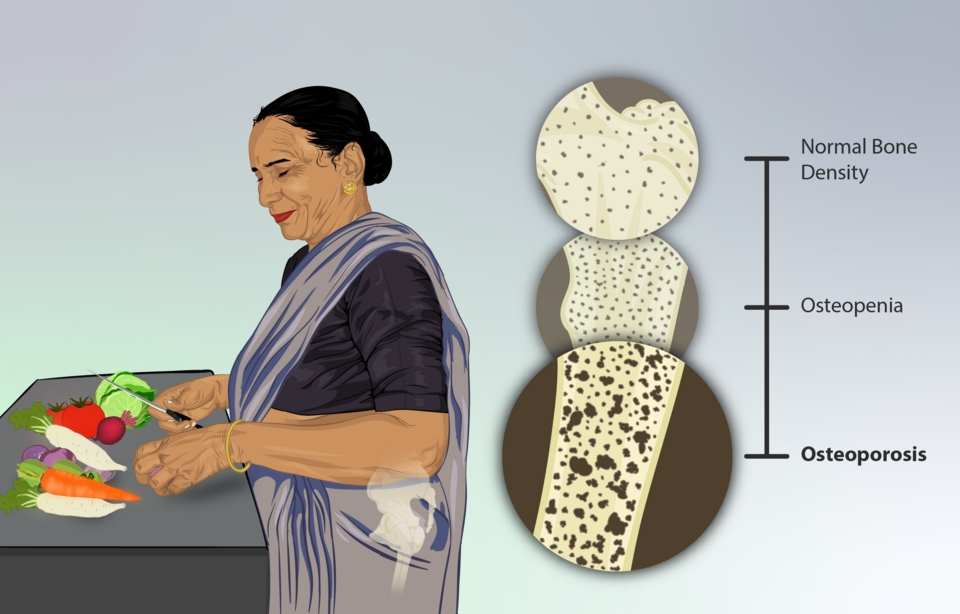
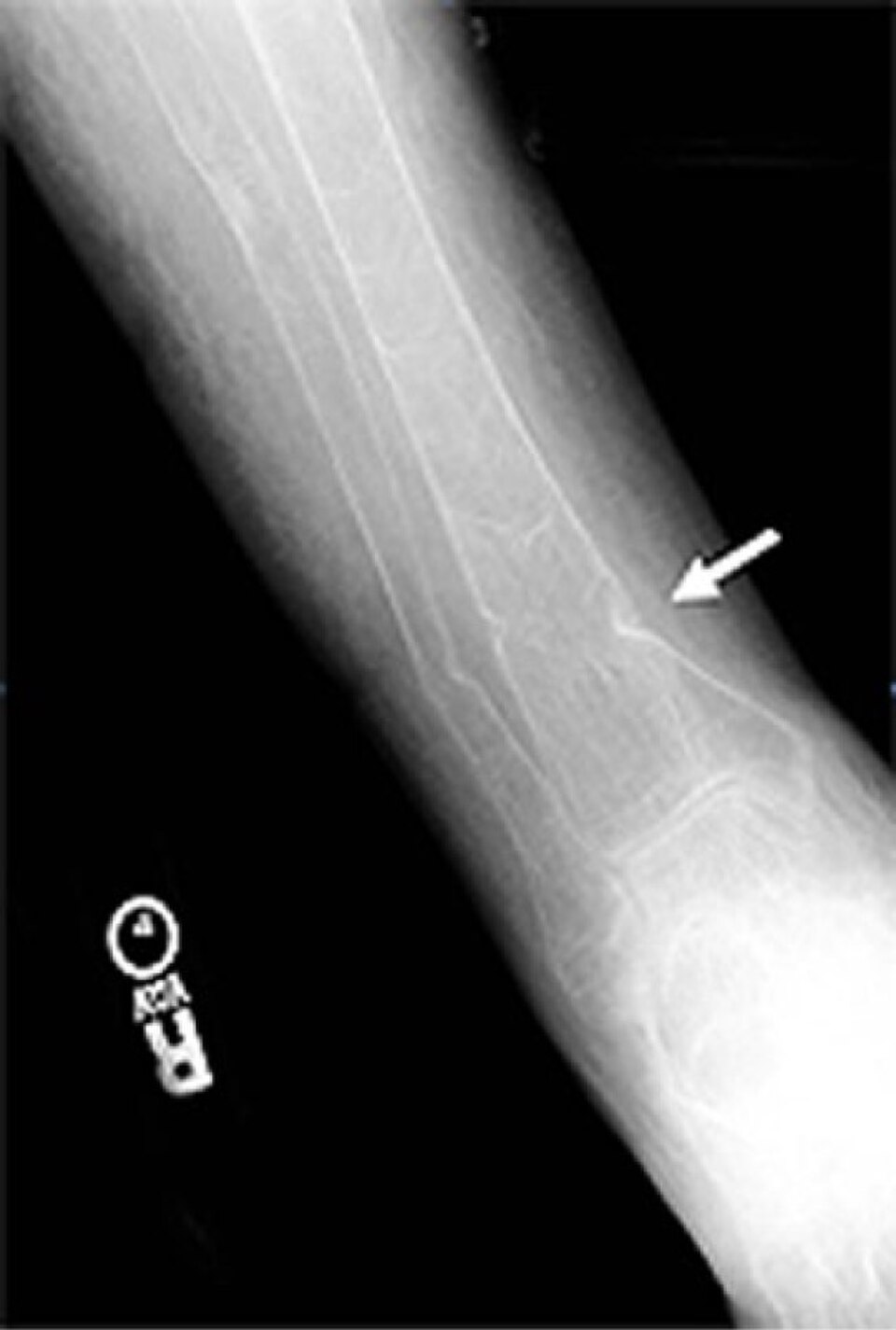
DXA measures mineral density at the lumbar spine, total hip, and femoral neck, and reports the T-score: standard deviations below the young adult mean. T-score at or below -2.5 is osteoporosis; -1.0 to -2.5 is osteopenia; above -1.0 is normal. The other route to the diagnosis is the fragility fracture: a fall from standing height or less that breaks bone that should have survived it, at the hip, vertebra, or distal radius. A fragility fracture plus any low T-score is osteoporosis regardless of the number, which is how a 58-year-old man with a fractured radius and a T-score of -2.6 lands in the treatment arm.
The ladder is a brake before a builder. Bisphosphonates (alendronate, then zoledronic acid) are first line: they inhibit farnesyl pyrophosphate synthase in the osteoclast, driving apoptosis and cutting resorption. Denosumab, the RANKL antibody, is the alternative when a bisphosphonate cannot be used, and it is the same drug class the jaws remember. Teriparatide is anabolic: intermittent recombinant PTH builds bone, but it is reserved for very high fracture risk, severe disease, or failure of antiresorptives. SERMs (raloxifene) are the niche alternative with vertebral-fracture benefit and a venous thromboembolism risk. Every drug rides on calcium and vitamin D repletion, and every woman with a fragility fracture deserves the secondary-cause workup boards grade: think glucocorticoids, hyperparathyroidism, and multiple myeloma when the story is young, atypical, or accompanied by anemia, renal disease, or hypercalcemia.
Read the T-score and pick the first-line drug.
A 66-year-old woman comes to the office 2 weeks after a fall from standing height caused a distal radius fracture. She went through menopause at age 50 and has no prior fractures. Vital signs are normal and examination is unremarkable. Serum calcium is 9.4 mg/dL, phosphate 3.4 mg/dL, alkaline phosphatase 82 U/L, 25-hydroxyvitamin D 36 ng/mL, and PTH 40 pg/mL. DXA of the hip shows a femoral neck T-score of -2.8. Which of the following is the most appropriate first-line treatment?
A. Alendronate. A fragility fracture plus a T-score of -2.8 is osteoporosis with a clean panel, and the bisphosphonate is the standard first-line antiresorptive. B. Teriparatide. Anabolic therapy is the escalation for very high fracture risk or bisphosphonate failure, not the opening move. C. Denosumab. The RANKL antibody is the alternative when bisphosphonates cannot be used, not the default first step. D. Calcium and vitamin D alone. Repletion is the substrate every drug needs, but a T-score of -2.8 with a fragility fracture is treated pharmacologically. Rule: fragility fracture plus T-score at or below -2.5 = osteoporosis, and osteoporosis starts with a bisphosphonate.
THE DISCRIMINATOR
Which of the Trio? The Two-Fork Discriminator
One enzyme decides the first fork, two minerals the second, and the film or the DXA locks the door.
Fork 1 is the ALP. Normal: neither osteomalacia nor Paget in the classic presentation, and the density story takes over on DXA. Elevated: check fork 2, the calcium and phosphate. If both are low with a low 25-OH-D, it is osteomalacia (or rickets in a child): replete vitamin D and calcium first, and never start a bisphosphonate before repletion, because suppressing resorption on an unmineralized skeleton does nothing and can deepen hypocalcemia. If both are normal with a normal 25-OH-D, it is Paget: the film locks it with cotton-wool skull or picture-frame vertebra (never Looser zones, which belong to osteomalacia), the bone scan shows hot uptake at the involved sites, and treatment is a bisphosphonate (zoledronic acid) with ALP as the response meter.
Down the normal-ALP arm, the DXA decides: T-score at or below -2.5 or a fragility fracture is osteoporosis and earns a bisphosphonate; -1.0 to -2.5 is osteopenia, managed with calcium, vitamin D, weight-bearing exercise, and fracture-risk reassessment before any drug.
Take each presentation through the forks and read the door it opens.
Which door does each panel open?
ON FILM
The Trio on Film: Looser Zones, Cotton-Wool, and the Bowed Tibia
The radiograph separates the two ALP diseases the way the labs cannot: pseudofractures belong to osteomalacia, mosaic thickening belongs to Paget.
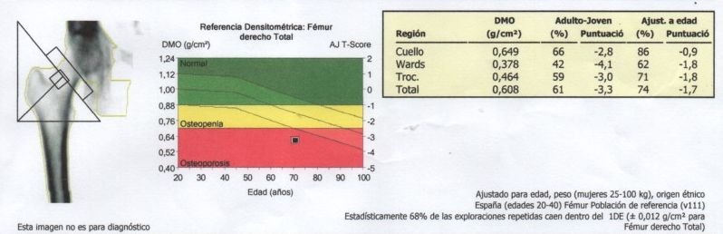
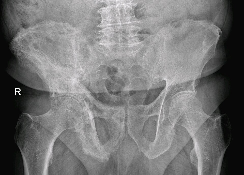
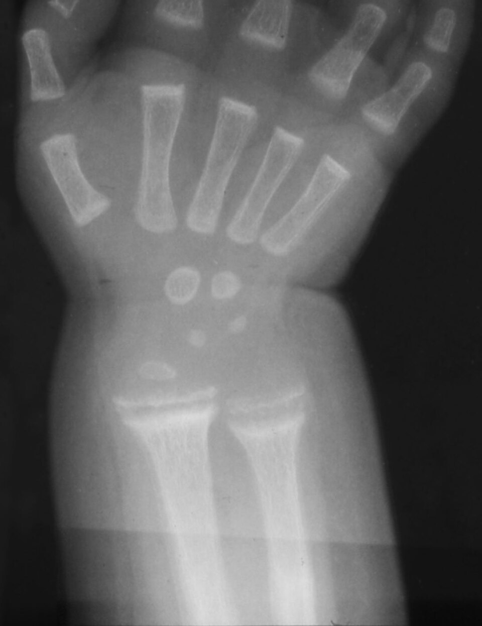
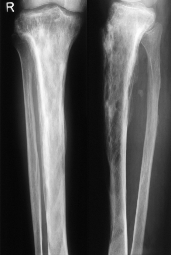
A Looser zone, also called a pseudofracture or Milkman fracture, is a transverse radiolucent band of unmineralized osteoid, classically along the medial femoral cortex, pubic rami, ribs, and scapulae. It is the film signature of osteomalacia, and it is never a Paget finding. The Paget film instead shows cotton-wool skull (the thickened calvarium that stops hats fitting), the picture-frame vertebra (a dense cortex outlining the vertebral body), and bowed, thickened long bones with coarse trabeculae. In rickets, the growth plate fails to mineralize and the metaphysis cups and frays, classically at the distal radius and distal femur.
The read order for a suspected Paget patient is a sequence boards grade: the enzyme first, then the film, then the scan, then the drug. The osteomalacia flip runs the same first step with a different ending: add a low 25-OH-D to the high-ALP panel and the repletion comes before any bisphosphonate, always.
Tap each letter of the panel lock.
LOWD PA
LLow calcium
OOsteoid without mineral: low phosphate, the second LOW
WWorking osteoblasts spill high ALP
DLow 25-OH-D, the starved fuel
PPaget keeps Phosphate (and calcium) normal
AALP high alone, everything else normal; tell by the film
Put the Paget read in order.
THE TRAPS
The Board Trap Cast
Three complications boards test over and over: the sarcoma hiding in a pagetic bone, the bisphosphonate given before repletion, and the femur that cracks under years of alendronate.
Long-standing Paget disease carries a small but board-favorite risk: sarcomatous transformation, most often osteosarcoma, in roughly 1% of patients after years of disease. The alarm is a change in character: new severe pain in a previously stable pagetic bone, a palpable enlarging mass, and night pain. That script is osteosarcoma until proven otherwise, and the next step is imaging plus biopsy, not another bisphosphonate course. The second trap is treatment order in osteomalacia: a bisphosphonate before vitamin D repletion suppresses resorption of bone that was never mineralized and can worsen hypocalcemia, so repletion always comes first. The third trap is the atypical femur fracture of long-term bisphosphonate therapy: lateral thigh pain after years of alendronate with a thickened lateral subtrochanteric cortex and a transverse lucent line means stop the drug, involve orthopedics, and consider prophylactic fixation before the incomplete crack completes.
Open each trap and hold the management.
Normal calcium, phosphate, ALP, vitamin D, and PTH with a T-score at or below -2.5, or a fragility fracture. Bisphosphonate first line with calcium and vitamin D repletion; denosumab, SERMs, and teriparatide are later-line or special-situation options. The trap: normal labs make it look like nothing is wrong.
Low calcium, low phosphate, high ALP, low 25-OH-D, reactive high PTH, with Looser zones on film (metaphyseal cupping in a child). Replete vitamin D and calcium first and fix the cause (diet, sun, malabsorption, renal loss). Never start a bisphosphonate before repletion.
Isolated high ALP with normal calcium and phosphate, mosaic bone, cotton-wool skull, picture-frame vertebra, bowed long bones, and skull-base involvement that can compress cranial nerves. Treat with zoledronic acid and monitor ALP; extensive disease can drive high-output heart failure.
Severe new pain, a palpable enlarging mass, or night pain in a bone with long-standing Paget is osteosarcoma until proven otherwise. Radiograph plus MRI, then biopsy. Do not treat a possible sarcoma as a Paget flare.
A patient labeled osteoporotic should keep a clean panel. A climbing ALP or PTH means a second disease (Paget, hyperparathyroidism, myeloma workup) or a healing fragility fracture raising ALP on its own. Re-read the labs instead of adding another drug.
Lateral thigh pain after 3 to 5 years of bisphosphonate therapy with a thickened lateral subtrochanteric cortex and a transverse lucent line: stop the bisphosphonate (drug holiday), weight-bearing precautions, and orthopedic consultation for possible prophylactic fixation. Reassess the need for continued therapy at the 3-to-5-year checkpoint.
Prove it
Walkthrough: split the trio under time pressure
Original practice scenarios, one at a time. Choose an answer, then open any option to work its reasoning.
Clinical walkthrough
