
Choose an answer, then open any option to work its reasoning.
MSK · Genetics and Metabolism
Hereditary Bone and Cartilage Disorders: OI, Osteopetrosis, Achondroplasia, MPS, Rickets
Every syndrome in this family is one gene, one skeletal signature, and one complication to screen. Osteogenesis imperfecta is a collagen defect with blue sclera and wormian bones, and its twin trap is child abuse. Osteopetrosis turns bone to marble and squeezes the nerves and marrow. Achondroplasia is a growth receptor stuck on, shortening the proximal segments and narrowing the foramen magnum. Learn the inheritance pattern, the gene, the signature, and the screen, because the board asks all four every time.
What this page makes you able to do
- Name the gene, inheritance pattern, skeletal signature, and screen for the hereditary bone family
- Discriminate osteogenesis imperfecta from nonaccidental trauma with the stigmata abuse cannot fake
- Screen the two narrowings in achondroplasia and the crowded structures in osteopetrosis
- Read the rickets lab pattern and the corneal clouding split
- Dr. Fatima Ali, DOPsychiatry residentPrimary reviewer
Last reviewed
The pearl
One gene, one signature, one screen. Blue sclera and wormian bones route fractures to OI; marble bone with cytopenias is osteopetrosis; rhizomelia with frontal bossing is achondroplasia, and its first screen is the foramen magnum; clouded cornea is Hurler, clear cornea in a boy with maternal uncles affected is Hunter; low phosphate with normal calcium is the renal phosphate leak, treated with phosphate plus calcitriol.
Prove it
Opening question
Answer before you read anything, then keep the gene, the signature, and the screen in mind through every section.
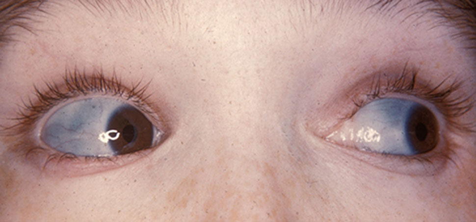
A 15-month-old girl is brought to the office because of a femur fracture after rolling off a low couch. Her mother reports two prior fractures with minor falls, and the family history includes a maternal aunt with early hearing loss. Examination shows blue-gray sclera and discolored, translucent teeth; the retinal examination is normal.Which of the following is the most appropriate next step?
- Why this is rightThree fractures from trivial forces, blue-gray sclera (thin collagen shows the choroid), translucent teeth (dentinogenesis imperfecta), and a family history of early hearing loss (otosclerosis) are the OI stigmata, and abuse cannot fake any of them. The fracture mechanism is out of proportion to the injury with a normal retinal exam, so the fractures route to osteogenesis imperfecta, and the treatment conversation includes bisphosphonates to reduce the fracture burden. Rule: blue sclera and translucent teeth route the fractures to OI; retinal hemorrhage and a changing story route them to abuse.
- Why this failsAbuse is the diagnosis when the OI checklist is empty: retinal hemorrhages, fractures at different healing stages, and a history that changes between tellings. This child carries blue sclera, translucent teeth, and a positive family history, and the retinal exam is normal, so the abuse workup would be aimed at the wrong disease. Rule: abuse cannot fake blue sclera, wormian bones, or dentinogenesis.
- Why this failsVitamin D and calcium support bone health, but they cannot repair a defective type I collagen scaffold, and a healing fracture with normal bone density is not the vitamin D deficiency pattern. The diagnosis is genetic, so the treatment conversation is genetic. Rule: vitamin D alone cannot rebuild a collagen defect.
- Why this failsTeriparatide is not the pediatric first-line agent for OI; bisphosphonates are the mainstay of medical treatment in children. A DXA scan adds nothing to the diagnosis, which is already written on the sclera and the teeth. Rule: in children with OI, the bisphosphonate conversation comes first.
- Why this failsThree fractures in 14 months is not reassurance material: this child has a diagnosable, treatable collagen disease, and the treatment discussion changes her fracture trajectory. Rule: recurrent fractures with OI stigmata are an active diagnosis, not a watch-and-wait.
Work the reasoning
The OI stigmata abuse cannot fake: blue-gray sclera, wormian bones on the skull film, dentinogenesis imperfecta, and a family history of early hearing loss. Abuse announces itself with retinal hemorrhages and fractures at different healing stages; neither is present.
COL1A1 and COL1A2 make type I collagen, the scaffold of bone. A defective scaffold fractures from forces a normal bone shrugs off, and the same collagen defect thins the sclera, disrupts the teeth, and later drives otosclerosis.
Bisphosphonate therapy reduces the fracture burden in OI. Vitamin D and calcium support the skeleton but cannot fix the scaffold, and teriparatide is not the pediatric first-line.
The answer is A: evaluate for osteogenesis imperfecta and refer for a bisphosphonate therapy discussion. Blue sclera and translucent teeth route the fractures to OI; retinal hemorrhage and a changing story route them to abuse.
THE FALLING CHILD
The Falling Child: Osteogenesis Imperfecta and the Abuse Trap
One gene pair makes the scaffold of bone, and when it fails the child fractures from a couch roll. Learn the stigmata abuse cannot fake, then the Sillence ladder that grades the severity.
Osteogenesis imperfecta is a type I collagen defect from COL1A1 and COL1A2 mutations, autosomal dominant in the classic forms with autosomal recessive forms in the severe types. Collagen is the bone scaffold, so the child fractures from minor trauma, the sclera thins and shows the blue choroid through it (blue-gray sclera), the teeth develop dentinogenesis imperfecta (gray-brown, translucent, easily worn), and the skull shows wormian bones on the film. Hearing loss from otosclerosis joins the picture in adolescence or young adulthood, which is why the family history question asks about early hearing aids.
The board pairs OI with abuse on purpose, and the discriminator is a checklist. Blue sclera, wormian bones, dentinogenesis imperfecta, and a positive family history belong to OI, and abuse cannot fake any of them. Retinal hemorrhages, fractures at different healing stages, posterior rib fractures, and a history that changes between tellings belong to abuse. Fractures at different healing stages occur in both, which is why the stem hands you the eyes and the family history. The OI fracture mechanism is out of proportion to the injury with a normal retinal examination, and that package routes the workup to the collagen, not to child protective services.
Flip between the two fracture children.
MARBLE BONE
Marble Bone: Osteopetrosis and the Osteoclast That Cannot Resorb
Dense bone is a resorption failure: the osteoclast cannot pump acid into the pit, so the skeleton keeps building and never remodels. The bone gets denser, the nerves and marrow get squeezed, and the film shows marble.
Osteopetrosis is a failure of bone resorption. The osteoclast attaches to bone but cannot acidify the resorption lacuna, so mineralized bone is deposited and never removed. The two defects the boards name are CLCN7, the chloride channel that keeps the acid compartment working, and carbonic anhydrase II, the enzyme that makes the hydrogen ion. The result is dense, brittle marble bone with the bone-within-bone appearance and the Erlenmeyer flask deformity of the metaphyses, where remodeling failure widens the bone ends.
The complications are a checklist that follows the crowding. Dense bone compresses the cranial nerves: blindness, facial palsy, and deafness. Dense bone crowds the marrow: anemia, thrombocytopenia, and leukopenia. And marrow failure drives hepatosplenomegaly from extramedullary hematopoiesis. The malignant infantile form is autosomal recessive and lethal without treatment; the adult form is autosomal dominant and mild, often an incidental finding on a film. The carbonic anhydrase II deficiency variant pairs marble bone with renal tubular acidosis, because the same enzyme makes hydrogen ions for the osteoclast pit and the renal tubule.
Name the form behind the infant with marble bone.
A 4-month-old boy is brought in because of poor feeding and progressive pallor. Examination shows frontal bossing, a distended abdomen with hepatosplenomegaly, and failure to track objects. Laboratory studies show anemia, thrombocytopenia, and leukopenia. A radiograph shows diffusely dense bones with a bone-within-bone appearance. Which of the following best describes this disorder?
A. Malignant infantile osteopetrosis. Dense bones plus pancytopenia plus hepatosplenomegaly plus cranial nerve findings in an infant is the autosomal recessive form, lethal without treatment; hematopoietic stem cell transplantation is the treatment conversation. B. Adult form. Autosomal dominant and mild, usually found incidentally on film, without the infant marrow-failure package. C. OI type II. OI is a collagen defect with blue sclera and fractures, not dense bones with cytopenias. D. Hypophosphatemic rickets. Rickets softens bone and bows the legs; it does not make bone denser. Rule: infant plus marble bone plus marrow failure is the malignant recessive form.
SHORT LIMBS
Short Limbs: Achondroplasia and the Growth Receptor Stuck On
FGFR3 is a brake on growth, and achondroplasia jams the brake on. The proximal segments suffer most, and the first screen is the foramen magnum, not the limb length.
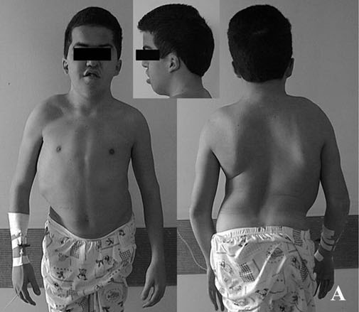
Achondroplasia is a gain-of-function FGFR3 mutation, autosomal dominant, and the most common form of short-limbed dwarfism. The constitutively active receptor inhibits chondrocyte proliferation, so endochondral ossification fails at the growth plates and the limbs shorten with rhizomelic predominance: the proximal segments, humerus and femur, suffer most because they depend on the growth plates that drive most of the bone length. The signature is macrocephaly with frontal bossing, a trident hand (the fingers spread with a gap between the third and fourth digits), lumbar lordosis, and normal intelligence.
The genetics are the board trap. More than 85 percent of cases are new mutations with a paternal age effect, so two average-statured parents commonly have an affected child, and germline mosaicism explains the rare family with more than one affected child. The complications to screen are the two narrowings: foramen magnum stenosis in infancy, with sleep apnea, hypotonia, and arching with stiffening from cord compression, and spinal stenosis in adulthood. Snoring plus arching plus long tract signs is the foramen magnum until imaging says otherwise.
Tap the segment that rhizomelia shortens, then the screen.
Tap a region.
THE SEVERITY LADDER
The Sillence Ladder and the Osteopetrosis Complications
Severity in this family is a ladder and a checklist. The Sillence types grade OI; the crowded-structure checklist grades osteopetrosis. Hold both orders and the two treatment conversations.
The Sillence types grade OI severity, and the boards ask the type number with the phenotype. Type I is mild: blue sclera, normal or near-normal stature, fractures from minor trauma. Type II is lethal perinatal: multiple in-utero fractures, severe limb deformities, and respiratory failure in the first days of life. Type III is progressively deforming, with fractures at birth and worsening deformity through childhood. Type IV is moderate, with white or light sclera and variable fracture burden. Across the types, bisphosphonate therapy reduces the fracture burden; vitamin D alone cannot fix the collagen scaffold, and teriparatide is not the pediatric first-line.
The osteopetrosis complications run a fixed order. Marble bone crowds the skull base first, compressing cranial nerves (blindness, facial palsy, deafness); then the marrow fails (anemia, thrombocytopenia, leukopenia); then hepatosplenomegaly appears from extramedullary hematopoiesis. The infantile form is autosomal recessive and lethal, the adult form autosomal dominant and mild, and the carbonic anhydrase II variant pairs marble bone with renal tubular acidosis.
Put the crowded-structure checklist in order.
Grade the newborn with fractures at birth.
A newborn girl is delivered at 37 weeks with multiple fractures present at birth, dark blue sclera, and severe limb deformities; she dies of respiratory failure in the first days of life. Which of the following best describes this presentation?
A. Sillence type II. Fractures at birth with lethal respiratory failure is the perinatal lethal form; in-utero fractures with severe deformity define it. B. Type I. Mild, with blue sclera, normal or near-normal stature, and survival. C. Type III. Progressively deforming but not lethal in the first days; the child survives with worsening deformity. D. Type IV. Moderate with white or light sclera and a variable fracture burden. Rule: lethal at birth with in-utero fractures is type II.
LYSOSOMES AND PHOSPHATE
Lysosomes and Phosphate: Mucopolysaccharidoses and the Rickets Variants
Two metabolic families, two questions: does the cornea cloud, and what does the phosphate do? The enzyme names the MPS; the lab pattern names the rickets.
The mucopolysaccharidoses are lysosomal enzyme defects that pile up glycosaminoglycans, producing coarse facies, hepatosplenomegaly, skeletal dysplasia, and developmental regression. The corneal clouding question splits the pair the boards use. Hurler (MPS I) is alpha-L-iduronidase deficiency, autosomal recessive, with corneal clouding. Hunter (MPS II) is iduronate sulfatase deficiency, X-linked, with clear corneas and the maternal-uncle family pattern. Treatment is enzyme replacement therapy plus, for eligible patients, hematopoietic stem cell transplantation, with the best results when started early.
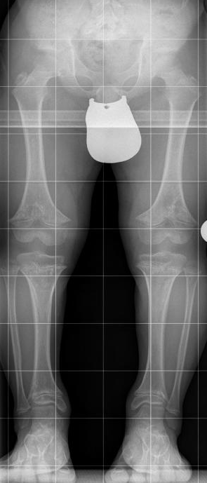
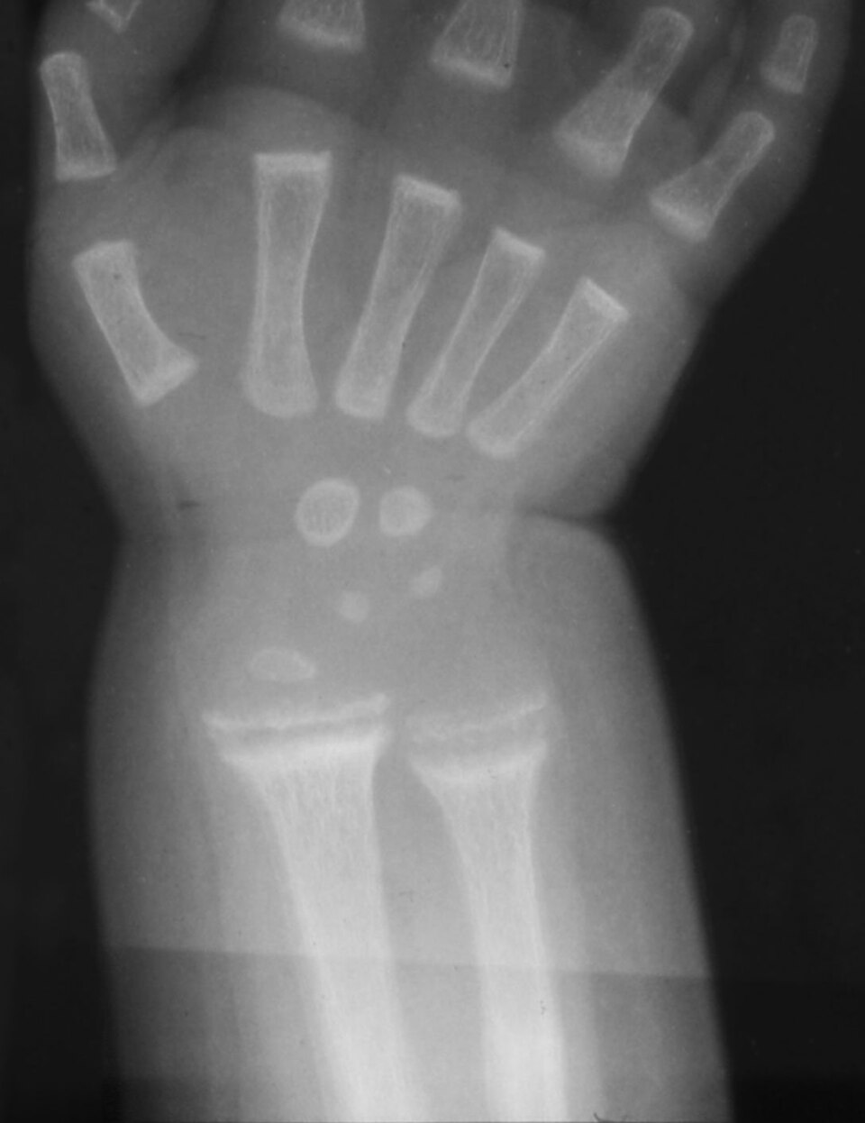
The rickets variants are mineralization failures, and the lab pattern names each one. Vitamin D dependent type I is 1-alpha-hydroxylase deficiency, autosomal recessive, with low calcium, low phosphate, and low calcitriol. Vitamin D dependent type II is a vitamin D receptor mutation with alopecia and a high calcitriol level: the receptor cannot respond, so the hormone accumulates. The board favorite is X-linked dominant hypophosphatemic rickets: a PHEX mutation raises FGF23, the kidney wastes phosphate, and the triad reads low phosphate, normal calcium, high alkaline phosphatase. Plain vitamin D cannot overcome the renal leak; the treatment is phosphate plus calcitriol.
Tick the labs you see, and read the diagnosis off the pattern.
Checked 0
THE ROUTER
The Broken or Short Child Router
The hereditary bone family announces itself with one of five headlines. The headline picks the gene family; the findings pick the diagnosis; the diagnosis picks the screen.
Run the headlines in order. Recurrent fractures from minor trauma is OI until the stigmata checklist is empty; blue sclera, wormian bones, dentinogenesis, and family history route it to the collagen, while retinal hemorrhages and a changing story route it to abuse, with a skeletal survey and a retinal exam either way. Diffusely dense bones is osteopetrosis: the infant with marrow failure gets the HSCT evaluation, the adult with incidental marble bone gets reassurance and monitoring, and marble bone plus a urine that will not acidify is carbonic anhydrase II deficiency. Short stature with rhizomelic limbs is achondroplasia until proven otherwise, and the screen is the foramen magnum in infancy and the spinal canal later. Coarse facies with hepatosplenomegaly is a mucopolysaccharidosis: clouded cornea is Hurler, clear cornea in a boy with maternal uncles affected is Hunter, and the enzyme assay names the subtype. Bowed legs with a low serum phosphate is the renal phosphate leak, and the therapy is phosphate plus calcitriol; add low calcium and low calcitriol for the 1-alpha-hydroxylase defect, and alopecia with a high calcitriol level for the receptor defect.
Route the child by headline, then commit to the screen.
Which headline is this child?
THE MUTATION MAP
The Mutation Map: Genes, Signatures, and the Tumor Syndromes
The family closes with cartilage and fibrous tissue run loose. Every gene pairs with a signature and a screen, and the tumor syndromes add the one risk to name: malignant transformation.
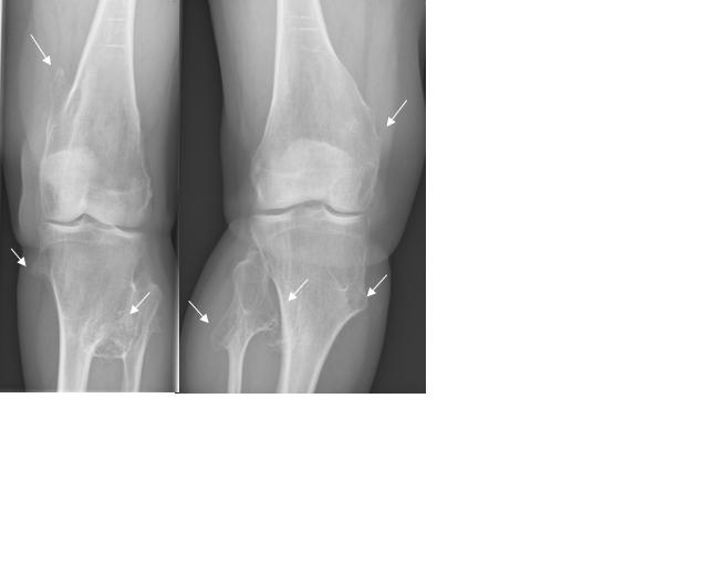
Hereditary multiple exostoses is an autosomal dominant EXT1 or EXT2 defect with sessile and pedunculated osteochondromas, and about 1 percent undergo malignant transformation to chondrosarcoma: an exostosis that grows or hurts after skeletal maturity is reimaged. Enchondromatosis (Ollier disease) is multiple enchondromas; Maffucci syndrome adds soft tissue hemangiomas, and the hemangioma is the entire split. McCune-Albright syndrome is an activating GNAS mutation present as somatic mosaicism: polyostotic fibrous dysplasia with the shepherd crook femoral deformity, coast of Maine cafe-au-lait spots (jagged borders), and precocious puberty. The board asks one question across the whole family: which gene, which inheritance, which signature, and what do you screen for?
Tap each letter of the cornea and coast mnemonics.
HH MX
HHurler has the H-aze: clouded cornea, MPS I, alpha-L-iduronidase, autosomal recessive
HHunter is H-azy-free: clear cornea, MPS II, iduronate sulfatase, X-linked, maternal uncles affected
MMaine: coast of Maine cafe-au-lait with jagged borders is McCune-Albright; coast of California with smooth borders is neurofibromatosis
XThe X-linked pair in this family: Hunter (MPS II) and X-linked hypophosphatemic rickets
Open each gene and hold the signature and the screen.
Osteogenesis imperfecta: type I collagen, autosomal dominant with autosomal recessive forms in the severe types. Signature: blue sclera, wormian bones, dentinogenesis imperfecta, hearing loss from otosclerosis. Screen: fracture burden, hearing; bisphosphonates reduce the fractures.
Osteopetrosis: osteoclast resorption failure. Signature: marble bone, bone-within-bone, Erlenmeyer flask deformity. Screen: cranial nerves, marrow, and the carbonic anhydrase II variant that pairs marble bone with renal tubular acidosis.
Achondroplasia: gain of function, autosomal dominant, more than 85 percent new mutations with a paternal age effect. Signature: rhizomelia, frontal bossing, trident hand, normal intelligence. Screen: foramen magnum stenosis in infancy, spinal stenosis in adulthood.
Hurler (MPS I, alpha-L-iduronidase, autosomal recessive, clouded cornea) and Hunter (MPS II, iduronate sulfatase, X-linked, clear cornea). Screen: enzyme replacement therapy plus hematopoietic stem cell transplant, best when started early.
X-linked hypophosphatemic rickets: FGF23 excess, renal phosphate wasting. Signature: bowed legs, low phosphate, normal calcium, high alkaline phosphatase. Treat with phosphate plus calcitriol; plain vitamin D cannot overcome the renal leak.
McCune-Albright syndrome: activating mutation, somatic mosaicism. Signature: polyostotic fibrous dysplasia with the shepherd crook femur, coast of Maine cafe-au-lait spots, precocious puberty. Screen: endocrinopathy and fracture risk in the fibrous dysplasia.
Hereditary multiple exostoses: autosomal dominant osteochondromas with about 1 percent malignant transformation to chondrosarcoma. Ollier is enchondromas alone; Maffucci adds hemangiomas. Screen: growth or pain in a stable bump after skeletal maturity is a reimaging trigger.
Prove it
Walkthrough: seven hereditary bone decisions
Original practice scenarios, one at a time. Choose an answer, then open any option to work its reasoning.
Clinical walkthrough
