
Choose an answer, then open any option to work its reasoning.
MSK · Orthopedic Anatomy
Limb Compartments and the Brachial Plexus: The Compartment-Nerve-Movement Chain
Every limb is a stack of fascial sleeves, and every sleeve carries one nerve and one set of movements. Compartment syndrome is the clock that makes the anatomy urgent: the anterior leg after a tibial shaft fracture, the volar forearm after a fracture or IV infiltration, and the silent deep posterior compartment that is missed until the toes claw. Then the plexus turns the upper limb into one wiring diagram, and Erb versus Klumpke becomes a pattern-matching game.
What this page makes you able to do
- Read the compartment-nerve-movement chain for the upper and lower limb
- Run the syndrome clock: recognize, measure, and decompress within 4 to 6 hours
- Separate the loud anterior leg from the silent deep posterior compartment
- Name the plexus level from the pattern: Erb, Klumpke, Horner, and the cord combinations
- Dr. Fatima Ali, DOPsychiatry residentPrimary reviewer
Last reviewed
The pearl
Every limb is a stack of fascial sleeves, and every sleeve carries one nerve and one set of movements. Run the chain compartment to nerve to movement, and remember the pulse is a late liar: pressure kills venous outflow long before arterial inflow, and irreversible damage begins around 4 to 6 hours.
Prove it
Opening question
Answer before you read anything, then keep the recognition quartet and the clock in mind through every section.
A 24-year-old man is brought to the emergency department 2 hours after a closed tibial shaft fracture from a motorcycle crash. He reports pain that is far worse than the fracture alone should cause. Examination shows a tense, swollen anterior leg, and passive toe extension reproduces the pain. He reports tingling over the dorsum of the foot. The dorsalis pedis pulse is palpable.Which of the following is the most appropriate next step in management?
- Why this is rightPain out of proportion, a tense compartment, pain with passive stretch of the muscles inside it, and new sensory change in the deep peroneal territory are the recognition quartet, and the palpable pulse is a trap: it survives until pressure exceeds arterial pressure, which is why pulselessness is a LATE sign. The next step is compartment pressure measurement (or fasciotomy when the exam is convincing), never elevation, ice, analgesia, or imaging. Rule: pain out of proportion plus pain on passive stretch in a high-risk fracture is compartment syndrome; measure the pressure and get to the operating room, never wait for the pulse to die.
- Why this failsElevation lowers the arteriovenous gradient and can actually reduce perfusion to a compartment already under pressure; it treats the symptom, never the pressure. A 4-hour recheck hands the sleeve the rest of the irreversibility window. Rule: elevation is for swelling without pressure; this is pressure.
- Why this failsCold and opioids mask the pain out of proportion that is the alarm itself; the pain is a sign of ischemia, and silencing it is how compartment syndromes get missed. Rule: never sedate the alarm system; open the compartment.
- Why this failsThe palpable pulse already tells you the artery is patent; the problem is the sleeve pressure, not the flow. Angiography answers the artery, never the fascia, and imaging time is dead time on the fasciotomy clock. Rule: the pulse is fine and the artery is fine; the compartment is not.
Work the reasoning
Pressure kills venous outflow before arterial inflow: the capillaries collapse and the muscle starves while the pulse still beats. The pulse dies only when pressure exceeds arterial pressure, which is why pulselessness is a LATE sign.
Pain out of proportion, a tense compartment, pain with passive stretch of the muscles inside it, and new paresthesias in the nerve that runs through it: the first three are the exam, the fourth is the nerve already complaining.
Measurement is for the uncertain exam. When the recognition quartet is complete, fasciotomy proceeds directly; the delta P number never outranks a convincing exam, and irreversible muscle and nerve damage begins around 4 to 6 hours of elevated pressure.
The answer is A: measure the compartment pressure and prepare for fasciotomy. Never wait for the pulse to die: irreversible muscle and nerve damage begins around 4 to 6 hours.
THE CHAIN
The Compartment Chain: Sleeve, Nerve, Movement
Every limb compartment has a one-line address: the sleeve, the nerve inside it, and the movement that dies when either one is compressed. Learn the chain and the board stem writes itself.
The upper limb runs on fascial sleeves. Each sleeve holds a muscle group, and each muscle group is owned by one nerve, so every compartment has a one-line address: the anterior arm sleeve (biceps, brachialis, coracobrachialis) is owned by the musculocutaneous nerve and dies as lost elbow flexion with lateral forearm numbness; the posterior arm sleeve (triceps, anconeus) is owned by the radial nerve and dies as lost elbow extension. The deltoid compartment belongs to the axillary nerve (C5-C6): lost shoulder abduction from 15 to 90 degrees with a numb deltoid patch, the injury story of an inferior glenohumeral dislocation or a surgical neck humerus fracture. Abduction is a relay, and the range is the nerve test: 0 to 15 degrees is supraspinatus (suprascapular), 15 to 90 degrees is deltoid (axillary), and finishing the arc above 90 belongs to scapular rotation (serratus anterior, long thoracic nerve, whose loss wings the scapula on a wall push).
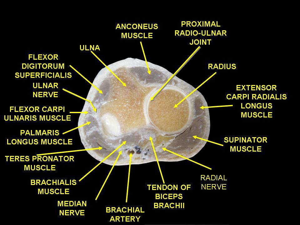
The forearm splits into two sleeves. The volar (flexor) sleeve holds the wrist and finger flexors and pronators with the median and ulnar nerves; the dorsal (extensor) sleeve holds the extensors with the radial nerve and its posterior interosseous branch. The hand adds ten small boxes: the thenar sleeve (recurrent median branch, lost thumb opposition), the hypothenar sleeve (deep ulnar branch, lost little finger abduction), and the interosseous sleeves (deep ulnar branch, lost finger abduction and adduction, the claw hand with Froment and Wartenberg signs). The humerus is a nerve map of its own: axillary at the surgical neck, radial at the midshaft spiral groove, median at the distal shaft.
Run the recognition sequence on a fresh fracture.
A 19-year-old man is brought to the emergency department 4 hours after a closed tibial shaft fracture sustained in a football game. He rates the leg pain 9 out of 10 and says it keeps getting worse despite the splint. Examination shows a tense anterior leg, pain with passive toe extension, and new tingling over the dorsum of the foot. The dorsalis pedis pulse is palpable. Which of the following is the most appropriate next step?
A. Measure the pressure and prepare for fasciotomy. Pain out of proportion, a tense sleeve, pain on passive toe extension (stretching the anterior compartment extensors), and new deep peroneal paresthesias are the recognition quartet, and the palpable pulse is a late-sign trap. B. Elevation. Elevation lowers the arteriovenous gradient and can reduce perfusion; it treats symptoms, not pressure, and wastes the window. C. CT angiogram. The pulse already proves the artery is patent; imaging answers the artery, never the fascia. D. Cold and opioids. They mask the pain that is the alarm. Rule: fracture plus stretch pain plus nerve paresthesias is fasciotomy, never observation; irreversible damage begins around 4 to 6 hours.
THE FOREARM SPLIT
The Forearm Split: Volar Flexors Versus Dorsal Extensors
Two fascial sleeves around two bones, and the stretch test names the one under pressure: passive finger EXTENSION indicts the volar flexors, passive finger FLEXION indicts the dorsal extensors.
The volar sleeve is the larger and more dangerous of the two: it holds the finger and wrist flexors, the pronators, and both major nerves, the median (with its anterior interosseous branch, the pure motor nerve whose death breaks the OK sign) and the ulnar. Bleeding from a distal radius or forearm fracture or fluid extravasation from an infiltrated IV in the antecubital fossa fills this tight sleeve, and in children the infiltrated IV is a leading cause of forearm compartment syndrome. The early exam is pain with passive finger extension (it stretches the flexors inside the sleeve) and median and ulnar paresthesias. A missed volar sleeve ends in the Volkmann ischemic contracture: a clawed, flexed wrist and fingers from dead flexors, the scar of a compartment syndrome that waited too long.
The dorsal sleeve holds the wrist and finger extensors with the radial nerve, which dives through the supinator as the posterior interosseous nerve (PIN), a pure motor branch. Its stretch test is the mirror image: pain with passive finger flexion stretches the extensors. The PIN distinction is a favorite board split: a full radial lesion drops the wrist and numbs the dorsoradial hand, while a PIN lesion drops only the fingers, keeps wrist extension (extensor carpi radialis longus is innervated above the arcade), and keeps sensation intact. The crush forearm is both sleeves until proven otherwise: stretch pain in both directions means both get released.
Flip between the two forearm sleeves.
THE SYNDROME CLOCK
The Syndrome Clock: Recognition and Fasciotomy Urgency
Compartment syndrome is a pressure story: the sleeve fills, venous outflow collapses, the nerves scream, then the muscles die. The boards test the clock: which injury, which signs, how fast, what cut.
The recognition ladder runs on pain. Pain out of proportion to the injury, pain with passive stretch of the muscles inside the sleeve, and a tense, swollen compartment in a high-risk setting (tibial shaft fracture, forearm fracture, supracondylar humerus fracture, crush, reperfusion, circumferential burn, tight cast) are the exam. The classic 5 Ps are pain, paresthesias, pallor, poikilothermia, and paralysis, and pulselessness is late: the pulse dies only when pressure exceeds arterial pressure, because the pressure kills venous outflow before arterial inflow. A palpable pulse is the norm in early compartment syndrome, and the stem will hand it to you as a trap.
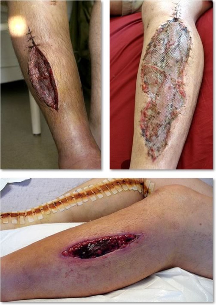
The numbers and the deadline: the classic fasciotomy threshold is a compartment pressure within 30 mm Hg of the diastolic blood pressure (delta P under 30 mm Hg), and an absolute pressure over 30 to 40 mm Hg in a tense, symptomatic compartment supports the call. The clock starts at the injury, not at the diagnosis: irreversible muscle and nerve damage begins around 4 to 6 hours of elevated pressure. The surgery is fasciotomy: full-length incisions through skin AND fascia to open every involved sleeve. The leg gets a two-incision, four-compartment release; the forearm gets volar and dorsal releases; the wounds stay open and close by delayed closure or graft. Elevation, ice, and analgesia treat symptoms, never pressure, and no medication opens a fascial sleeve.
Put the fasciotomy response in order before the deadline.
Tally how many fasciotomy triggers this limb has.
Checked 0
Three swollen limbs, three doors: which one is the operating room?
A swollen, painful limb. Which door fits which presentation?
THE LOWER LIMB
The Lower Limb: Four Leg Sleeves, Three Thigh Sleeves, One Gluteal Sleeve
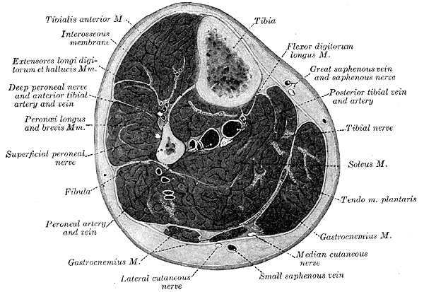
The leg is four compartments, the thigh is three, and the buttock is one big one. The chain runs the same way, and the stretch test names the sleeve.
The anterior leg is the fracture classic: tibialis anterior, extensor hallucis longus, and extensor digitorum longus with the deep peroneal nerve, and it is the compartment most commonly involved in tibial shaft fracture. Its loss is loud and unmistakable: foot drop with first web space numbness, provoked by passive toe extension. The lateral leg (peroneus longus and brevis, superficial peroneal nerve) fails as lost eversion with dorsum numbness that spares the first web space, and its stretch test is passive ankle inversion. The superficial posterior sleeve is the calf: gastrocnemius and soleus with the sural nerve, failing as lost plantar flexion with a faded ankle reflex (S1), provoked by passive ankle dorsiflexion, and a tense calf here is the DVT trap. The deep posterior sleeve is the one the boards set up to miss: tibialis posterior, flexor digitorum longus, and flexor hallucis longus tucked against the bones with the tibial nerve inside. It is called the silent compartment because there is no surface swelling to see and no dramatic posture to photograph: the motor loss (toe flexion) is subtle, and the first reliable clue is pain with passive toe extension stretching the toe flexors, with sole paresthesias from the tibial nerve. A missed deep posterior compartment leaves clawed toes, intrinsic wasting, and a numb sole.
The thigh and buttock run the same chain with bigger muscles and bigger nerves: anterior thigh (quadriceps, femoral nerve, lost knee extension and a dead patellar reflex), medial thigh (adductors, obturator nerve, lost adduction), posterior thigh (hamstrings, sciatic nerve, lost knee flexion), and the gluteal sleeve, where a gluteal compartment syndrome crushes the sciatic nerve with the glutei: lost hip extension and everything below the knee. The stretch-test map is the memory hook: toe extension = anterior AND deep posterior, inversion = lateral, dorsiflexion = superficial posterior.
THE PLEXUS
The Brachial Plexus: From the Roots to the Pattern
Five roots, three trunks, six divisions, three cords, five terminal branches, and every board question is a pattern of lost movements looking for a level.
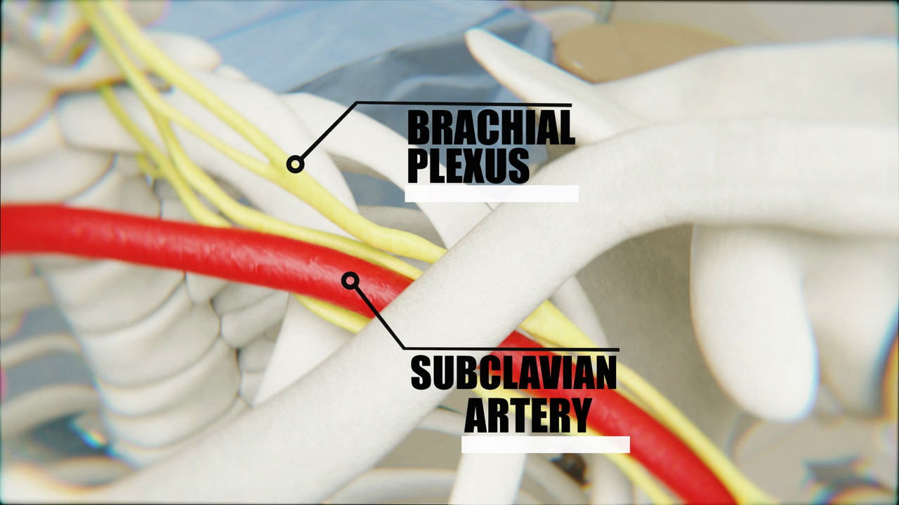
The plexus is the wiring diagram of the upper limb, and the order is the mnemonic: roots, trunks, divisions, cords, branches. The C5 through T1 roots emerge between the scalene muscles (the phrenic nerve, C3-C5, rides the front of the anterior scalene at this level). They merge into the upper trunk (C5-C6), middle trunk (C7), and lower trunk (C8-T1) in the posterior triangle, split behind the clavicle into anterior and posterior divisions (anterior divisions feed the flexor side of the limb, posterior divisions the extensor side), and recombine into the three cords around the axillary artery. The median nerve is the hybrid: born from the lateral cord (C5-C7 fibers) and the medial cord (C8-T1 fibers), which is why a cord lesion only weakens half its territory.
The cord questions are always combination questions. Lateral cord = musculocutaneous plus the lateral half of the median: weak elbow flexion with lateral forearm numbness plus the thumb-side median story. Posterior cord = axillary plus radial, the extensor side: deltoid AND wrist drop together, with latissimus and subscapularis weakness. Medial cord = ulnar plus the medial half of the median, the flexor side: the claw with weak finger flexion and medial forearm numbness. Two named nerves dying together means the lesion is at the plexus; one nerve dying alone means that nerve. The named branches keep their own fingerprints: the long thoracic nerve wings the scapula on a wall push (serratus anterior, after axillary surgery), the suprascapular nerve kills the first 15 degrees of abduction and external rotation with fossa wasting and no sensory loss, and the axillary nerve kills 15 to 90 degrees with the deltoid patch numb. Parsonage-Turner syndrome, the idiopathic brachial neuritis, starts with acute severe shoulder pain and days later leaves patchy weakness that often includes the long thoracic or suprascapular nerves: the pain-then-palsy sequence separates it from trauma.
Click an injury site on the plexus and read the pattern it produces.
Click an injury site to light the pattern.
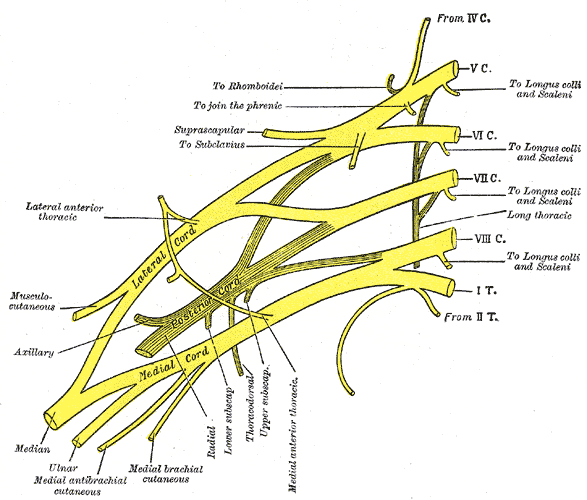
THE NAMED PALSIES
Erb Versus Klumpke: The Two Named Palsies
The upper trunk is the shoulder story; the lower trunk is the hand story plus Horner. Learn the posture, the mechanism, and the sympathetic flag.
Erb-Duchenne palsy is the upper trunk (C5-C6) torn or stretched by shoulder dystocia at birth, a fall on the shoulder, or lateral neck traction. The arm hangs in the waiter's tip posture: adducted and internally rotated with the elbow extended and the wrist flexed, from loss of the shoulder abductors and external rotators (deltoid, supraspinatus, infraspinatus), the elbow flexors (biceps, brachialis), and the forearm supinators. The hand is the rule-out: grasp, finger movement, and hand sensation stay intact because C8-T1 is untouched, and the pupils stay symmetric because the sympathetic fibers ride T1, not C5-C6. Erb never carries Horner.
Klumpke palsy is the lower trunk (C8-T1) from upward traction on the arm or an apical lung tumor (Pancoast): the hand claws from intrinsic loss, the medial hand and forearm go numb, and shoulder function stays because the upper trunk is intact. The signature flag is the Horner syndrome: when T1 is avulsed or destroyed, the ipsilateral face shows ptosis, miosis, and anhidrosis because the sympathetic fibers travel with the T1 root. The 58-year-old smoker with a Pancoast tumor and the newborn with a difficult breech delivery share the same pattern: claw hand plus Horner is Klumpke; waiter's tip with an intact hand is Erb. The cord lesions split the difference below the clavicle: a posterior cord lesion is deltoid plus wrist drop (axillary plus radial), a lateral cord lesion is weak elbow flexion plus the thumb-side median story, and a medial cord lesion is the claw plus the medial median story, none of them ever carrying Horner.
Tap each letter of the plexus order and the two named palsies.
RTDCB RUL K
RRoots: C5-T1 emerge between the scalenes
TTrunks: upper (C5-C6), middle (C7), lower (C8-T1)
DDivisions: anterior feeds the flexor side, posterior the extensor side
CCords: lateral, posterior, medial around the axillary artery
BBranches: musculocutaneous, axillary, radial, median, ulnar
RRemember Upper Lateral: Erb is C5-C6, the shoulder story
UUpper trunk: waiter's tip, adducted internally rotated arm, elbow extended, wrist flexed
LLateral: the hand lives and the pupils stay symmetric, Erb never carries Horner
KKlumpke is C8-T1: K for claw hand, plus Horner (ptosis, miosis, anhidrosis) when T1 is avulsed
THE ONE-SCREEN CHAIN
The One-Screen Chain: Compartments and Plexus
The whole page is one table: sleeve, nerve, movement, and the plexus level that owns it. Open every row until the map is memorized.
Run the rows in order and the board stems write themselves. The upper limb is deltoid (axillary, abduction 15 to 90), anterior arm (musculocutaneous, elbow flexion), posterior arm (radial, elbow extension, spiral groove sparing the triceps), volar forearm (median and ulnar, wrist and finger flexion and pronation with the OK sign), dorsal forearm (radial and PIN, wrist and finger extension), and the hand boxes (thenar, hypothenar, interosseous). The lower limb is the four leg sleeves (deep peroneal foot drop, superficial peroneal eversion, sural calf, tibial sole), the three thigh sleeves (femoral, obturator, sciatic), and the gluteal sleeve that owns the sciatic nerve. The plexus level cards close the loop: upper trunk Erb, lower trunk Klumpke, lateral cord musculocutaneous plus lateral median, posterior cord axillary plus radial, medial cord ulnar plus medial median. The median is the hinge of the whole map: its two halves come from two cords, so a cord lesion only ever weakens half the median story.
Open the rows that are still fuzzy.
Nerve: axillary (C5-C6), posterior cord. Lost movement: shoulder abduction 15 to 90 degrees, flattened deltoid. Sensory patch: deltoid patch, upper lateral arm. Story: inferior glenohumeral dislocation, surgical neck humerus fracture, quadrilateral space compression.
Nerve: musculocutaneous (C5-C7), lateral cord. Lost movement: elbow flexion, faded biceps reflex. Sensory patch: lateral forearm (lateral antebrachial cutaneous). Story: humeral shaft injury, shoulder surgery, heavy exertion.
Nerve: radial (C5-T1), posterior cord. Lost movement: elbow extension, dead triceps reflex; a spiral groove lesion spares the triceps, an axillary-level lesion drops it. Story: crutch palsy above the branches, Saturday night palsy at the groove.
Nerve: median (with the anterior interosseous branch) and ulnar. Lost movements: wrist and finger flexion, pronation, broken OK sign. Stretch test: pain with passive finger extension. Story: forearm fracture, IV infiltration, crush; Volkmann contracture if missed.
Nerve: radial and posterior interosseous (PIN). Lost movements: wrist and finger extension, wrist drop; a PIN lesion drops the fingers while wrist extension and sensation survive. Stretch test: pain with passive finger flexion.
Nerve: recurrent branch of the median. Lost movement: thumb opposition and abduction, thenar wasting. Stretch test: pain with passive thumb abduction. Story: carpal tunnel compression, thenar crush, wrist lacerations.
Nerve: deep branch of the ulnar. Lost movements: little finger abduction and opposition, finger abduction and adduction, claw hand with Froment and Wartenberg signs. Stretch test: pain with passive little finger abduction (hypothenar) or finger adduction (interosseous). Story: Guyon canal compression, hand crush, metacarpal fractures.
Nerve: deep peroneal. Lost movement: ankle dorsiflexion and toe extension, foot drop. Sensory patch: first web space. Stretch test: pain with passive toe extension. Story: the classic tibial shaft fracture site, the most commonly involved compartment.
Nerve: superficial peroneal. Lost movement: ankle eversion. Sensory patch: dorsum of the foot except the first web space. Stretch test: pain with passive ankle inversion.
Nerve: tibial branches to the triceps surae, sural nerve through the sleeve. Lost movement: plantar flexion, faded ankle reflex (S1). Sensory patch: posterolateral calf and lateral foot (sural). Stretch test: pain with passive ankle dorsiflexion; the tense calf here is the DVT trap.
Nerve: tibial nerve. Lost movement: toe flexion, then intrinsic clawing; sole numbness. Stretch test: pain with passive toe extension, the test everyone forgets. Story: no surface swelling, subtle motor loss, missed while the anterior sleeve gets the attention.
Anterior thigh: femoral nerve (L2-L4), lost knee extension and patellar reflex. Medial thigh: obturator nerve (L2-L4), lost adduction. Posterior thigh: sciatic nerve (L4-S3), lost knee flexion and everything below the knee. Gluteal: the sciatic nerve with the glutei: lost hip extension and the whole leg below the knee.
Upper trunk (C5-C6): Erb, waiter's tip, hand intact, no Horner. Lower trunk (C8-T1): Klumpke, claw hand, Horner with T1 avulsion. Lateral cord: musculocutaneous plus the lateral half of the median. Posterior cord: axillary plus radial, the extensor side. Medial cord: ulnar plus the medial half of the median, the flexor side. The median is the hinge: two cords, two halves, and a cord lesion only ever weakens half the story.
Prove it
Walkthrough: race the fasciotomy clock
Original practice scenarios, one at a time. Choose an answer, then open any option to work its reasoning.
Clinical walkthrough
