Lower Extremity Nerve Injuries: Localize by Function
Trace lower-extremity nerve lesions from branch anatomy to urgent workup, recovery monitoring, rehabilitation, referral and real-world symptom control.
Read this lesson in four passes. Level 1 explains branch anatomy and injury physiology. Level 2 turns that map into localization, testing and urgent workup. Level 3 follows recovery, function and referral over time. The Beyond layer adds practical electrodiagnostic timing, counseling and representative adult symptom-control dosing without confusing pain relief with nerve repair.
Read a deficit as a branching map
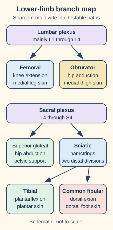
Begin proximal, then narrow. The lumbar plexus is formed mainly by L1 through L4 ventral rami, often with a T12 contribution; the sacral plexus receives L4 through S4. The memory phrase "I twice get late on Fridays" cues iliohypogastric, ilioinguinal, genitofemoral, lateral femoral cutaneous, obturator and femoral nerves. A root sends fibers into several named nerves, while a mononeuropathy follows one smaller branch set. [1][4]
Trace each route from shared roots to a named nerve and then to its main bedside action. The branch widths are schematic and do not represent axon counts.
The femoral path separates knee extension from adduction. Femoral fibers from L2 through L4 supply quadriceps, the patellar reflex arc, anterior thigh sensation and medial leg sensation through the saphenous branch. The nerve also supplies iliacus. Psoas major receives direct lumbar ventral ramus innervation, so partial hip flexion can remain after a femoral lesion. Iliacus compartment hematoma, pelvic trauma and regional infection can compress the nerve; sudden groin pain, falling hemoglobin and new weakness require urgent assessment for bleeding. [5][13]
The obturator path tests a different L2 through L4 route. It supplies adductor longus, adductor brevis, gracilis and the adductor portion of adductor magnus, with variable medial-thigh sensation. The hamstring portion of adductor magnus receives tibial division supply, so one muscle name can contain two neural territories. Weak adduction after pelvic surgery with preserved quadriceps strength and patellar response favors obturator injury, while abnormalities crossing femoral and obturator territories raise concern for plexus or root disease. [4]
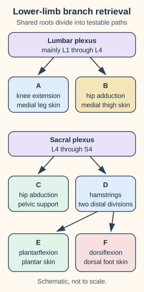
Retrieval view: identify A through F before opening the answer key. Root overlap is preserved while the nerve names are hidden.Reveal the branch-map key
The hidden labels run from proximal function to distal branch. A is femoral, B is obturator, C is superior gluteal, D is sciatic, E is tibial and F is common fibular. Rebuild the map from action first, then confirm with sensation and reflexes.
Use a cross-nerve comparator. If knee extension is weak and medial-leg sensation is reduced but adduction is strong, the femoral route is the economical explanation. If quadriceps, adductors and ankle dorsiflexors are all abnormal after pelvic trauma, one isolated distal nerve cannot account for the pattern. The lesson throughout is to test a function that leaves the suspected pathway before naming the lesion.
Try it here · Checkpoint 1 of 3
Make your prediction before reading the choices. A first attempt is just a starting point.
Case 3
Show answer and explanations for case 3
A. Abnormal iliacus, psoas major and adductor longus (Why this does not fit)
Involvement of all three would cross femoral, direct lumbar and obturator pathways. That would be broader than the focal nerve injury supported by the supplied pattern.
Reasoning steps for option A
Why can "Abnormal iliacus, psoas major and adductor longus" seem attractive before the full pattern is checked in the 72-year-old man?
Its partial appeal is that involvement of all three would cross femoral, direct lumbar and obturator pathways. That connection alone does not account for every impaired and spared pathway, so "Abnormal iliacus, psoas major and adductor longus" still requires a complete boundary check in this presentation.
Which discriminator removes "Abnormal iliacus, psoas major and adductor longus" from first place for the 72-year-old man?
The decisive mismatch is that that would be broader than the focal nerve injury supported by the supplied pattern. The case requires one explanation for the full distribution, so that mismatch outweighs the initial appeal of "Abnormal iliacus, psoas major and adductor longus".
B. Normal iliacus; abnormal psoas major plus adductor longus (Why this does not fit)
This emphasizes direct lumbar and obturator pathways while sparing a proximal femoral muscle. It is not the expected extension of the quadriceps and saphenous deficit.
Reasoning steps for option B
Why can "Normal iliacus; abnormal psoas major plus adductor longus" seem attractive before the full pattern is checked in the 72-year-old man?
Its partial appeal is that this emphasizes direct lumbar and obturator pathways while sparing a proximal femoral muscle. That connection alone does not account for every impaired and spared pathway, so "Normal iliacus; abnormal psoas major plus adductor longus" still requires a complete boundary check in this presentation.
Which discriminator removes "Normal iliacus; abnormal psoas major plus adductor longus" from first place for the 72-year-old man?
The decisive mismatch is that it is not the expected extension of the quadriceps and saphenous deficit. The case requires one explanation for the full distribution, so that mismatch outweighs the initial appeal of "Normal iliacus; abnormal psoas major plus adductor longus".
C. Normal iliacus and psoas major; abnormal adductor longus (Why this does not fit)
That pattern would favor obturator rather than femoral involvement. It does not explain the reduced patellar response and knee-extension deficit.
Reasoning steps for option C
Why can "Normal iliacus and psoas major; abnormal adductor longus" seem attractive before the full pattern is checked in the 72-year-old man?
Its partial appeal is that that pattern would favor obturator rather than femoral involvement. That connection alone does not account for every impaired and spared pathway, so "Normal iliacus and psoas major; abnormal adductor longus" still requires a complete boundary check in this presentation.
Which discriminator removes "Normal iliacus and psoas major; abnormal adductor longus" from first place for the 72-year-old man?
The decisive mismatch is that it does not explain the reduced patellar response and knee-extension deficit. The case requires one explanation for the full distribution, so that mismatch outweighs the initial appeal of "Normal iliacus and psoas major; abnormal adductor longus".
D. Abnormal iliacus with normal psoas major and adductor longus (Best answer)
The motor, reflex and sensory pattern first identifies femoral involvement. Iliacus lies downstream of the stated proximal compression, while direct lumbar supply to psoas and obturator supply to adductors remain separate, preserving some hip-flexion force.
Reasoning steps for option D
Which two observations make "Abnormal iliacus with normal psoas major and adductor longus" coherent in the 72-year-old man presentation?
First, localize knee-extension, patellar and saphenous findings to femoral supply. Next, apply the stated branch level to iliacus while separating direct lumbar psoas and obturator supply. That paired reasoning keeps "Abnormal iliacus with normal psoas major and adductor longus" consistent with the same anatomic pattern in this 72-year-old man.
Why does "Abnormal iliacus with normal psoas major and adductor longus" outrank the nearest alternative for the 72-year-old man?
For the objective "Explain residual hip flexion after proximal femoral compression", this option accounts for both impaired and preserved pathways. A rival level would require an additional deficit not supplied by the vignette, so "Abnormal iliacus with normal psoas major and adductor longus" remains the most internally consistent choice.
E. Abnormal iliacus and adductor longus; psoas major remains normal (Why this does not fit)
This would suggest combined femoral and obturator involvement, such as a broader plexus process. The focal imaging and strong adduction instead support restriction to the femoral pathway.
Reasoning steps for option E
Why can "Abnormal iliacus and adductor longus; psoas major remains normal" seem attractive before the full pattern is checked in the 72-year-old man?
Its partial appeal is that this would suggest combined femoral and obturator involvement, such as a broader plexus process. That connection alone does not account for every impaired and spared pathway, so "Abnormal iliacus and adductor longus; psoas major remains normal" still requires a complete boundary check in this presentation.
Which discriminator removes "Abnormal iliacus and adductor longus; psoas major remains normal" from first place for the 72-year-old man?
The decisive mismatch is that the focal imaging and strong adduction instead support restriction to the femoral pathway. The case requires one explanation for the full distribution, so that mismatch outweighs the initial appeal of "Abnormal iliacus and adductor longus; psoas major remains normal".
Takeaway: Determine the injured pathway before predicting what remains above or outside it; psoas is not a femoral-innervated synonym for iliacus.
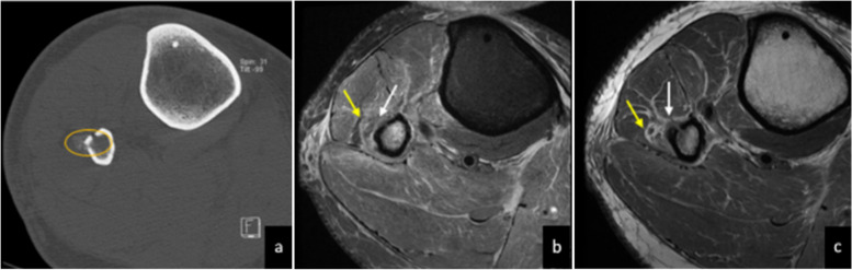
Foot drop means inadequate ankle dorsiflexion during swing. It often produces a high-stepping gait, but it does not identify the injured level. Near the fibular neck, the common fibular nerve divides into deep fibular fibers that dorsiflex, extend the toes and sense the first dorsal web space, plus superficial fibular fibers that evert and sense most of the dorsal foot. Weakness of both branches after a tight cast, leg crossing, prolonged squatting, rapid weight loss or proximal fibular injury supports a common fibular lesion. [1][2]
Kumar S et al., Insights into Imaging (2023), Figure 7, panels a through c. Computed tomography shows proximal fibular fracture-related new bone; magnetic resonance images show adjacent swollen deep fibular nerve (white arrow) and superficial fibular nerve (yellow arrow). Published annotations are unmodified. Original figure, CC BY 4.0.
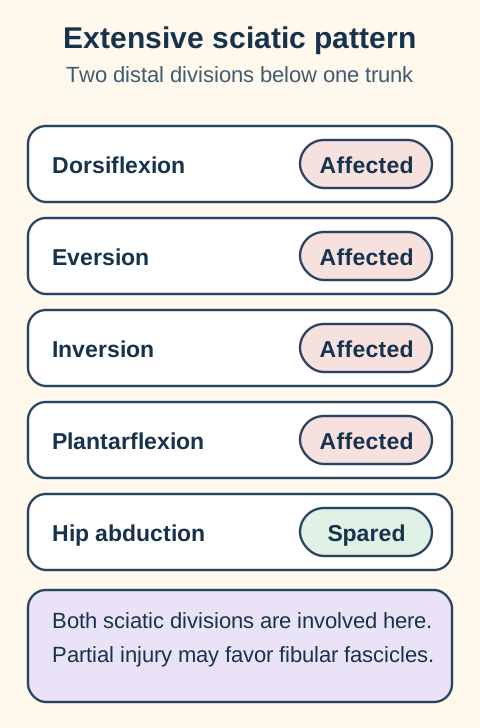
The short memory aid is PED and TIP. Peroneal, the older term for fibular, everts and dorsiflexes; tibial inverts and plantarflexes. Use the phrase as a first pass, not as proof. Deep-branch injury can spare eversion, superficial-branch injury can spare dorsiflexion, and a partial sciatic lesion can favor fibular fascicles while preserving much of the tibial division. [2][8]
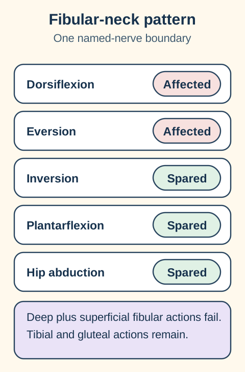
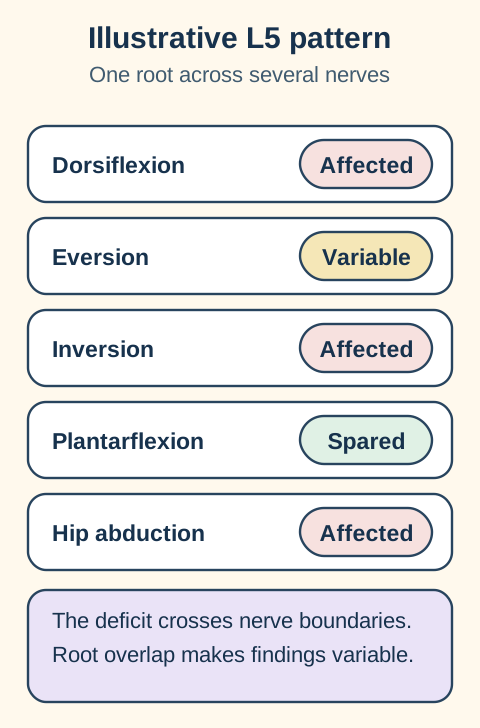
Predict the spared route before naming the lesion. Dorsiflexion and eversion weakness with strong inversion and plantarflexion respects the common fibular boundary. Dorsiflexion weakness plus weak tibialis posterior inversion or gluteus medius abduction crosses that boundary through L5-weighted fibers and favors a root-level process. Hamstring or calf involvement, plantar sensory loss or short-head biceps femoris denervation points above the fibular neck toward sciatic disease.
A fibular-neck pattern stops at the fibular boundary. Deep and superficial fibular actions can fail together while tibial-powered plantarflexion and tibialis posterior inversion remain. Focal tenderness, conduction slowing across the neck or a matching structural lesion adds support.
An L5 pattern crosses named nerves. Tibialis anterior, tibialis posterior and gluteus medius share important L5 input while traveling through fibular, tibial and superior gluteal pathways. Back or radiating pain can support radiculopathy, but its absence cannot exclude it.
A sciatic pattern starts above both distal divisions. Extensive injury can impair hamstrings, dorsiflexion, eversion, inversion, plantarflexion and sole sensation. Real sciatic injuries may be fascicular and uneven, so preserved calf strength lowers rather than eliminates the probability. [8]
Trace the fibular-neck pattern
A complete fibular-neck example affects both fibular branches while the tested tibial and superior gluteal actions remain available. Severity can vary.
Test the branches separately. Great-toe extension and first-web sensation emphasize the deep branch; eversion and central dorsal-foot sensation emphasize the superficial branch. Normal tibialis posterior and gluteus medius provide nonfibular L5 comparators.
Trace the L5 pattern
An L5 pattern crosses fibular, tibial and superior gluteal pathways. Root overlap and lesion severity make the exact examination variable.
Crossing named nerves is the key idea. A root lesion can weaken dorsiflexion, inversion and hip abduction together. Preserved distal sensory nerve action potentials and paraspinal denervation can add support when the study timing and technique are appropriate.
Trace the sciatic pattern
This extensive example reaches both sciatic divisions. A fibular-predominant partial sciatic injury can look more distal.
Sample above the knee when the distal pattern is misleading. Short-head biceps femoris belongs to the fibular division before the common fibular nerve reaches the knee. Its denervation with normal gluteus medius and paraspinals shifts the site toward sciatic rather than L5 disease.
Transfer the comparison to a common history. After rapid weight loss and prolonged leg crossing, weak dorsiflexion and eversion with intact inversion, plantarflexion and sole sensation supports fibular-neck compression. The history raises probability, but the spared routes make the localization defensible. [2]
The tibial path changes meaning with the level
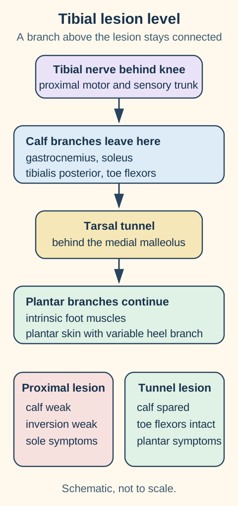
Follow the tibial nerve downstream before interpreting sole symptoms. The tibial division of the sciatic nerve supplies the posterior leg, including calf plantarflexors and tibialis posterior, before it enters the retromalleolar tunnel. Distal to that tunnel it divides into plantar branches for intrinsic foot muscles and plantar skin. A lesion below a branch cannot denervate the muscle that branch already reached.
Compare the two interruption sites. Proximal tibial injury reaches calf and plantar functions; a tunnel lesion occurs after the principal calf motor branches have departed.
A proximal tibial lesion reaches the calf. A popliteal mass, trauma or a more proximal sciatic process can weaken repeated heel rises, toe flexion and inversion, reduce the Achilles response and alter plantar sensation. Eversion should remain available in an isolated tibial lesion because its principal muscles use the superficial fibular nerve. A reduced sural response may indicate a lesion above the ankle or another postganglionic process rather than an isolated distal tunnel syndrome. [1]
A retromalleolar lesion spares branches that already left. Tarsal tunnel disease can produce burning or numb plantar pain, symptoms behind the medial malleolus and intrinsic-foot weakness while calf plantarflexion, long toe flexion and the Achilles response remain intact. Rheumatoid flexor tenosynovitis, trauma, deformity or a space-occupying lesion can narrow the tunnel. Heel sensation varies because calcaneal branching can occur proximal to or within the tunnel. [7]
A local percussion response is supporting evidence. Reproduction of tingling behind the medial malleolus fits tarsal tunnel disease, but a positive response is not diagnostic and a negative response does not exclude it. Electrodiagnostic testing can also be falsely negative. Match the symptom field, intrinsic-foot examination and spared proximal branches before accepting the site. [7]
Reassess the proposed level when function changes. New inability to perform repeated heel rises, expanding sensory loss or progressive weakness should trigger a new proximal examination rather than a stronger commitment to the original tunnel diagnosis. Pain, balance and effort can alter a heel-rise test, so compare sides and isolate individual muscles when possible.
Longitudinal care protects function while localization matures. Record strength by branch, walking safety, skin integrity, range of motion and pain at each visit. An ankle-foot orthosis can reduce trips in dorsiflexion weakness; stretching and therapy help prevent equinus contracture while recovery is assessed. Serial examinations matter because an early picture may reflect conduction block, evolving axon loss or pain-limited effort. [14]
A falling pelvis identifies the stance-side support system
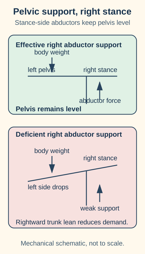
Name the stance leg before naming the weak side. When the left foot is lifted, the right gluteus medius and minimus must hold the unsupported left hemipelvis level. If the left pelvis falls during right single-leg stance, the failing support system is on the right. The superior gluteal nerve from L4 through S1 also supplies tensor fasciae latae and has no cutaneous bedside field.
The right stance abductors oppose the body-weight turning effect. When their effective force falls, the opposite unsupported pelvis drops.
Trunk lean is compensation, not proof of recovery. Leaning toward the weak stance hip shortens the body-weight moment arm and lowers the abductor force required to balance the pelvis. Painful hip disease, tendon disruption and mechanical instability can produce the same observed pelvic drop, so confirm true abductor weakness and review structural context. [11][12]
Inferior gluteal injury produces a different functional split. Gluteus maximus supplies powerful hip extension for rising from a low chair or climbing stairs. The inferior gluteal nerve from L5 through S2 can be injured during pelvic or posterior hip procedures. Hamstrings can still extend the hip, especially from a flexed position, and the lesion does not create a cutaneous sensory patch.
Meralgia paresthetica is sensory only. Lateral femoral cutaneous nerve entrapment near the anterior superior iliac spine and inguinal ligament produces burning or numb anterolateral thigh symptoms without weakness or reflex change. Tight garments, pregnancy and increased abdominal pressure can contribute. A small lumbar magnetic resonance imaging abnormality does not replace the clinical field; reduce local pressure first when the presentation is otherwise uncomplicated. [6]
Separate neighboring patterns instead of forcing one explanation. Groin and proximal medial-thigh symptoms fit ilioinguinal territory better than lateral femoral cutaneous territory. Quadriceps weakness or a reduced patellar response requires a femoral or root explanation. True hip-abductor weakness needs a superior gluteal, L5, plexus or structural hip assessment rather than attribution to a pure sensory nerve.
Roots, reflexes and sensory studies answer different questions
A root pattern crosses peripheral-nerve borders. L2 through L4 contribute to hip flexion, knee extension and adduction through different named nerves. L4 often contributes medial-leg sensation and the patellar reflex; L5 commonly contributes great-toe extension, inversion and hip abduction; S1 contributes plantarflexion, eversion and the Achilles response. Dermatomes overlap, reflexes draw from more than one root and pain fields vary, so no single stripe or tendon response is absolute. [9]
Working root pattern, not exclusive territories
Roots
Typical sensory region
Representative action
Reflex contribution
RootsL2 through L3
Typical sensory regionAnterior and medial thigh
Representative actionHip flexion and some adduction
Reflex contributionAdductor response can involve L2 through L4
RootsL4
Typical sensory regionMedial leg and ankle
Representative actionKnee extension and ankle dorsiflexion
Reflex contributionPatellar, chiefly L3 through L4
RootsL5
Typical sensory regionLateral leg, dorsal foot and great toe
Representative actionGreat-toe extension, inversion and hip abduction
Reflex contributionNo dependable routine deep tendon reflex
RootsS1
Typical sensory regionPosterior calf and lateral foot
Representative actionPlantarflexion and eversion
Reflex contributionAchilles, chiefly S1 with S2 contribution
RootsS2 through S4
Typical sensory regionPosterior thigh at S2, then perineal and perianal regions
Representative actionPelvic-floor and sphincter function
Reflex contributionBulbocavernosus response
Posterior-thigh skin is not a direct sciatic motor map. The posterior femoral cutaneous nerve supplies much of that skin, while the sciatic nerve supplies hamstrings and then divides into tibial and fibular components. A disc lesion produces radiculopathy rather than automatically producing sciatic mononeuropathy. Deep-gluteal symptoms require comparison with spinal, hip and pelvic causes. [8]
The dorsal root ganglion predicts the sensory study. A typical intraspinal root lesion lies proximal to the sensory cell body, so the distal sensory nerve action potential often remains preserved. A reduced superficial fibular, saphenous or sural sensory response on the symptomatic side supports a postganglionic lesion such as mononeuropathy or plexopathy, provided technical factors and coexisting polyneuropathy have been addressed. [8]
Needle sampling should cross named nerves that share a root. For suspected L5 disease, compare tibialis anterior with tibialis posterior and gluteus medius; for S1, compare gastrocnemius with gluteus maximus and other suitable muscles. Paraspinal abnormalities can support radiculopathy, but timing, sampling, prior surgery and local trauma affect interpretation. The clinical pattern chooses the muscles; the test should not substitute for the examination.
Injury physiology explains why the same site can have different severity. Neurapraxia is conduction failure with preserved axonal continuity and often favorable recovery. Axonotmesis disrupts axons while connective scaffolding is variably preserved, producing Wallerian degeneration and slower regrowth. Neurotmesis disrupts the nerve and supporting architecture, making spontaneous recovery unlikely without repair. Clinical and electrodiagnostic follow-up distinguishes these trajectories more reliably than the initial mechanism alone. [14]
A partial sciatic lesion can imitate a distal palsy. After posterior hip dislocation, marked dorsiflexion and eversion weakness with preserved calf strength may reflect fibular-predominant sciatic injury. Short-head biceps femoris denervation places the lesion above its branch, while normal gluteus medius and paraspinals argue against a broad L5 process. Test proximal sciatic muscles and both distal divisions before accepting a fibular-neck site. [8]
Try it here · Checkpoint 2 of 3
Make your prediction before reading the choices. A first attempt is just a starting point.
Case 23
Show answer and explanations for case 23
A. An isolated L5 root lesion with selective distal weakness (Why this does not fit)
A root lesion can be patchy and normal paraspinals do not exclude it. However, the asymmetric sensory response, above-knee fibular-division abnormality and hip-trauma context together favor a postganglionic peripheral injury.
Reasoning steps for option A
Why can "An isolated L5 root lesion with selective distal weakness" seem attractive before the full pattern is checked in the 26-year-old man?
Its partial appeal is that a root lesion can be patchy and normal paraspinals do not exclude it. That connection alone does not account for every impaired and spared pathway, so "An isolated L5 root lesion with selective distal weakness" still requires a complete boundary check in this presentation.
Which discriminator removes "An isolated L5 root lesion with selective distal weakness" from first place for the 26-year-old man?
The decisive mismatch is that however, the asymmetric sensory response, above-knee fibular-division abnormality and hip-trauma context together favor a postganglionic peripheral injury. The case requires one explanation for the full distribution, so that mismatch outweighs the initial appeal of "An isolated L5 root lesion with selective distal weakness".
B. An isolated deep fibular lesion below the fibular neck (Why this does not fit)
That lesion cannot explain short-head biceps femoris abnormalities above the knee or the eversion deficit.
Reasoning steps for option B
Why can "An isolated deep fibular lesion below the fibular neck" seem attractive before the full pattern is checked in the 26-year-old man?
Its partial appeal is that that lesion cannot explain short-head biceps femoris abnormalities above the knee or the eversion deficit. That connection alone does not account for every impaired and spared pathway, so "An isolated deep fibular lesion below the fibular neck" still requires a complete boundary check in this presentation.
Which discriminator removes "An isolated deep fibular lesion below the fibular neck" from first place for the 26-year-old man?
The decisive mismatch is that that lesion cannot explain short-head biceps femoris abnormalities above the knee or the eversion deficit. The case requires one explanation for the full distribution, so that mismatch outweighs the initial appeal of "An isolated deep fibular lesion below the fibular neck".
C. A broad pelvic plexus injury involving femoral and superior gluteal fibers (Why this does not fit)
A broad plexus lesion could affect several distal nerves, but the supplied normal quadriceps and gluteus medius do not support the additional pathways named in this option. The abnormalities are concentrated in the fibular division of sciatic supply.
Reasoning steps for option C
Why can "A broad pelvic plexus injury involving femoral and superior gluteal fibers" seem attractive before the full pattern is checked in the 26-year-old man?
Its partial appeal is that a broad plexus lesion could affect several distal nerves, but the supplied normal quadriceps and gluteus medius do not support the additional pathways named in this option. That connection alone does not account for every impaired and spared pathway, so "A broad pelvic plexus injury involving femoral and superior gluteal fibers" still requires a complete boundary check in this presentation.
Which discriminator removes "A broad pelvic plexus injury involving femoral and superior gluteal fibers" from first place for the 26-year-old man?
The decisive mismatch is that the abnormalities are concentrated in the fibular division of sciatic supply. The case requires one explanation for the full distribution, so that mismatch outweighs the initial appeal of "A broad pelvic plexus injury involving femoral and superior gluteal fibers".
D. An isolated common fibular lesion at the fibular neck (Why this does not fit)
The short-head biceps femoris branch leaves proximally, so its abnormality argues against a lesion confined to the neck.
Reasoning steps for option D
Why can "An isolated common fibular lesion at the fibular neck" seem attractive before the full pattern is checked in the 26-year-old man?
Its partial appeal is that the short-head biceps femoris branch leaves proximally, so its abnormality argues against a lesion confined to the neck. That connection alone does not account for every impaired and spared pathway, so "An isolated common fibular lesion at the fibular neck" still requires a complete boundary check in this presentation.
Which discriminator removes "An isolated common fibular lesion at the fibular neck" from first place for the 26-year-old man?
The decisive mismatch is that the short-head biceps femoris branch leaves proximally, so its abnormality argues against a lesion confined to the neck. The case requires one explanation for the full distribution, so that mismatch outweighs the initial appeal of "An isolated common fibular lesion at the fibular neck".
E. A fibular-predominant partial sciatic injury above the short-head branch (Best answer)
The hip trauma and proximal fibular-division muscle abnormality place the injury above the knee. Selective fascicular involvement explains preserved tibial functions; normal paraspinals alone do not exclude root disease.
Reasoning steps for option E
Which two observations make "A fibular-predominant partial sciatic injury above the short-head branch" coherent in the 26-year-old man presentation?
First, use above-knee muscle involvement and an abnormal sensory response to favor a proximal postganglionic fibular-division injury. Next, explain preserved tibial functions by partial fascicular rather than complete sciatic involvement. That paired reasoning keeps "A fibular-predominant partial sciatic injury above the short-head branch" consistent with the same anatomic pattern in this 26-year-old man.
Why does "A fibular-predominant partial sciatic injury above the short-head branch" outrank the nearest alternative for the 26-year-old man?
For the objective "Distinguish partial sciatic injury from a distal fibular lesion", this option accounts for both impaired and preserved pathways. A rival level would require an additional deficit not supplied by the vignette, so "A fibular-predominant partial sciatic injury above the short-head branch" remains the most internally consistent choice.
Takeaway: A partial sciatic injury can resemble common fibular neuropathy; an above-knee fibular-division muscle can expose the difference.
Danger outranks fine localization. New urinary retention, saddle sensory change, rapidly progressive bilateral weakness or severe back pain with sacral findings can indicate cauda equina compression. Emergency spinal assessment and lumbar magnetic resonance imaging take priority over routine nerve-conduction testing. Magnetic resonance imaging without or with contrast is usually appropriate when cauda equina syndrome is suspected. [10][16]
Acute limb threats also require escalation. Open laceration near a nerve, fracture or dislocation with a new neurologic deficit, expanding hematoma, vascular compromise, compartment findings or rapidly increasing weakness needs urgent specialty assessment. A suspected external pressure palsy from a cast or position requires safe relief of compression and a documented neurovascular examination rather than passive observation.
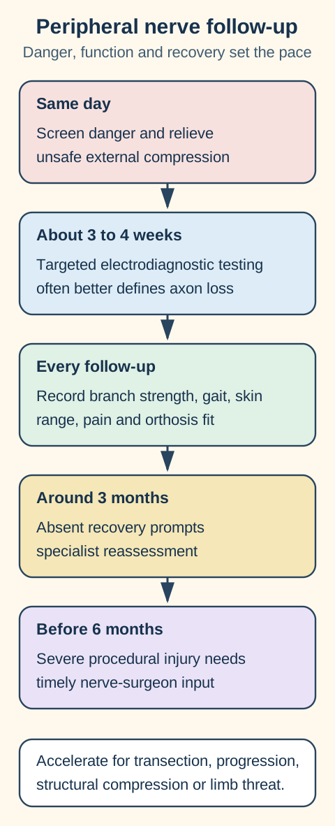
A practical sequence, not a rigid clock. The mechanism, severity, open injury, progression and functional risk can accelerate every step.
Electrodiagnostic timing depends on the question. Nerve-conduction studies can identify conduction block early, but needle evidence of active denervation develops over time. For many axonal injuries, a targeted study around 3 to 4 weeks is more informative than an immediate study; procedure-related reviews often recommend assessment at about 4 weeks. An early normal study cannot settle a recent axonal lesion, and serial testing can document reinnervation or persistent loss. [14][15]
Protect gait, skin and range while the diagnosis matures. Use an ankle-foot orthosis when dorsiflexion weakness creates toe drag, confirm that pressure points are safe in sensory loss, and pair bracing with stretching and strengthening. Therapy should maintain joint range, prevent contracture, retrain safe walking and preserve unaffected muscles. Recheck branch-specific strength rather than recording only "foot drop present." [14]
Trajectory determines referral intensity. Suspected transection or open injury warrants early surgical input. Progressive deficit or a compressive mass requires prompt structural evaluation. Lack of clinical or electrodiagnostic recovery by roughly 3 months should trigger specialist reassessment rather than automatic waiting; severe peri-procedural injuries merit surgical consultation well before 6 months. These ranges guide decisions and do not override the mechanism or examination. [14][15]
Beyond the examination: symptom control without false promises
No medication reconnects a transected nerve. There is no approved pharmacologic treatment that regenerates a severed peripheral nerve or removes a structural compression. Medication can reduce neuropathic pain while decompression, protection, rehabilitation and recovery surveillance address the lesion itself. [14]
Duloxetine dosing depends on the diagnosed pain syndrome. For the labeled indication of diabetic peripheral neuropathic pain in adults, the usual dose is 60 mg once daily; a lower starting dose may be considered for tolerability. Higher doses have not shown added benefit for that indication and are less well tolerated. Avoid use in chronic liver disease or cirrhosis and in severe renal impairment, and taper rather than stop abruptly. Use for traumatic mononeuropathic pain may be off-label. Verify current labeling before prescribing. [17][18]
Pregabalin requires renal adjustment and sedation counseling. For the labeled indication of diabetic peripheral neuropathic pain in an adult with creatinine clearance at least 60 mL per minute, a representative start is 50 mg three times daily, with increase to 100 mg three times daily within 1 week when needed and tolerated. Reduce the dose for renal impairment, taper over at least 1 week, and warn about dizziness, somnolence and respiratory depression risk with opioids or other central nervous system depressants. Use outside a labeled diagnosis may be off-label. Verify current labeling before prescribing. [17][19]
Medication choice follows patient risk, not nerve location alone. Review falls, driving, renal and hepatic function, interacting sedatives, pregnancy, mood history and the pain phenotype. A numb weak foot without painful paresthesia may benefit more from protection and therapy than from a sedating drug. Reassess benefit, adverse effects and function rather than continuing treatment because the diagnosis sounds neuropathic.
Counsel with a range rather than a guarantee. Conduction block can improve over weeks, axonal regrowth is slower and complete disruption may not recover without repair. Explain what is being monitored, which findings should prompt urgent contact and how the orthosis or exercise plan protects function during uncertainty. A specific patient timeline depends on distance to the target muscle, severity, age, comorbidity and whether the compressive cause has been corrected.
Try it here · Checkpoint 3 of 3
Make your prediction before reading the choices. A first attempt is just a starting point.
Case 27
Show answer and explanations for case 27
A. Urgent spinal assessment and lumbar MRI now (Best answer)
New sacral sensory change with progressing weakness raises concern for cauda equina compression even while voiding is possible. Assessment should not wait for established retention.
Reasoning steps for option A
Which two observations make "Urgent spinal assessment and lumbar MRI now" coherent in the 69-year-old woman presentation?
First, recognize that evolving perianal and motor changes require urgent assessment despite preserved voiding. Next, confirm that the spared functions respect the proposed boundary. That paired reasoning keeps "Urgent spinal assessment and lumbar MRI now" consistent with the same anatomic pattern in this 69-year-old woman.
Why does "Urgent spinal assessment and lumbar MRI now" outrank the nearest alternative for the 69-year-old woman?
For the objective "Interpret incomplete spinal warning symptoms without waiting for retention", this option accounts for both impaired and preserved pathways. A rival level would require an additional deficit not supplied by the vignette, so "Urgent spinal assessment and lumbar MRI now" remains the most internally consistent choice.
B. Urgent pelvic MRI focused on a unilateral plexus lesion before spinal assessment (Why this does not fit)
Plexus disease can affect several nerves, but the acute back pain with bilateral and perianal findings makes spinal compression the priority rather than an isolated unilateral plexus study.
Reasoning steps for option B
Why can "Urgent pelvic MRI focused on a unilateral plexus lesion before spinal assessment" seem attractive before the full pattern is checked in the 69-year-old woman?
Its partial appeal is that plexus disease can affect several nerves, but the acute back pain with bilateral and perianal findings makes spinal compression the priority rather than an isolated unilateral plexus study. That connection alone does not account for every impaired and spared pathway, so "Urgent pelvic MRI focused on a unilateral plexus lesion before spinal assessment" still requires a complete boundary check in this presentation.
Which discriminator removes "Urgent pelvic MRI focused on a unilateral plexus lesion before spinal assessment" from first place for the 69-year-old woman?
The decisive mismatch is that plexus disease can affect several nerves, but the acute back pain with bilateral and perianal findings makes spinal compression the priority rather than an isolated unilateral plexus study. The case requires one explanation for the full distribution, so that mismatch outweighs the initial appeal of "Urgent pelvic MRI focused on a unilateral plexus lesion before spinal assessment".
C. Urgent brain MRI before spinal assessment (Why this does not fit)
A central lesion can cause weakness, but this combination of back pain, sacral sensory change and lower-limb deficits directs urgent assessment to the lumbosacral canal.
Reasoning steps for option C
Why can "Urgent brain MRI before spinal assessment" seem attractive before the full pattern is checked in the 69-year-old woman?
Its partial appeal is that a central lesion can cause weakness, but this combination of back pain, sacral sensory change and lower-limb deficits directs urgent assessment to the lumbosacral canal. That connection alone does not account for every impaired and spared pathway, so "Urgent brain MRI before spinal assessment" still requires a complete boundary check in this presentation.
Which discriminator removes "Urgent brain MRI before spinal assessment" from first place for the 69-year-old woman?
The decisive mismatch is that a central lesion can cause weakness, but this combination of back pain, sacral sensory change and lower-limb deficits directs urgent assessment to the lumbosacral canal. The case requires one explanation for the full distribution, so that mismatch outweighs the initial appeal of "Urgent brain MRI before spinal assessment".
D. Same-day electrodiagnostic testing before spinal imaging (Why this does not fit)
Electrodiagnosis can refine localization later but does not promptly exclude a time-sensitive compressive canal lesion and should not delay imaging.
Reasoning steps for option D
Why can "Same-day electrodiagnostic testing before spinal imaging" seem attractive before the full pattern is checked in the 69-year-old woman?
Its partial appeal is that electrodiagnosis can refine localization later but does not promptly exclude a time-sensitive compressive canal lesion and should not delay imaging. That connection alone does not account for every impaired and spared pathway, so "Same-day electrodiagnostic testing before spinal imaging" still requires a complete boundary check in this presentation.
Which discriminator removes "Same-day electrodiagnostic testing before spinal imaging" from first place for the 69-year-old woman?
The decisive mismatch is that electrodiagnosis can refine localization later but does not promptly exclude a time-sensitive compressive canal lesion and should not delay imaging. The case requires one explanation for the full distribution, so that mismatch outweighs the initial appeal of "Same-day electrodiagnostic testing before spinal imaging".
E. Close outpatient reassessment after a bladder emptying measurement (Why this does not fit)
A bladder measurement may inform the assessment but should not override the supplied progressive motor and sacral sensory warnings. Preserved voiding is not reassurance enough for routine outpatient observation.
Reasoning steps for option E
Why can "Close outpatient reassessment after a bladder emptying measurement" seem attractive before the full pattern is checked in the 69-year-old woman?
Its partial appeal is that a bladder measurement may inform the assessment but should not override the supplied progressive motor and sacral sensory warnings. That connection alone does not account for every impaired and spared pathway, so "Close outpatient reassessment after a bladder emptying measurement" still requires a complete boundary check in this presentation.
Which discriminator removes "Close outpatient reassessment after a bladder emptying measurement" from first place for the 69-year-old woman?
The decisive mismatch is that preserved voiding is not reassurance enough for routine outpatient observation. The case requires one explanation for the full distribution, so that mismatch outweighs the initial appeal of "Close outpatient reassessment after a bladder emptying measurement".
Takeaway: New perianal sensory change and progressive leg weakness require urgent spinal assessment even while voiding remains possible.
Use the same sequence in every case. Name the failed action, identify its named nerve, test a second action outside that nerve, compare sensation and reflexes, then decide whether urgency or recovery trajectory changes the next step. The cases span mechanism, diagnosis, testing, protection, monitoring and escalation.
Case 1
Show answer and explanations for case 1
A. Abnormal tibialis posterior and gluteus medius (Why this does not fit)
These muscles receive L5 fibers through different nonfibular nerves. Their combined abnormalities would challenge restriction to a local fibular pathway.
Reasoning steps for option A
Why can "Abnormal tibialis posterior and gluteus medius" seem attractive before the full pattern is checked in the 32-year-old man?
Its partial appeal is that these muscles receive L5 fibers through different nonfibular nerves. That connection alone does not account for every impaired and spared pathway, so "Abnormal tibialis posterior and gluteus medius" still requires a complete boundary check in this presentation.
Which discriminator removes "Abnormal tibialis posterior and gluteus medius" from first place for the 32-year-old man?
The decisive mismatch is that their combined abnormalities would challenge restriction to a local fibular pathway. The case requires one explanation for the full distribution, so that mismatch outweighs the initial appeal of "Abnormal tibialis posterior and gluteus medius".
B. Normal tibialis anterior with abnormal tibialis posterior (Why this does not fit)
This would emphasize the tibial inversion pathway while sparing a principal dorsiflexor. It does not match the observed combined dorsiflexion and eversion loss.
Reasoning steps for option B
Why can "Normal tibialis anterior with abnormal tibialis posterior" seem attractive before the full pattern is checked in the 32-year-old man?
Its partial appeal is that this would emphasize the tibial inversion pathway while sparing a principal dorsiflexor. That connection alone does not account for every impaired and spared pathway, so "Normal tibialis anterior with abnormal tibialis posterior" still requires a complete boundary check in this presentation.
Which discriminator removes "Normal tibialis anterior with abnormal tibialis posterior" from first place for the 32-year-old man?
The decisive mismatch is that it does not match the observed combined dorsiflexion and eversion loss. The case requires one explanation for the full distribution, so that mismatch outweighs the initial appeal of "Normal tibialis anterior with abnormal tibialis posterior".
C. Abnormal tibialis anterior with normal fibularis longus (Why this does not fit)
This would fit a predominantly deep-branch pattern better than the observed combined deep and superficial deficits. It does not supply a nonfibular L5 comparator.
Reasoning steps for option C
Why can "Abnormal tibialis anterior with normal fibularis longus" seem attractive before the full pattern is checked in the 32-year-old man?
Its partial appeal is that this would fit a predominantly deep-branch pattern better than the observed combined deep and superficial deficits. That connection alone does not account for every impaired and spared pathway, so "Abnormal tibialis anterior with normal fibularis longus" still requires a complete boundary check in this presentation.
Which discriminator removes "Abnormal tibialis anterior with normal fibularis longus" from first place for the 32-year-old man?
The decisive mismatch is that it does not supply a nonfibular L5 comparator. The case requires one explanation for the full distribution, so that mismatch outweighs the initial appeal of "Abnormal tibialis anterior with normal fibularis longus".
D. Abnormal tibialis anterior; tibialis posterior remains normal (Best answer)
The clinical pattern first implicates both fibular divisions. Comparing an affected deep fibular muscle with a spared tibial L5 muscle then supports a peripheral nerve boundary rather than a shared-root pattern.
Reasoning steps for option D
Which two observations make "Abnormal tibialis anterior; tibialis posterior remains normal" coherent in the 32-year-old man presentation?
First, infer involvement of both fibular divisions from dorsiflexion and eversion loss. Next, use a tibial L5 muscle as the nonfibular comparator rather than relying on absence of back pain. That paired reasoning keeps "Abnormal tibialis anterior; tibialis posterior remains normal" consistent with the same anatomic pattern in this 32-year-old man.
Why does "Abnormal tibialis anterior; tibialis posterior remains normal" outrank the nearest alternative for the 32-year-old man?
For the objective "Distinguish a fibular-pattern lesion from shared-root dysfunction", this option accounts for both impaired and preserved pathways. A rival level would require an additional deficit not supplied by the vignette, so "Abnormal tibialis anterior; tibialis posterior remains normal" remains the most internally consistent choice.
E. Abnormal tibialis posterior with lumbar paraspinal fibrillations (Why this does not fit)
This combines a nonfibular L5 muscle and paraspinals. It favors reconsidering a root process rather than strengthening the focal knee hypothesis.
Reasoning steps for option E
Why can "Abnormal tibialis posterior with lumbar paraspinal fibrillations" seem attractive before the full pattern is checked in the 32-year-old man?
Its partial appeal is that this combines a nonfibular L5 muscle and paraspinals. That connection alone does not account for every impaired and spared pathway, so "Abnormal tibialis posterior with lumbar paraspinal fibrillations" still requires a complete boundary check in this presentation.
Which discriminator removes "Abnormal tibialis posterior with lumbar paraspinal fibrillations" from first place for the 32-year-old man?
The decisive mismatch is that it favors reconsidering a root process rather than strengthening the focal knee hypothesis. The case requires one explanation for the full distribution, so that mismatch outweighs the initial appeal of "Abnormal tibialis posterior with lumbar paraspinal fibrillations".
Takeaway: After identifying a fibular-pattern deficit, test an L5 muscle outside that nerve rather than using absent back pain as an exclusion.
A. Abnormal adductor longus with preserved hamstring part of adductor magnus (Best answer)
Adduction and medial-thigh findings first favor obturator injury. Its supply to longus differs from the tibial-division supply of the hamstring part of magnus, explaining the predicted dissociation.
Reasoning steps for option A
Which two observations make "Abnormal adductor longus with preserved hamstring part of adductor magnus" coherent in the 49-year-old woman presentation?
First, integrate adduction and medial-thigh deficits with preserved femoral function. Next, predict a dissociation between obturator-supplied longus and sciatic-supplied hamstring magnus. That paired reasoning keeps "Abnormal adductor longus with preserved hamstring part of adductor magnus" consistent with the same anatomic pattern in this 49-year-old woman.
Why does "Abnormal adductor longus with preserved hamstring part of adductor magnus" outrank the nearest alternative for the 49-year-old woman?
For the objective "Predict split adductor magnus supply after an inferred obturator lesion", this option accounts for both impaired and preserved pathways. A rival level would require an additional deficit not supplied by the vignette, so "Abnormal adductor longus with preserved hamstring part of adductor magnus" remains the most internally consistent choice.
B. Preserved adductor longus with abnormal hamstring part of adductor magnus (Why this does not fit)
This reverses the expected obturator-versus-sciatic dissociation. It would not explain the main adductor pattern as well.
Reasoning steps for option B
Why can "Preserved adductor longus with abnormal hamstring part of adductor magnus" seem attractive before the full pattern is checked in the 49-year-old woman?
Its partial appeal is that this reverses the expected obturator-versus-sciatic dissociation. That connection alone does not account for every impaired and spared pathway, so "Preserved adductor longus with abnormal hamstring part of adductor magnus" still requires a complete boundary check in this presentation.
Which discriminator removes "Preserved adductor longus with abnormal hamstring part of adductor magnus" from first place for the 49-year-old woman?
The decisive mismatch is that it would not explain the main adductor pattern as well. The case requires one explanation for the full distribution, so that mismatch outweighs the initial appeal of "Preserved adductor longus with abnormal hamstring part of adductor magnus".
C. Abnormal adductor longus with abnormal vastus medialis (Why this does not fit)
These muscles belong to obturator and femoral pathways. Their joint involvement would widen the localization beyond the single nerve suggested by preserved quadriceps testing.
Reasoning steps for option C
Why can "Abnormal adductor longus with abnormal vastus medialis" seem attractive before the full pattern is checked in the 49-year-old woman?
Its partial appeal is that these muscles belong to obturator and femoral pathways. That connection alone does not account for every impaired and spared pathway, so "Abnormal adductor longus with abnormal vastus medialis" still requires a complete boundary check in this presentation.
Which discriminator removes "Abnormal adductor longus with abnormal vastus medialis" from first place for the 49-year-old woman?
The decisive mismatch is that their joint involvement would widen the localization beyond the single nerve suggested by preserved quadriceps testing. The case requires one explanation for the full distribution, so that mismatch outweighs the initial appeal of "Abnormal adductor longus with abnormal vastus medialis".
D. Abnormal adductor longus with lumbar paraspinal fibrillations (Why this does not fit)
Paraspinal abnormalities would prompt consideration of upper lumbar root involvement rather than an isolated obturator lesion.
Reasoning steps for option D
Why can "Abnormal adductor longus with lumbar paraspinal fibrillations" seem attractive before the full pattern is checked in the 49-year-old woman?
Its partial appeal is that paraspinal abnormalities would prompt consideration of upper lumbar root involvement rather than an isolated obturator lesion. That connection alone does not account for every impaired and spared pathway, so "Abnormal adductor longus with lumbar paraspinal fibrillations" still requires a complete boundary check in this presentation.
Which discriminator removes "Abnormal adductor longus with lumbar paraspinal fibrillations" from first place for the 49-year-old woman?
The decisive mismatch is that paraspinal abnormalities would prompt consideration of upper lumbar root involvement rather than an isolated obturator lesion. The case requires one explanation for the full distribution, so that mismatch outweighs the initial appeal of "Abnormal adductor longus with lumbar paraspinal fibrillations".
E. Preserved adductor longus with abnormal vastus medialis (Why this does not fit)
This would favor femoral motor involvement, the opposite of weak adduction with normal knee extension.
Reasoning steps for option E
Why can "Preserved adductor longus with abnormal vastus medialis" seem attractive before the full pattern is checked in the 49-year-old woman?
Its partial appeal is that this would favor femoral motor involvement, the opposite of weak adduction with normal knee extension. That connection alone does not account for every impaired and spared pathway, so "Preserved adductor longus with abnormal vastus medialis" still requires a complete boundary check in this presentation.
Which discriminator removes "Preserved adductor longus with abnormal vastus medialis" from first place for the 49-year-old woman?
The decisive mismatch is that this would favor femoral motor involvement, the opposite of weak adduction with normal knee extension. The case requires one explanation for the full distribution, so that mismatch outweighs the initial appeal of "Preserved adductor longus with abnormal vastus medialis".
Takeaway: A shared root contribution does not prevent a peripheral lesion from separating adductor longus from femoral muscles and the hamstring part of magnus.
A. Tensor fasciae latae denervation with preserved tibialis posterior (Why this does not fit)
This emphasizes superior gluteal rather than sciatic supply. It does not extend the supplied hamstring and distal-division pattern in the expected way.
Reasoning steps for option A
Why can "Tensor fasciae latae denervation with preserved tibialis posterior" seem attractive before the full pattern is checked in the 28-year-old woman?
Its partial appeal is that this emphasizes superior gluteal rather than sciatic supply. That connection alone does not account for every impaired and spared pathway, so "Tensor fasciae latae denervation with preserved tibialis posterior" still requires a complete boundary check in this presentation.
Which discriminator removes "Tensor fasciae latae denervation with preserved tibialis posterior" from first place for the 28-year-old woman?
The decisive mismatch is that it does not extend the supplied hamstring and distal-division pattern in the expected way. The case requires one explanation for the full distribution, so that mismatch outweighs the initial appeal of "Tensor fasciae latae denervation with preserved tibialis posterior".
B. Abnormal tibialis posterior with normal flexor digitorum longus (Why this does not fit)
Both muscles receive proximal tibial branches downstream of a high sciatic lesion. Their dissociation is less supportive of the broad motor pattern than sparing a muscle outside sciatic supply.
Reasoning steps for option B
Why can "Abnormal tibialis posterior with normal flexor digitorum longus" seem attractive before the full pattern is checked in the 28-year-old woman?
Its partial appeal is that both muscles receive proximal tibial branches downstream of a high sciatic lesion. That connection alone does not account for every impaired and spared pathway, so "Abnormal tibialis posterior with normal flexor digitorum longus" still requires a complete boundary check in this presentation.
Which discriminator removes "Abnormal tibialis posterior with normal flexor digitorum longus" from first place for the 28-year-old woman?
The decisive mismatch is that their dissociation is less supportive of the broad motor pattern than sparing a muscle outside sciatic supply. The case requires one explanation for the full distribution, so that mismatch outweighs the initial appeal of "Abnormal tibialis posterior with normal flexor digitorum longus".
C. Abnormal tibialis posterior with normal tensor fasciae latae (Best answer)
Hamstring and both distal divisions first favor a high sciatic site. Tibialis posterior is another downstream tibial muscle, whereas tensor fasciae latae uses the separate superior gluteal pathway and can remain normal.
Reasoning steps for option C
Which two observations make "Abnormal tibialis posterior with normal tensor fasciae latae" coherent in the 28-year-old woman presentation?
First, use hamstring and both distal-division findings to infer a proximal sciatic site. Next, predict another downstream tibial muscle and a separately supplied superior gluteal comparator. That paired reasoning keeps "Abnormal tibialis posterior with normal tensor fasciae latae" consistent with the same anatomic pattern in this 28-year-old woman.
Why does "Abnormal tibialis posterior with normal tensor fasciae latae" outrank the nearest alternative for the 28-year-old woman?
For the objective "Predict neighboring pathways spared by a proximal sciatic lesion", this option accounts for both impaired and preserved pathways. A rival level would require an additional deficit not supplied by the vignette, so "Abnormal tibialis posterior with normal tensor fasciae latae" remains the most internally consistent choice.
D. Abnormal flexor digitorum longus with normal tibialis anterior (Why this does not fit)
This emphasizes tibial involvement while sparing a principal dorsiflexor. It fits the supplied combined dorsiflexion and plantarflexion deficit less well than the keyed cross-pathway comparison.
Reasoning steps for option D
Why can "Abnormal flexor digitorum longus with normal tibialis anterior" seem attractive before the full pattern is checked in the 28-year-old woman?
Its partial appeal is that this emphasizes tibial involvement while sparing a principal dorsiflexor. That connection alone does not account for every impaired and spared pathway, so "Abnormal flexor digitorum longus with normal tibialis anterior" still requires a complete boundary check in this presentation.
Which discriminator removes "Abnormal flexor digitorum longus with normal tibialis anterior" from first place for the 28-year-old woman?
The decisive mismatch is that it fits the supplied combined dorsiflexion and plantarflexion deficit less well than the keyed cross-pathway comparison. The case requires one explanation for the full distribution, so that mismatch outweighs the initial appeal of "Abnormal flexor digitorum longus with normal tibialis anterior".
E. Abnormal gluteus maximus with normal rectus femoris (Why this does not fit)
That pair compares inferior gluteal and femoral supply. It does not demonstrate the expected extension of a sciatic pattern into another tibial muscle.
Reasoning steps for option E
Why can "Abnormal gluteus maximus with normal rectus femoris" seem attractive before the full pattern is checked in the 28-year-old woman?
Its partial appeal is that that pair compares inferior gluteal and femoral supply. That connection alone does not account for every impaired and spared pathway, so "Abnormal gluteus maximus with normal rectus femoris" still requires a complete boundary check in this presentation.
Which discriminator removes "Abnormal gluteus maximus with normal rectus femoris" from first place for the 28-year-old woman?
The decisive mismatch is that it does not demonstrate the expected extension of a sciatic pattern into another tibial muscle. The case requires one explanation for the full distribution, so that mismatch outweighs the initial appeal of "Abnormal gluteus maximus with normal rectus femoris".
Takeaway: Localize the shared proximal trunk, then predict an untested muscle within it and another supplied by a separate neighboring pathway.
The falling left pelvis identifies deficient right stance-side support. A superior gluteal lesion can affect right tensor fasciae latae along with the hip abductors, linking laterality to a second muscle prediction.
Reasoning steps for option A
Which two observations make "Right tensor fasciae latae" coherent in the 66-year-old woman presentation?
First, infer deficient right stance support from a falling left hemipelvis. Next, use intact tendons and the neural assessment to predict another right superior gluteal muscle. That paired reasoning keeps "Right tensor fasciae latae" consistent with the same anatomic pattern in this 66-year-old woman.
Why does "Right tensor fasciae latae" outrank the nearest alternative for the 66-year-old woman?
For the objective "Determine stance laterality and another muscle on the affected gluteal pathway", this option accounts for both impaired and preserved pathways. A rival level would require an additional deficit not supplied by the vignette, so "Right tensor fasciae latae" remains the most internally consistent choice.
B. Right quadriceps femoris (Why this does not fit)
Quadriceps receives femoral supply. Normal knee extension argues against predicting quadriceps denervation from the suspected right abductor dysfunction.
Reasoning steps for option B
Why can "Right quadriceps femoris" seem attractive before the full pattern is checked in the 66-year-old woman?
Its partial appeal is that quadriceps receives femoral supply. That connection alone does not account for every impaired and spared pathway, so "Right quadriceps femoris" still requires a complete boundary check in this presentation.
Which discriminator removes "Right quadriceps femoris" from first place for the 66-year-old woman?
The decisive mismatch is that normal knee extension argues against predicting quadriceps denervation from the suspected right abductor dysfunction. The case requires one explanation for the full distribution, so that mismatch outweighs the initial appeal of "Right quadriceps femoris".
C. Right adductor longus (Why this does not fit)
Adductor longus receives obturator supply. It does not share the superior gluteal pathway implicated by the suspected right abductor dysfunction.
Reasoning steps for option C
Why can "Right adductor longus" seem attractive before the full pattern is checked in the 66-year-old woman?
Its partial appeal is that adductor longus receives obturator supply. That connection alone does not account for every impaired and spared pathway, so "Right adductor longus" still requires a complete boundary check in this presentation.
Which discriminator removes "Right adductor longus" from first place for the 66-year-old woman?
The decisive mismatch is that it does not share the superior gluteal pathway implicated by the suspected right abductor dysfunction. The case requires one explanation for the full distribution, so that mismatch outweighs the initial appeal of "Right adductor longus".
D. Right gluteus maximus (Why this does not fit)
Gluteus maximus receives inferior gluteal supply. Its denervation would suggest additional injury rather than the selective superior gluteal pattern.
Reasoning steps for option D
Why can "Right gluteus maximus" seem attractive before the full pattern is checked in the 66-year-old woman?
Its partial appeal is that gluteus maximus receives inferior gluteal supply. That connection alone does not account for every impaired and spared pathway, so "Right gluteus maximus" still requires a complete boundary check in this presentation.
Which discriminator removes "Right gluteus maximus" from first place for the 66-year-old woman?
The decisive mismatch is that its denervation would suggest additional injury rather than the selective superior gluteal pattern. The case requires one explanation for the full distribution, so that mismatch outweighs the initial appeal of "Right gluteus maximus".
E. Left tensor fasciae latae (Why this does not fit)
The unsupported left pelvis drops because the right stance abductors fail. That observation does not predict denervation in the left tensor fasciae latae.
Reasoning steps for option E
Why can "Left tensor fasciae latae" seem attractive before the full pattern is checked in the 66-year-old woman?
Its partial appeal is that the unsupported left pelvis drops because the right stance abductors fail. That connection alone does not account for every impaired and spared pathway, so "Left tensor fasciae latae" still requires a complete boundary check in this presentation.
Which discriminator removes "Left tensor fasciae latae" from first place for the 66-year-old woman?
The decisive mismatch is that that observation does not predict denervation in the left tensor fasciae latae. The case requires one explanation for the full distribution, so that mismatch outweighs the initial appeal of "Left tensor fasciae latae".
Takeaway: Tensor fasciae latae shares superior gluteal supply with the stance-side abductors; determine the side before predicting its findings.
A. Abnormal flexor hallucis longus with normal abductor hallucis (Why this does not fit)
This emphasizes a leg muscle while sparing a downstream intrinsic muscle. It is the reverse of the expected distal tunnel pattern.
Reasoning steps for option A
Why can "Abnormal flexor hallucis longus with normal abductor hallucis" seem attractive before the full pattern is checked in the 54-year-old woman?
Its partial appeal is that this emphasizes a leg muscle while sparing a downstream intrinsic muscle. That connection alone does not account for every impaired and spared pathway, so "Abnormal flexor hallucis longus with normal abductor hallucis" still requires a complete boundary check in this presentation.
Which discriminator removes "Abnormal flexor hallucis longus with normal abductor hallucis" from first place for the 54-year-old woman?
The decisive mismatch is that it is the reverse of the expected distal tunnel pattern. The case requires one explanation for the full distribution, so that mismatch outweighs the initial appeal of "Abnormal flexor hallucis longus with normal abductor hallucis".
B. Abnormal flexor hallucis longus with abnormal medial gastrocnemius (Why this does not fit)
Both muscles receive branches above the ankle. Their joint denervation would argue for more proximal motor involvement.
Reasoning steps for option B
Why can "Abnormal flexor hallucis longus with abnormal medial gastrocnemius" seem attractive before the full pattern is checked in the 54-year-old woman?
Its partial appeal is that both muscles receive branches above the ankle. That connection alone does not account for every impaired and spared pathway, so "Abnormal flexor hallucis longus with abnormal medial gastrocnemius" still requires a complete boundary check in this presentation.
Which discriminator removes "Abnormal flexor hallucis longus with abnormal medial gastrocnemius" from first place for the 54-year-old woman?
The decisive mismatch is that their joint denervation would argue for more proximal motor involvement. The case requires one explanation for the full distribution, so that mismatch outweighs the initial appeal of "Abnormal flexor hallucis longus with abnormal medial gastrocnemius".
C. Normal abductor hallucis with abnormal tibialis anterior (Why this does not fit)
This implicates a deep fibular muscle rather than the plantar tibial pathway suggested by the symptoms.
Reasoning steps for option C
Why can "Normal abductor hallucis with abnormal tibialis anterior" seem attractive before the full pattern is checked in the 54-year-old woman?
Its partial appeal is that this implicates a deep fibular muscle rather than the plantar tibial pathway suggested by the symptoms. That connection alone does not account for every impaired and spared pathway, so "Normal abductor hallucis with abnormal tibialis anterior" still requires a complete boundary check in this presentation.
Which discriminator removes "Normal abductor hallucis with abnormal tibialis anterior" from first place for the 54-year-old woman?
The decisive mismatch is that this implicates a deep fibular muscle rather than the plantar tibial pathway suggested by the symptoms. The case requires one explanation for the full distribution, so that mismatch outweighs the initial appeal of "Normal abductor hallucis with abnormal tibialis anterior".
D. Normal flexor hallucis longus with abnormal abductor hallucis (Best answer)
The clinical pattern suggests a distal tibial site. The long flexor branch has already left in the leg, whereas the intrinsic medial plantar supply continues through the ankle region.
Reasoning steps for option D
Which two observations make "Normal flexor hallucis longus with abnormal abductor hallucis" coherent in the 54-year-old woman presentation?
First, integrate plantar symptoms, local provocation and proximal motor sparing to infer a distal tibial interval. Next, predict a normal long-flexor branch with abnormal downstream intrinsic plantar supply. That paired reasoning keeps "Normal flexor hallucis longus with abnormal abductor hallucis" consistent with the same anatomic pattern in this 54-year-old woman.
Why does "Normal flexor hallucis longus with abnormal abductor hallucis" outrank the nearest alternative for the 54-year-old woman?
For the objective "Differentiate distal tibial entrapment by long-flexor and intrinsic supply", this option accounts for both impaired and preserved pathways. A rival level would require an additional deficit not supplied by the vignette, so "Normal flexor hallucis longus with abnormal abductor hallucis" remains the most internally consistent choice.
E. Abnormal tibialis posterior with abnormal lumbar paraspinals (Why this does not fit)
This would extend the process to a non-distal muscle and a root-related sample. It would not support restriction to an ankle tunnel.
Reasoning steps for option E
Why can "Abnormal tibialis posterior with abnormal lumbar paraspinals" seem attractive before the full pattern is checked in the 54-year-old woman?
Its partial appeal is that this would extend the process to a non-distal muscle and a root-related sample. That connection alone does not account for every impaired and spared pathway, so "Abnormal tibialis posterior with abnormal lumbar paraspinals" still requires a complete boundary check in this presentation.
Which discriminator removes "Abnormal tibialis posterior with abnormal lumbar paraspinals" from first place for the 54-year-old woman?
The decisive mismatch is that it would not support restriction to an ankle tunnel. The case requires one explanation for the full distribution, so that mismatch outweighs the initial appeal of "Abnormal tibialis posterior with abnormal lumbar paraspinals".
Takeaway: Use the relation of long-flexor and intrinsic-foot branches to the lesion, not sole symptoms alone, to distinguish levels.
A. Weak inversion with weak hip abduction (Why this does not fit)
That adds superior gluteal involvement to a tibial action. It would suggest a wider root or plexus pattern rather than a focal tibial explanation near the cyst.
Reasoning steps for option A
Why can "Weak inversion with weak hip abduction" seem attractive before the full pattern is checked in the 61-year-old man?
Its partial appeal is that that adds superior gluteal involvement to a tibial action. That connection alone does not account for every impaired and spared pathway, so "Weak inversion with weak hip abduction" still requires a complete boundary check in this presentation.
Which discriminator removes "Weak inversion with weak hip abduction" from first place for the 61-year-old man?
The decisive mismatch is that it would suggest a wider root or plexus pattern rather than a focal tibial explanation near the cyst. The case requires one explanation for the full distribution, so that mismatch outweighs the initial appeal of "Weak inversion with weak hip abduction".
B. Weak inversion with preserved eversion (Best answer)
The abnormal calf and plantar muscles with an abnormal distal sensory response favor proximal postganglionic tibial involvement over a typical root lesion or distal tunnel disease. Tibialis posterior can then be affected while superficial fibular evertors remain supplied.
Reasoning steps for option B
Which two observations make "Weak inversion with preserved eversion" coherent in the 61-year-old man presentation?
First, use calf and plantar denervation plus an asymmetric sensory response to favor a proximal postganglionic tibial pattern. Next, predict affected tibialis posterior inversion while the separate superficial fibular evertor route is preserved. That paired reasoning keeps "Weak inversion with preserved eversion" consistent with the same anatomic pattern in this 61-year-old man.
Why does "Weak inversion with preserved eversion" outrank the nearest alternative for the 61-year-old man?
For the objective "Combine sensory physiology with branch-level tibial motor prediction", this option accounts for both impaired and preserved pathways. A rival level would require an additional deficit not supplied by the vignette, so "Weak inversion with preserved eversion" remains the most internally consistent choice.
C. Preserved inversion with weak eversion (Why this does not fit)
This instead emphasizes the superficial fibular pathway. It does not extend the supplied tibial motor abnormalities in the expected direction.
Reasoning steps for option C
Why can "Preserved inversion with weak eversion" seem attractive before the full pattern is checked in the 61-year-old man?
Its partial appeal is that this instead emphasizes the superficial fibular pathway. That connection alone does not account for every impaired and spared pathway, so "Preserved inversion with weak eversion" still requires a complete boundary check in this presentation.
Which discriminator removes "Preserved inversion with weak eversion" from first place for the 61-year-old man?
The decisive mismatch is that it does not extend the supplied tibial motor abnormalities in the expected direction. The case requires one explanation for the full distribution, so that mismatch outweighs the initial appeal of "Preserved inversion with weak eversion".
D. Weak dorsiflexion with weak eversion (Why this does not fit)
These are fibular actions and would broaden the localization beyond isolated tibial involvement. The normal tibialis anterior sample already argues against that pattern.
Reasoning steps for option D
Why can "Weak dorsiflexion with weak eversion" seem attractive before the full pattern is checked in the 61-year-old man?
Its partial appeal is that these are fibular actions and would broaden the localization beyond isolated tibial involvement. That connection alone does not account for every impaired and spared pathway, so "Weak dorsiflexion with weak eversion" still requires a complete boundary check in this presentation.
Which discriminator removes "Weak dorsiflexion with weak eversion" from first place for the 61-year-old man?
The decisive mismatch is that the normal tibialis anterior sample already argues against that pattern. The case requires one explanation for the full distribution, so that mismatch outweighs the initial appeal of "Weak dorsiflexion with weak eversion".
E. Weak hip abduction with weak knee extension (Why this does not fit)
That combination involves superior gluteal and femoral pathways. It does not follow from the focal calf and plantar abnormalities.
Reasoning steps for option E
Why can "Weak hip abduction with weak knee extension" seem attractive before the full pattern is checked in the 61-year-old man?
Its partial appeal is that that combination involves superior gluteal and femoral pathways. That connection alone does not account for every impaired and spared pathway, so "Weak hip abduction with weak knee extension" still requires a complete boundary check in this presentation.
Which discriminator removes "Weak hip abduction with weak knee extension" from first place for the 61-year-old man?
The decisive mismatch is that it does not follow from the focal calf and plantar abnormalities. The case requires one explanation for the full distribution, so that mismatch outweighs the initial appeal of "Weak hip abduction with weak knee extension".
Takeaway: Use sensory physiology and sampled muscle levels to choose the pathway, then predict a different action supplied along it.
A. Normal short-head biceps femoris with normal gluteus medius (Why this does not fit)
These results would not require abandoning the focal knee hypothesis. They fail to demonstrate an above-knee fibular-division abnormality.
Reasoning steps for option A
Why can "Normal short-head biceps femoris with normal gluteus medius" seem attractive before the full pattern is checked in the 37-year-old woman?
Its partial appeal is that these results would not require abandoning the focal knee hypothesis. That connection alone does not account for every impaired and spared pathway, so "Normal short-head biceps femoris with normal gluteus medius" still requires a complete boundary check in this presentation.
Which discriminator removes "Normal short-head biceps femoris with normal gluteus medius" from first place for the 37-year-old woman?
The decisive mismatch is that they fail to demonstrate an above-knee fibular-division abnormality. The case requires one explanation for the full distribution, so that mismatch outweighs the initial appeal of "Normal short-head biceps femoris with normal gluteus medius".
B. Normal short-head biceps femoris with abnormal gluteus medius (Why this does not fit)
An abnormal non-sciatic L5 muscle would direct attention toward a root or plexus pattern. It does not support restriction outside the superior gluteal pathway.
Reasoning steps for option B
Why can "Normal short-head biceps femoris with abnormal gluteus medius" seem attractive before the full pattern is checked in the 37-year-old woman?
Its partial appeal is that an abnormal non-sciatic L5 muscle would direct attention toward a root or plexus pattern. That connection alone does not account for every impaired and spared pathway, so "Normal short-head biceps femoris with abnormal gluteus medius" still requires a complete boundary check in this presentation.
Which discriminator removes "Normal short-head biceps femoris with abnormal gluteus medius" from first place for the 37-year-old woman?
The decisive mismatch is that it does not support restriction outside the superior gluteal pathway. The case requires one explanation for the full distribution, so that mismatch outweighs the initial appeal of "Normal short-head biceps femoris with abnormal gluteus medius".
C. Abnormal short-head biceps femoris with abnormal gluteus medius (Why this does not fit)
The proximal fibular-division abnormality extends above the knee, but gluteus medius involvement also extends outside sciatic supply.
Reasoning steps for option C
Why can "Abnormal short-head biceps femoris with abnormal gluteus medius" seem attractive before the full pattern is checked in the 37-year-old woman?
Its partial appeal is that the proximal fibular-division abnormality extends above the knee, but gluteus medius involvement also extends outside sciatic supply. That connection alone does not account for every impaired and spared pathway, so "Abnormal short-head biceps femoris with abnormal gluteus medius" still requires a complete boundary check in this presentation.
Which discriminator removes "Abnormal short-head biceps femoris with abnormal gluteus medius" from first place for the 37-year-old woman?
The decisive mismatch is that the proximal fibular-division abnormality extends above the knee, but gluteus medius involvement also extends outside sciatic supply. The case requires one explanation for the full distribution, so that mismatch outweighs the initial appeal of "Abnormal short-head biceps femoris with abnormal gluteus medius".
D. Normal short-head biceps femoris with normal lumbar paraspinals (Why this does not fit)
These normal proximal samples do not demonstrate a lesion above the knee and would not require revising the original focal knee hypothesis. Normal paraspinals also do not independently exclude radiculopathy.
Reasoning steps for option D
Why can "Normal short-head biceps femoris with normal lumbar paraspinals" seem attractive before the full pattern is checked in the 37-year-old woman?
Its partial appeal is that these normal proximal samples do not demonstrate a lesion above the knee and would not require revising the original focal knee hypothesis. That connection alone does not account for every impaired and spared pathway, so "Normal short-head biceps femoris with normal lumbar paraspinals" still requires a complete boundary check in this presentation.
Which discriminator removes "Normal short-head biceps femoris with normal lumbar paraspinals" from first place for the 37-year-old woman?
The decisive mismatch is that normal paraspinals also do not independently exclude radiculopathy. The case requires one explanation for the full distribution, so that mismatch outweighs the initial appeal of "Normal short-head biceps femoris with normal lumbar paraspinals".
E. Abnormal short-head biceps femoris with normal gluteus medius (Best answer)
The short-head branch leaves above the fibular neck. Its involvement requires a more proximal site, while sparing a superior gluteal muscle supports, without proving, a sciatic rather than broader plexus pattern.
Reasoning steps for option E
Which two observations make "Abnormal short-head biceps femoris with normal gluteus medius" coherent in the 37-year-old woman presentation?
First, recognize that abnormal short-head biceps femoris places fibular-division injury above the knee. Next, compare superior gluteal muscle sparing to judge whether the proximal pattern remains sciatic rather than broader. That paired reasoning keeps "Abnormal short-head biceps femoris with normal gluteus medius" consistent with the same anatomic pattern in this 37-year-old woman.
Why does "Abnormal short-head biceps femoris with normal gluteus medius" outrank the nearest alternative for the 37-year-old woman?
For the objective "Revise a knee-level hypothesis with proximal and adjacent-nerve sampling", this option accounts for both impaired and preserved pathways. A rival level would require an additional deficit not supplied by the vignette, so "Abnormal short-head biceps femoris with normal gluteus medius" remains the most internally consistent choice.
Takeaway: A new abnormality above a branch point changes the proposed level; compare a neighboring nerve to judge how broad that proximal lesion is.
A. Reduced superficial fibular sensory amplitude with focal motor block across the neck (Why this does not fit)
That combination would support postganglionic fibular disease at the knee. It does not explain the cross-nerve and paraspinal pattern as a single typical root lesion.
Reasoning steps for option A
Why can "Reduced superficial fibular sensory amplitude with focal motor block across the neck" seem attractive before the full pattern is checked in the 45-year-old man?
Its partial appeal is that that combination would support postganglionic fibular disease at the knee. That connection alone does not account for every impaired and spared pathway, so "Reduced superficial fibular sensory amplitude with focal motor block across the neck" still requires a complete boundary check in this presentation.
Which discriminator removes "Reduced superficial fibular sensory amplitude with focal motor block across the neck" from first place for the 45-year-old man?
The decisive mismatch is that it does not explain the cross-nerve and paraspinal pattern as a single typical root lesion. The case requires one explanation for the full distribution, so that mismatch outweighs the initial appeal of "Reduced superficial fibular sensory amplitude with focal motor block across the neck".
B. Preserved superficial fibular sensory amplitude with focal motor block across the neck (Why this does not fit)
A preserved sensory response can fit root disease, but a focal motor block across the neck would be additional evidence for a peripheral lesion rather than the isolated root pattern.
Reasoning steps for option B
Why can "Preserved superficial fibular sensory amplitude with focal motor block across the neck" seem attractive before the full pattern is checked in the 45-year-old man?
Its partial appeal is that a preserved sensory response can fit root disease, but a focal motor block across the neck would be additional evidence for a peripheral lesion rather than the isolated root pattern. That connection alone does not account for every impaired and spared pathway, so "Preserved superficial fibular sensory amplitude with focal motor block across the neck" still requires a complete boundary check in this presentation.
Which discriminator removes "Preserved superficial fibular sensory amplitude with focal motor block across the neck" from first place for the 45-year-old man?
The decisive mismatch is that a preserved sensory response can fit root disease, but a focal motor block across the neck would be additional evidence for a peripheral lesion rather than the isolated root pattern. The case requires one explanation for the full distribution, so that mismatch outweighs the initial appeal of "Preserved superficial fibular sensory amplitude with focal motor block across the neck".
C. Preserved superficial fibular sensory amplitude without focal motor block across the neck (Best answer)
Cross-nerve L5 and paraspinal abnormalities first favor a root lesion. Typical preganglionic injury preserves distal sensory responses and does not create a focal neck conduction block.
Reasoning steps for option C
Which two observations make "Preserved superficial fibular sensory amplitude without focal motor block across the neck" coherent in the 45-year-old man presentation?
First, infer a root process from nonfibular L5 muscle and paraspinal involvement. Next, predict distal sensory preservation without a separate focal fibular-neck motor block. That paired reasoning keeps "Preserved superficial fibular sensory amplitude without focal motor block across the neck" consistent with the same anatomic pattern in this 45-year-old man.
Why does "Preserved superficial fibular sensory amplitude without focal motor block across the neck" outrank the nearest alternative for the 45-year-old man?
For the objective "Predict sensory and focal conduction results after root localization", this option accounts for both impaired and preserved pathways. A rival level would require an additional deficit not supplied by the vignette, so "Preserved superficial fibular sensory amplitude without focal motor block across the neck" remains the most internally consistent choice.
D. Reduced superficial fibular sensory amplitude without focal motor block across the neck (Why this does not fit)
This can occur with a postganglionic axonal lesion, including sciatic or fibular disease. It is not the most expected sensory physiology for the supplied isolated root pattern.
Reasoning steps for option D
Why can "Reduced superficial fibular sensory amplitude without focal motor block across the neck" seem attractive before the full pattern is checked in the 45-year-old man?
Its partial appeal is that this can occur with a postganglionic axonal lesion, including sciatic or fibular disease. That connection alone does not account for every impaired and spared pathway, so "Reduced superficial fibular sensory amplitude without focal motor block across the neck" still requires a complete boundary check in this presentation.
Which discriminator removes "Reduced superficial fibular sensory amplitude without focal motor block across the neck" from first place for the 45-year-old man?
The decisive mismatch is that it is not the most expected sensory physiology for the supplied isolated root pattern. The case requires one explanation for the full distribution, so that mismatch outweighs the initial appeal of "Reduced superficial fibular sensory amplitude without focal motor block across the neck".
Takeaway: Interpret motor distribution first, then predict both distal sensory preservation and the absence of a separate focal peripheral block.
A. A postganglionic plexus lesion affecting several nerve territories (Best answer)
Multiple named nerves and abnormal sensory responses favor a postganglionic plexus injury. Normal paraspinals support, but do not independently prove, that localization.
Reasoning steps for option A
Which two observations make "A postganglionic plexus lesion affecting several nerve territories" coherent in the 34-year-old woman presentation?
First, recognize that femoral, obturator and fibular abnormalities exceed one distal nerve. Next, use asymmetric sensory abnormalities and the trauma pattern to favor a postganglionic plexus site. That paired reasoning keeps "A postganglionic plexus lesion affecting several nerve territories" consistent with the same anatomic pattern in this 34-year-old woman.
Why does "A postganglionic plexus lesion affecting several nerve territories" outrank the nearest alternative for the 34-year-old woman?
For the objective "Integrate cross-nerve motor distribution and sensory physiology", this option accounts for both impaired and preserved pathways. A rival level would require an additional deficit not supplied by the vignette, so "A postganglionic plexus lesion affecting several nerve territories" remains the most internally consistent choice.
B. An isolated common fibular lesion with referred proximal weakness (Why this does not fit)
A fibular lesion explains foot drop, but not objective femoral and obturator denervation or reduced saphenous sensory amplitude.
Reasoning steps for option B
Why can "An isolated common fibular lesion with referred proximal weakness" seem attractive before the full pattern is checked in the 34-year-old woman?
Its partial appeal is that a fibular lesion explains foot drop, but not objective femoral and obturator denervation or reduced saphenous sensory amplitude. That connection alone does not account for every impaired and spared pathway, so "An isolated common fibular lesion with referred proximal weakness" still requires a complete boundary check in this presentation.
Which discriminator removes "An isolated common fibular lesion with referred proximal weakness" from first place for the 34-year-old woman?
The decisive mismatch is that a fibular lesion explains foot drop, but not objective femoral and obturator denervation or reduced saphenous sensory amplitude. The case requires one explanation for the full distribution, so that mismatch outweighs the initial appeal of "An isolated common fibular lesion with referred proximal weakness".
C. An isolated femoral lesion with overlapping cutaneous territories (Why this does not fit)
Femoral injury explains quadriceps and saphenous findings, but not the independently tested obturator and fibular motor abnormalities.
Reasoning steps for option C
Why can "An isolated femoral lesion with overlapping cutaneous territories" seem attractive before the full pattern is checked in the 34-year-old woman?
Its partial appeal is that femoral injury explains quadriceps and saphenous findings, but not the independently tested obturator and fibular motor abnormalities. That connection alone does not account for every impaired and spared pathway, so "An isolated femoral lesion with overlapping cutaneous territories" still requires a complete boundary check in this presentation.
Which discriminator removes "An isolated femoral lesion with overlapping cutaneous territories" from first place for the 34-year-old woman?
The decisive mismatch is that femoral injury explains quadriceps and saphenous findings, but not the independently tested obturator and fibular motor abnormalities. The case requires one explanation for the full distribution, so that mismatch outweighs the initial appeal of "An isolated femoral lesion with overlapping cutaneous territories".
D. A typical preganglionic multilevel radiculopathy with intact ganglia (Why this does not fit)
Multilevel roots could affect many muscles, but distal sensory responses are usually preserved with intact ganglia. The asymmetric abnormalities in two sensory nerves favor postganglionic disease.
Reasoning steps for option D
Why can "A typical preganglionic multilevel radiculopathy with intact ganglia" seem attractive before the full pattern is checked in the 34-year-old woman?
Its partial appeal is that multilevel roots could affect many muscles, but distal sensory responses are usually preserved with intact ganglia. That connection alone does not account for every impaired and spared pathway, so "A typical preganglionic multilevel radiculopathy with intact ganglia" still requires a complete boundary check in this presentation.
Which discriminator removes "A typical preganglionic multilevel radiculopathy with intact ganglia" from first place for the 34-year-old woman?
The decisive mismatch is that the asymmetric abnormalities in two sensory nerves favor postganglionic disease. The case requires one explanation for the full distribution, so that mismatch outweighs the initial appeal of "A typical preganglionic multilevel radiculopathy with intact ganglia".
E. A generalized length-dependent neuropathy unmasked by trauma (Why this does not fit)
Multiple sensory abnormalities can occur in polyneuropathy, but the unilateral proximal and distal pattern with normal contralateral testing and pelvic trauma favors a focal plexus injury.
Reasoning steps for option E
Why can "A generalized length-dependent neuropathy unmasked by trauma" seem attractive before the full pattern is checked in the 34-year-old woman?
Its partial appeal is that multiple sensory abnormalities can occur in polyneuropathy, but the unilateral proximal and distal pattern with normal contralateral testing and pelvic trauma favors a focal plexus injury. That connection alone does not account for every impaired and spared pathway, so "A generalized length-dependent neuropathy unmasked by trauma" still requires a complete boundary check in this presentation.
Which discriminator removes "A generalized length-dependent neuropathy unmasked by trauma" from first place for the 34-year-old woman?
The decisive mismatch is that multiple sensory abnormalities can occur in polyneuropathy, but the unilateral proximal and distal pattern with normal contralateral testing and pelvic trauma favors a focal plexus injury. The case requires one explanation for the full distribution, so that mismatch outweighs the initial appeal of "A generalized length-dependent neuropathy unmasked by trauma".
Takeaway: Combine the number of involved nerves with sensory response location; a normal paraspinal sample alone cannot exclude root disease.
A. Deep fibular nerve in the proximal anterior leg (Why this does not fit)
A lesion before the anterior-leg motor branches would threaten tibialis anterior and long toe extensors, which are strong.
Reasoning steps for option A
Why can "Deep fibular nerve in the proximal anterior leg" seem attractive before the full pattern is checked in the 42-year-old man?
Its partial appeal is that a lesion before the anterior-leg motor branches would threaten tibialis anterior and long toe extensors, which are strong. That connection alone does not account for every impaired and spared pathway, so "Deep fibular nerve in the proximal anterior leg" still requires a complete boundary check in this presentation.
Which discriminator removes "Deep fibular nerve in the proximal anterior leg" from first place for the 42-year-old man?
The decisive mismatch is that a lesion before the anterior-leg motor branches would threaten tibialis anterior and long toe extensors, which are strong. The case requires one explanation for the full distribution, so that mismatch outweighs the initial appeal of "Deep fibular nerve in the proximal anterior leg".
B. Common fibular nerve at the fibular neck (Why this does not fit)
This is proximal to both major divisions and is less consistent with isolated distal deep-branch findings while long dorsiflexors, long toe extensors and evertors are strong.
Reasoning steps for option B
Why can "Common fibular nerve at the fibular neck" seem attractive before the full pattern is checked in the 42-year-old man?
Its partial appeal is that this is proximal to both major divisions and is less consistent with isolated distal deep-branch findings while long dorsiflexors, long toe extensors and evertors are strong. That connection alone does not account for every impaired and spared pathway, so "Common fibular nerve at the fibular neck" still requires a complete boundary check in this presentation.
Which discriminator removes "Common fibular nerve at the fibular neck" from first place for the 42-year-old man?
The decisive mismatch is that this is proximal to both major divisions and is less consistent with isolated distal deep-branch findings while long dorsiflexors, long toe extensors and evertors are strong. The case requires one explanation for the full distribution, so that mismatch outweighs the initial appeal of "Common fibular nerve at the fibular neck".
C. Superficial fibular nerve at its fascial exit (Why this does not fit)
That nerve supplies most dorsal skin but not the first web space or extensor digitorum brevis.
Reasoning steps for option C
Why can "Superficial fibular nerve at its fascial exit" seem attractive before the full pattern is checked in the 42-year-old man?
Its partial appeal is that that nerve supplies most dorsal skin but not the first web space or extensor digitorum brevis. That connection alone does not account for every impaired and spared pathway, so "Superficial fibular nerve at its fascial exit" still requires a complete boundary check in this presentation.
Which discriminator removes "Superficial fibular nerve at its fascial exit" from first place for the 42-year-old man?
The decisive mismatch is that that nerve supplies most dorsal skin but not the first web space or extensor digitorum brevis. The case requires one explanation for the full distribution, so that mismatch outweighs the initial appeal of "Superficial fibular nerve at its fascial exit".
D. Deep fibular nerve at the anterior ankle tunnel (Best answer)
The focal lesion is after the branches to tibialis anterior and long extensors but before distal deep-branch supply. Thus first-web sensation and a short toe extensor can be affected without ankle foot drop.
Reasoning steps for option D
Which two observations make "Deep fibular nerve at the anterior ankle tunnel" coherent in the 42-year-old man presentation?
First, identify the combined first-web and short-extensor pattern as deep fibular involvement. Next, place the lesion distal to the spared anterior-leg motor branches. That paired reasoning keeps "Deep fibular nerve at the anterior ankle tunnel" consistent with the same anatomic pattern in this 42-year-old man.
Why does "Deep fibular nerve at the anterior ankle tunnel" outrank the nearest alternative for the 42-year-old man?
For the objective "Localize distal deep fibular injury using proximal muscle sparing", this option accounts for both impaired and preserved pathways. A rival level would require an additional deficit not supplied by the vignette, so "Deep fibular nerve at the anterior ankle tunnel" remains the most internally consistent choice.
E. Tibial nerve at the medial ankle tunnel (Why this does not fit)
The tibial tunnel pathway supplies plantar, not first dorsal web, structures.
Reasoning steps for option E
Why can "Tibial nerve at the medial ankle tunnel" seem attractive before the full pattern is checked in the 42-year-old man?
Its partial appeal is that the tibial tunnel pathway supplies plantar, not first dorsal web, structures. That connection alone does not account for every impaired and spared pathway, so "Tibial nerve at the medial ankle tunnel" still requires a complete boundary check in this presentation.
Which discriminator removes "Tibial nerve at the medial ankle tunnel" from first place for the 42-year-old man?
The decisive mismatch is that the tibial tunnel pathway supplies plantar, not first dorsal web, structures. The case requires one explanation for the full distribution, so that mismatch outweighs the initial appeal of "Tibial nerve at the medial ankle tunnel".
Takeaway: A distal deep fibular lesion need not cause ankle dorsiflexion weakness: locate the lesion relative to departing motor branches.
A. Reduced first-web sensation with preserved plantar-sole sensation (Why this does not fit)
First-web loss would add deep fibular sensory involvement. That would make the lesion broader than the isolated branch implied by evertor weakness with normal dorsiflexion.
Reasoning steps for option A
Why can "Reduced first-web sensation with preserved plantar-sole sensation" seem attractive before the full pattern is checked in the 29-year-old woman?
Its partial appeal is that first-web loss would add deep fibular sensory involvement. That connection alone does not account for every impaired and spared pathway, so "Reduced first-web sensation with preserved plantar-sole sensation" still requires a complete boundary check in this presentation.
Which discriminator removes "Reduced first-web sensation with preserved plantar-sole sensation" from first place for the 29-year-old woman?
The decisive mismatch is that that would make the lesion broader than the isolated branch implied by evertor weakness with normal dorsiflexion. The case requires one explanation for the full distribution, so that mismatch outweighs the initial appeal of "Reduced first-web sensation with preserved plantar-sole sensation".
B. Preserved first-web sensation with preserved plantar-sole sensation (Best answer)
The motor split favors superficial fibular involvement. Deep fibular first-web supply and tibial plantar supply can remain intact because both lie outside that branch.
Reasoning steps for option B
Which two observations make "Preserved first-web sensation with preserved plantar-sole sensation" coherent in the 29-year-old woman presentation?
First, infer superficial rather than common fibular involvement from weak eversion with intact dorsiflexion. Next, predict preservation of deep fibular web-space and tibial plantar territories. That paired reasoning keeps "Preserved first-web sensation with preserved plantar-sole sensation" consistent with the same anatomic pattern in this 29-year-old woman.
Why does "Preserved first-web sensation with preserved plantar-sole sensation" outrank the nearest alternative for the 29-year-old woman?
For the objective "Predict sensory sparing after selective evertor weakness", this option accounts for both impaired and preserved pathways. A rival level would require an additional deficit not supplied by the vignette, so "Preserved first-web sensation with preserved plantar-sole sensation" remains the most internally consistent choice.
C. Reduced first-web sensation with reduced plantar-sole sensation (Why this does not fit)
This would add both deep fibular and tibial territories. It would require a substantially broader localization than the selective evertor pattern.
Reasoning steps for option C
Why can "Reduced first-web sensation with reduced plantar-sole sensation" seem attractive before the full pattern is checked in the 29-year-old woman?
Its partial appeal is that this would add both deep fibular and tibial territories. That connection alone does not account for every impaired and spared pathway, so "Reduced first-web sensation with reduced plantar-sole sensation" still requires a complete boundary check in this presentation.
Which discriminator removes "Reduced first-web sensation with reduced plantar-sole sensation" from first place for the 29-year-old woman?
The decisive mismatch is that it would require a substantially broader localization than the selective evertor pattern. The case requires one explanation for the full distribution, so that mismatch outweighs the initial appeal of "Reduced first-web sensation with reduced plantar-sole sensation".
D. Preserved first-web sensation with reduced plantar-sole sensation (Why this does not fit)
The preserved web fits, but plantar loss adds a tibial territory not explained by an isolated superficial fibular lesion.
Reasoning steps for option D
Why can "Preserved first-web sensation with reduced plantar-sole sensation" seem attractive before the full pattern is checked in the 29-year-old woman?
Its partial appeal is that the preserved web fits, but plantar loss adds a tibial territory not explained by an isolated superficial fibular lesion. That connection alone does not account for every impaired and spared pathway, so "Preserved first-web sensation with reduced plantar-sole sensation" still requires a complete boundary check in this presentation.
Which discriminator removes "Preserved first-web sensation with reduced plantar-sole sensation" from first place for the 29-year-old woman?
The decisive mismatch is that the preserved web fits, but plantar loss adds a tibial territory not explained by an isolated superficial fibular lesion. The case requires one explanation for the full distribution, so that mismatch outweighs the initial appeal of "Preserved first-web sensation with reduced plantar-sole sensation".
Takeaway: First use motor sparing to identify the branch, then predict sensory sparing in its neighboring pathways.
A. Weak plantarflexion and a reduced Achilles response (Why this does not fit)
These belong to the tibial motor-reflex pathway, not the femoral parent of saphenous.
Reasoning steps for option A
Why can "Weak plantarflexion and a reduced Achilles response" seem attractive before the full pattern is checked in the 57-year-old woman?
Its partial appeal is that these belong to the tibial motor-reflex pathway, not the femoral parent of saphenous. That connection alone does not account for every impaired and spared pathway, so "Weak plantarflexion and a reduced Achilles response" still requires a complete boundary check in this presentation.
Which discriminator removes "Weak plantarflexion and a reduced Achilles response" from first place for the 57-year-old woman?
The decisive mismatch is that these belong to the tibial motor-reflex pathway, not the femoral parent of saphenous. The case requires one explanation for the full distribution, so that mismatch outweighs the initial appeal of "Weak plantarflexion and a reduced Achilles response".
B. Weak adduction and medial-thigh sensory loss (Why this does not fit)
This predominantly describes the obturator pathway rather than the femoral parent nerve.
Reasoning steps for option B
Why can "Weak adduction and medial-thigh sensory loss" seem attractive before the full pattern is checked in the 57-year-old woman?
Its partial appeal is that this predominantly describes the obturator pathway rather than the femoral parent nerve. That connection alone does not account for every impaired and spared pathway, so "Weak adduction and medial-thigh sensory loss" still requires a complete boundary check in this presentation.
Which discriminator removes "Weak adduction and medial-thigh sensory loss" from first place for the 57-year-old woman?
The decisive mismatch is that this predominantly describes the obturator pathway rather than the femoral parent nerve. The case requires one explanation for the full distribution, so that mismatch outweighs the initial appeal of "Weak adduction and medial-thigh sensory loss".
C. Weak eversion and first-web-space sensory loss (Why this does not fit)
This combines the two fibular divisions and does not follow the saphenous parent.
Reasoning steps for option C
Why can "Weak eversion and first-web-space sensory loss" seem attractive before the full pattern is checked in the 57-year-old woman?
Its partial appeal is that this combines the two fibular divisions and does not follow the saphenous parent. That connection alone does not account for every impaired and spared pathway, so "Weak eversion and first-web-space sensory loss" still requires a complete boundary check in this presentation.
Which discriminator removes "Weak eversion and first-web-space sensory loss" from first place for the 57-year-old woman?
The decisive mismatch is that this combines the two fibular divisions and does not follow the saphenous parent. The case requires one explanation for the full distribution, so that mismatch outweighs the initial appeal of "Weak eversion and first-web-space sensory loss".
D. Weak hip abduction and contralateral pelvic drop (Why this does not fit)
These findings implicate the superior gluteal support system rather than the saphenous parent.
Reasoning steps for option D
Why can "Weak hip abduction and contralateral pelvic drop" seem attractive before the full pattern is checked in the 57-year-old woman?
Its partial appeal is that these findings implicate the superior gluteal support system rather than the saphenous parent. That connection alone does not account for every impaired and spared pathway, so "Weak hip abduction and contralateral pelvic drop" still requires a complete boundary check in this presentation.
Which discriminator removes "Weak hip abduction and contralateral pelvic drop" from first place for the 57-year-old woman?
The decisive mismatch is that these findings implicate the superior gluteal support system rather than the saphenous parent. The case requires one explanation for the full distribution, so that mismatch outweighs the initial appeal of "Weak hip abduction and contralateral pelvic drop".
E. Weak knee extension and a reduced patellar response (Best answer)
Saphenous is the sensory continuation of the femoral pathway. A more proximal femoral lesion can add quadriceps weakness and patellar arc impairment.
Reasoning steps for option E
Which two observations make "Weak knee extension and a reduced patellar response" coherent in the 57-year-old woman presentation?
First, identify the distal sensory pattern as saphenous rather than a mixed motor nerve lesion. Next, trace its parent proximally and predict new quadriceps and patellar involvement. That paired reasoning keeps "Weak knee extension and a reduced patellar response" consistent with the same anatomic pattern in this 57-year-old woman.
Why does "Weak knee extension and a reduced patellar response" outrank the nearest alternative for the 57-year-old woman?
For the objective "Predict added motor and reflex findings at the parent-nerve level", this option accounts for both impaired and preserved pathways. A rival level would require an additional deficit not supplied by the vignette, so "Weak knee extension and a reduced patellar response" remains the most internally consistent choice.
Takeaway: A sensory branch lesion can spare the motor and reflex functions that would be affected farther proximally in its parent nerve.
A. Favor lateral femoral cutaneous entrapment; reduce waist pressure and reassess the sensory field (Best answer)
The focal anterolateral-thigh sensory pattern, pressure exposure and preserved motor examination favor meralgia paresthetica. Reducing external pressure is reasonable while reassessing any extension beyond the expected field; normal strength alone does not exclude sensory radicular disease.
Reasoning steps for option A
Which two observations make "Favor lateral femoral cutaneous entrapment; reduce waist pressure and reassess the sensory field" coherent in the 38-year-old man presentation?
First, use the restricted lateral-thigh sensory pattern and belt pressure to choose a reasonable initial peripheral approach. Next, confirm that the spared functions respect the proposed boundary. That paired reasoning keeps "Favor lateral femoral cutaneous entrapment; reduce waist pressure and reassess the sensory field" consistent with the same anatomic pattern in this 38-year-old man.
Why does "Favor lateral femoral cutaneous entrapment; reduce waist pressure and reassess the sensory field" outrank the nearest alternative for the 38-year-old man?
For the objective "Correlate a sensory thigh pattern with pressure risk rather than incidental imaging", this option accounts for both impaired and preserved pathways. A rival level would require an additional deficit not supplied by the vignette, so "Favor lateral femoral cutaneous entrapment; reduce waist pressure and reassess the sensory field" remains the most internally consistent choice.
B. Favor L2-L3 radicular pain; use the MRI as the main basis for a spine-directed program (Why this does not fit)
Upper lumbar radicular symptoms can involve the thigh, so this is a relevant alternative. However, the study does not document root compression, and a concordant pressure-related cutaneous field is stronger evidence than the small protrusion alone.
Reasoning steps for option B
Why can "Favor L2-L3 radicular pain; use the MRI as the main basis for a spine-directed program" seem attractive before the full pattern is checked in the 38-year-old man?
Its partial appeal is that upper lumbar radicular symptoms can involve the thigh, so this is a relevant alternative. That connection alone does not account for every impaired and spared pathway, so "Favor L2-L3 radicular pain; use the MRI as the main basis for a spine-directed program" still requires a complete boundary check in this presentation.
Which discriminator removes "Favor L2-L3 radicular pain; use the MRI as the main basis for a spine-directed program" from first place for the 38-year-old man?
The decisive mismatch is that however, the study does not document root compression, and a concordant pressure-related cutaneous field is stronger evidence than the small protrusion alone. The case requires one explanation for the full distribution, so that mismatch outweighs the initial appeal of "Favor L2-L3 radicular pain; use the MRI as the main basis for a spine-directed program".
C. Favor femoral neuropathy; direct rehabilitation toward quadriceps and track the patellar response (Why this does not fit)
Femoral neuropathy is a relevant proximal differential, but the preserved knee motor function and restricted lateral sensory field do not support it as well as a cutaneous entrapment.
Reasoning steps for option C
Why can "Favor femoral neuropathy; direct rehabilitation toward quadriceps and track the patellar response" seem attractive before the full pattern is checked in the 38-year-old man?
Its partial appeal is that femoral neuropathy is a relevant proximal differential, but the preserved knee motor function and restricted lateral sensory field do not support it as well as a cutaneous entrapment. That connection alone does not account for every impaired and spared pathway, so "Favor femoral neuropathy; direct rehabilitation toward quadriceps and track the patellar response" still requires a complete boundary check in this presentation.
Which discriminator removes "Favor femoral neuropathy; direct rehabilitation toward quadriceps and track the patellar response" from first place for the 38-year-old man?
The decisive mismatch is that femoral neuropathy is a relevant proximal differential, but the preserved knee motor function and restricted lateral sensory field do not support it as well as a cutaneous entrapment. The case requires one explanation for the full distribution, so that mismatch outweighs the initial appeal of "Favor femoral neuropathy; direct rehabilitation toward quadriceps and track the patellar response".
D. Favor ilioinguinal entrapment; reduce groin pressure and reassess genital and upper medial-thigh sensation (Why this does not fit)
Pressure-related groin neuropathies can produce sensory symptoms, but the ilioinguinal field is genital and upper medial thigh rather than the supplied anterolateral field.
Reasoning steps for option D
Why can "Favor ilioinguinal entrapment; reduce groin pressure and reassess genital and upper medial-thigh sensation" seem attractive before the full pattern is checked in the 38-year-old man?
Its partial appeal is that pressure-related groin neuropathies can produce sensory symptoms, but the ilioinguinal field is genital and upper medial thigh rather than the supplied anterolateral field. That connection alone does not account for every impaired and spared pathway, so "Favor ilioinguinal entrapment; reduce groin pressure and reassess genital and upper medial-thigh sensation" still requires a complete boundary check in this presentation.
Which discriminator removes "Favor ilioinguinal entrapment; reduce groin pressure and reassess genital and upper medial-thigh sensation" from first place for the 38-year-old man?
The decisive mismatch is that pressure-related groin neuropathies can produce sensory symptoms, but the ilioinguinal field is genital and upper medial thigh rather than the supplied anterolateral field. The case requires one explanation for the full distribution, so that mismatch outweighs the initial appeal of "Favor ilioinguinal entrapment; reduce groin pressure and reassess genital and upper medial-thigh sensation".
E. Favor obturator neuropathy; direct rehabilitation toward adduction and medial-thigh symptoms (Why this does not fit)
Obturator disease can produce thigh symptoms, but its principal motor action and medial sensory territory do not match this isolated lateral cutaneous pattern.
Reasoning steps for option E
Why can "Favor obturator neuropathy; direct rehabilitation toward adduction and medial-thigh symptoms" seem attractive before the full pattern is checked in the 38-year-old man?
Its partial appeal is that obturator disease can produce thigh symptoms, but its principal motor action and medial sensory territory do not match this isolated lateral cutaneous pattern. That connection alone does not account for every impaired and spared pathway, so "Favor obturator neuropathy; direct rehabilitation toward adduction and medial-thigh symptoms" still requires a complete boundary check in this presentation.
Which discriminator removes "Favor obturator neuropathy; direct rehabilitation toward adduction and medial-thigh symptoms" from first place for the 38-year-old man?
The decisive mismatch is that obturator disease can produce thigh symptoms, but its principal motor action and medial sensory territory do not match this isolated lateral cutaneous pattern. The case requires one explanation for the full distribution, so that mismatch outweighs the initial appeal of "Favor obturator neuropathy; direct rehabilitation toward adduction and medial-thigh symptoms".
Takeaway: Correlate imaging with the clinical territory; reducing a clear external pressure source does not eliminate the need to reassess an evolving pattern.
A. Abnormal gastrocnemius and gluteus maximus (Best answer)
The examination and lateral-recess level favor S1 involvement. Gastrocnemius receives tibial supply while gluteus maximus receives inferior gluteal supply, so this pair tests S1 across different peripheral routes.
Reasoning steps for option A
Which two observations make "Abnormal gastrocnemius and gluteus maximus" coherent in the 51-year-old man presentation?
First, integrate the calf/reflex pattern with the L5-S1 lateral-recess location to infer S1 involvement. Next, select muscles sharing that contribution through tibial and inferior gluteal pathways rather than repeating one nerve. That paired reasoning keeps "Abnormal gastrocnemius and gluteus maximus" consistent with the same anatomic pattern in this 51-year-old man.
Why does "Abnormal gastrocnemius and gluteus maximus" outrank the nearest alternative for the 51-year-old man?
For the objective "Choose cross-nerve muscle sampling after S1 localization", this option accounts for both impaired and preserved pathways. A rival level would require an additional deficit not supplied by the vignette, so "Abnormal gastrocnemius and gluteus maximus" remains the most internally consistent choice.
B. Abnormal gastrocnemius and soleus (Why this does not fit)
Both calf muscles receive tibial supply. Their shared abnormality supports calf involvement but does not establish spread across different named peripheral nerves.
Reasoning steps for option B
Why can "Abnormal gastrocnemius and soleus" seem attractive before the full pattern is checked in the 51-year-old man?
Its partial appeal is that both calf muscles receive tibial supply. That connection alone does not account for every impaired and spared pathway, so "Abnormal gastrocnemius and soleus" still requires a complete boundary check in this presentation.
Which discriminator removes "Abnormal gastrocnemius and soleus" from first place for the 51-year-old man?
The decisive mismatch is that their shared abnormality supports calf involvement but does not establish spread across different named peripheral nerves. The case requires one explanation for the full distribution, so that mismatch outweighs the initial appeal of "Abnormal gastrocnemius and soleus".
C. Abnormal abductor hallucis and flexor digitorum brevis (Why this does not fit)
Both receive medial plantar supply downstream of the tibial pathway. This pair would not demonstrate root-level involvement across named nerves.
Reasoning steps for option C
Why can "Abnormal abductor hallucis and flexor digitorum brevis" seem attractive before the full pattern is checked in the 51-year-old man?
Its partial appeal is that both receive medial plantar supply downstream of the tibial pathway. That connection alone does not account for every impaired and spared pathway, so "Abnormal abductor hallucis and flexor digitorum brevis" still requires a complete boundary check in this presentation.
Which discriminator removes "Abnormal abductor hallucis and flexor digitorum brevis" from first place for the 51-year-old man?
The decisive mismatch is that this pair would not demonstrate root-level involvement across named nerves. The case requires one explanation for the full distribution, so that mismatch outweighs the initial appeal of "Abnormal abductor hallucis and flexor digitorum brevis".
D. Abnormal vastus medialis plus adductor longus (Why this does not fit)
This crosses femoral and obturator pathways but principally tests upper lumbar contributions. It does not match the supplied lower-root reflex and calf pattern.
Reasoning steps for option D
Why can "Abnormal vastus medialis plus adductor longus" seem attractive before the full pattern is checked in the 51-year-old man?
Its partial appeal is that this crosses femoral and obturator pathways but principally tests upper lumbar contributions. That connection alone does not account for every impaired and spared pathway, so "Abnormal vastus medialis plus adductor longus" still requires a complete boundary check in this presentation.
Which discriminator removes "Abnormal vastus medialis plus adductor longus" from first place for the 51-year-old man?
The decisive mismatch is that it does not match the supplied lower-root reflex and calf pattern. The case requires one explanation for the full distribution, so that mismatch outweighs the initial appeal of "Abnormal vastus medialis plus adductor longus".
E. Abnormal tibialis anterior and extensor hallucis longus (Why this does not fit)
Both travel through the deep fibular pathway and emphasize dorsiflexor/toe-extensor function. They neither demonstrate the requested cross-nerve S1 comparison nor match the principal calf deficit.
Reasoning steps for option E
Why can "Abnormal tibialis anterior and extensor hallucis longus" seem attractive before the full pattern is checked in the 51-year-old man?
Its partial appeal is that both travel through the deep fibular pathway and emphasize dorsiflexor/toe-extensor function. That connection alone does not account for every impaired and spared pathway, so "Abnormal tibialis anterior and extensor hallucis longus" still requires a complete boundary check in this presentation.
Which discriminator removes "Abnormal tibialis anterior and extensor hallucis longus" from first place for the 51-year-old man?
The decisive mismatch is that they neither demonstrate the requested cross-nerve S1 comparison nor match the principal calf deficit. The case requires one explanation for the full distribution, so that mismatch outweighs the initial appeal of "Abnormal tibialis anterior and extensor hallucis longus".
Takeaway: After identifying a likely root, sample muscles that share that root through different peripheral nerves rather than several muscles within one distal pathway.
A. Emergency lumbar MRI and immediate spine-service assessment (Best answer)
The combination of new urinary retention, saddle sensory loss and bilateral weakness raises concern for cauda equina compression. Emergency spinal imaging and urgent spine assessment should not be delayed for peripheral localization.
Reasoning steps for option A
Which two observations make "Emergency lumbar MRI and immediate spine-service assessment" coherent in the 46-year-old woman presentation?
First, identify new sphincter and saddle dysfunction with bilateral weakness as requiring emergency spinal assessment. Next, confirm that the spared functions respect the proposed boundary. That paired reasoning keeps "Emergency lumbar MRI and immediate spine-service assessment" consistent with the same anatomic pattern in this 46-year-old woman.
Why does "Emergency lumbar MRI and immediate spine-service assessment" outrank the nearest alternative for the 46-year-old woman?
For the objective "Recognize a time-sensitive cauda equina presentation", this option accounts for both impaired and preserved pathways. A rival level would require an additional deficit not supplied by the vignette, so "Emergency lumbar MRI and immediate spine-service assessment" remains the most internally consistent choice.
B. Emergency brain MRI and stroke assessment (Why this does not fit)
Brain disease can cause weakness but does not coherently explain this acute lower spinal sensory and sphincter pattern beside a lumbar extrusion. The immediate imaging priority is the lumbar canal.
Reasoning steps for option B
Why can "Emergency brain MRI and stroke assessment" seem attractive before the full pattern is checked in the 46-year-old woman?
Its partial appeal is that brain disease can cause weakness but does not coherently explain this acute lower spinal sensory and sphincter pattern beside a lumbar extrusion. That connection alone does not account for every impaired and spared pathway, so "Emergency brain MRI and stroke assessment" still requires a complete boundary check in this presentation.
Which discriminator removes "Emergency brain MRI and stroke assessment" from first place for the 46-year-old woman?
The decisive mismatch is that the immediate imaging priority is the lumbar canal. The case requires one explanation for the full distribution, so that mismatch outweighs the initial appeal of "Emergency brain MRI and stroke assessment".
C. Urgent pelvic CT to assess a compressive pelvic hematoma (Why this does not fit)
A pelvic hematoma can injure plexus elements, but the supplied lumbar extrusion with bilateral and saddle symptoms makes canal compression the priority. Pelvic CT should not replace urgent spinal assessment.
Reasoning steps for option C
Why can "Urgent pelvic CT to assess a compressive pelvic hematoma" seem attractive before the full pattern is checked in the 46-year-old woman?
Its partial appeal is that a pelvic hematoma can injure plexus elements, but the supplied lumbar extrusion with bilateral and saddle symptoms makes canal compression the priority. That connection alone does not account for every impaired and spared pathway, so "Urgent pelvic CT to assess a compressive pelvic hematoma" still requires a complete boundary check in this presentation.
Which discriminator removes "Urgent pelvic CT to assess a compressive pelvic hematoma" from first place for the 46-year-old woman?
The decisive mismatch is that pelvic CT should not replace urgent spinal assessment. The case requires one explanation for the full distribution, so that mismatch outweighs the initial appeal of "Urgent pelvic CT to assess a compressive pelvic hematoma".
D. Expedited outpatient lumbar MRI within one week (Why this does not fit)
New retention and progressive neurologic symptoms are not appropriate for a one-week outpatient wait. Delay risks missing time-sensitive compression.
Reasoning steps for option D
Why can "Expedited outpatient lumbar MRI within one week" seem attractive before the full pattern is checked in the 46-year-old woman?
Its partial appeal is that new retention and progressive neurologic symptoms are not appropriate for a one-week outpatient wait. That connection alone does not account for every impaired and spared pathway, so "Expedited outpatient lumbar MRI within one week" still requires a complete boundary check in this presentation.
Which discriminator removes "Expedited outpatient lumbar MRI within one week" from first place for the 46-year-old woman?
The decisive mismatch is that delay risks missing time-sensitive compression. The case requires one explanation for the full distribution, so that mismatch outweighs the initial appeal of "Expedited outpatient lumbar MRI within one week".
E. Urgent lower-extremity nerve-conduction studies (Why this does not fit)
Nerve-conduction studies may help later localization but cannot promptly exclude a compressive canal lesion. They should not precede the emergency spinal pathway.
Reasoning steps for option E
Why can "Urgent lower-extremity nerve-conduction studies" seem attractive before the full pattern is checked in the 46-year-old woman?
Its partial appeal is that nerve-conduction studies may help later localization but cannot promptly exclude a compressive canal lesion. That connection alone does not account for every impaired and spared pathway, so "Urgent lower-extremity nerve-conduction studies" still requires a complete boundary check in this presentation.
Which discriminator removes "Urgent lower-extremity nerve-conduction studies" from first place for the 46-year-old woman?
The decisive mismatch is that they should not precede the emergency spinal pathway. The case requires one explanation for the full distribution, so that mismatch outweighs the initial appeal of "Urgent lower-extremity nerve-conduction studies".
Takeaway: New sphincter dysfunction and saddle anesthesia with bilateral weakness require emergency spinal evaluation rather than peripheral-nerve workup.
A. Femoral neuropathy; iliacus may show corresponding denervation (Why this does not fit)
Iliacus is an appropriate additional femoral muscle, but a femoral lesion cannot explain adductor longus and paraspinal abnormalities together. The valid nerve-muscle association is insufficient for the supplied whole pattern.
Reasoning steps for option A
Why can "Femoral neuropathy; iliacus may show corresponding denervation" seem attractive before the full pattern is checked in the 63-year-old man?
Its partial appeal is that iliacus is an appropriate additional femoral muscle, but a femoral lesion cannot explain adductor longus and paraspinal abnormalities together. That connection alone does not account for every impaired and spared pathway, so "Femoral neuropathy; iliacus may show corresponding denervation" still requires a complete boundary check in this presentation.
Which discriminator removes "Femoral neuropathy; iliacus may show corresponding denervation" from first place for the 63-year-old man?
The decisive mismatch is that the valid nerve-muscle association is insufficient for the supplied whole pattern. The case requires one explanation for the full distribution, so that mismatch outweighs the initial appeal of "Femoral neuropathy; iliacus may show corresponding denervation".
B. Upper lumbar radiculopathy; psoas major may show corresponding denervation (Best answer)
Cross-nerve proximal motor abnormalities with paraspinal involvement and preserved sensory responses favor a preganglionic root process. Psoas has direct lumbar branches and provides another way to assess that proximal contribution.
Reasoning steps for option B
Which two observations make "Upper lumbar radiculopathy; psoas major may show corresponding denervation" coherent in the 63-year-old man presentation?
First, integrate femoral-obturator motor abnormalities, paraspinal findings and preserved sensory responses. Next, predict psoas involvement through direct lumbar branches rather than assuming a single peripheral motor nerve. That paired reasoning keeps "Upper lumbar radiculopathy; psoas major may show corresponding denervation" consistent with the same anatomic pattern in this 63-year-old man.
Why does "Upper lumbar radiculopathy; psoas major may show corresponding denervation" outrank the nearest alternative for the 63-year-old man?
For the objective "Infer upper lumbar root involvement and predict a direct lumbar muscle finding", this option accounts for both impaired and preserved pathways. A rival level would require an additional deficit not supplied by the vignette, so "Upper lumbar radiculopathy; psoas major may show corresponding denervation" remains the most internally consistent choice.
C. Obturator neuropathy; gracilis may show corresponding denervation (Why this does not fit)
Gracilis shares obturator supply with adductor longus, but this would not explain quadriceps and paraspinal abnormalities. The broader pattern is not restricted to that peripheral nerve.
Reasoning steps for option C
Why can "Obturator neuropathy; gracilis may show corresponding denervation" seem attractive before the full pattern is checked in the 63-year-old man?
Its partial appeal is that gracilis shares obturator supply with adductor longus, but this would not explain quadriceps and paraspinal abnormalities. That connection alone does not account for every impaired and spared pathway, so "Obturator neuropathy; gracilis may show corresponding denervation" still requires a complete boundary check in this presentation.
Which discriminator removes "Obturator neuropathy; gracilis may show corresponding denervation" from first place for the 63-year-old man?
The decisive mismatch is that the broader pattern is not restricted to that peripheral nerve. The case requires one explanation for the full distribution, so that mismatch outweighs the initial appeal of "Obturator neuropathy; gracilis may show corresponding denervation".
D. Upper lumbar plexopathy; sartorius may show corresponding denervation (Why this does not fit)
Sartorius can be involved through the femoral part of an upper lumbar plexus lesion. However, the paraspinal abnormalities and preserved distal sensory responses favor a root process in this case; neither sensory preservation nor one normal muscle alone would exclude plexopathy.
Reasoning steps for option D
Why can "Upper lumbar plexopathy; sartorius may show corresponding denervation" seem attractive before the full pattern is checked in the 63-year-old man?
Its partial appeal is that sartorius can be involved through the femoral part of an upper lumbar plexus lesion. That connection alone does not account for every impaired and spared pathway, so "Upper lumbar plexopathy; sartorius may show corresponding denervation" still requires a complete boundary check in this presentation.
Which discriminator removes "Upper lumbar plexopathy; sartorius may show corresponding denervation" from first place for the 63-year-old man?
The decisive mismatch is that however, the paraspinal abnormalities and preserved distal sensory responses favor a root process in this case; neither sensory preservation nor one normal muscle alone would exclude plexopathy. The case requires one explanation for the full distribution, so that mismatch outweighs the initial appeal of "Upper lumbar plexopathy; sartorius may show corresponding denervation".
E. Sciatic neuropathy; short-head biceps femoris may show corresponding denervation (Why this does not fit)
The muscle association is anatomically valid, but the observed quadriceps and adductor abnormalities lie outside sciatic supply. The normal distal muscles further weaken this explanation.
Reasoning steps for option E
Why can "Sciatic neuropathy; short-head biceps femoris may show corresponding denervation" seem attractive before the full pattern is checked in the 63-year-old man?
Its partial appeal is that the muscle association is anatomically valid, but the observed quadriceps and adductor abnormalities lie outside sciatic supply. That connection alone does not account for every impaired and spared pathway, so "Sciatic neuropathy; short-head biceps femoris may show corresponding denervation" still requires a complete boundary check in this presentation.
Which discriminator removes "Sciatic neuropathy; short-head biceps femoris may show corresponding denervation" from first place for the 63-year-old man?
The decisive mismatch is that the normal distal muscles further weaken this explanation. The case requires one explanation for the full distribution, so that mismatch outweighs the initial appeal of "Sciatic neuropathy; short-head biceps femoris may show corresponding denervation".
Takeaway: Use cross-nerve motor findings and preserved distal sensory responses to infer a root process, then sample a directly innervated lumbar muscle rather than another distal branch alone.
A. Right abductors: 1800 N initially, then 900 N (Why this does not fit)
The force calculation is appropriate, but the right limb is unsupported. The left stance abductors oppose the body-weight torque.
Reasoning steps for option A
Why can "Right abductors: 1800 N initially, then 900 N" seem attractive before the full pattern is checked in the 35-year-old woman?
Its partial appeal is that the force calculation is appropriate, but the right limb is unsupported. That connection alone does not account for every impaired and spared pathway, so "Right abductors: 1800 N initially, then 900 N" still requires a complete boundary check in this presentation.
Which discriminator removes "Right abductors: 1800 N initially, then 900 N" from first place for the 35-year-old woman?
The decisive mismatch is that the left stance abductors oppose the body-weight torque. The case requires one explanation for the full distribution, so that mismatch outweighs the initial appeal of "Right abductors: 1800 N initially, then 900 N".
B. Right abductors: required force rises from 900 N to 1800 N (Why this does not fit)
The side is correct, but shortening the body-weight arm reduces rather than increases demand. The initial torque is larger than the final torque.
Reasoning steps for option B
Why can "Right abductors: required force rises from 900 N to 1800 N" seem attractive before the full pattern is checked in the 35-year-old woman?
Its partial appeal is that the side is correct, but shortening the body-weight arm reduces rather than increases demand. That connection alone does not account for every impaired and spared pathway, so "Right abductors: required force rises from 900 N to 1800 N" still requires a complete boundary check in this presentation.
Which discriminator removes "Right abductors: required force rises from 900 N to 1800 N" from first place for the 35-year-old woman?
The decisive mismatch is that the initial torque is larger than the final torque. The case requires one explanation for the full distribution, so that mismatch outweighs the initial appeal of "Right abductors: required force rises from 900 N to 1800 N".
C. Right abductors: 900 N before the shift, 1800 N after (Why this does not fit)
This selects the unsupported side and reverses the force change. Both stance laterality and the ratio of moment arms must be considered.
Reasoning steps for option C
Why can "Right abductors: 900 N before the shift, 1800 N after" seem attractive before the full pattern is checked in the 35-year-old woman?
Its partial appeal is that this selects the unsupported side and reverses the force change. That connection alone does not account for every impaired and spared pathway, so "Right abductors: 900 N before the shift, 1800 N after" still requires a complete boundary check in this presentation.
Which discriminator removes "Right abductors: 900 N before the shift, 1800 N after" from first place for the 35-year-old woman?
The decisive mismatch is that both stance laterality and the ratio of moment arms must be considered. The case requires one explanation for the full distribution, so that mismatch outweighs the initial appeal of "Right abductors: 900 N before the shift, 1800 N after".
D. Left abductors: unchanged at 1800 N (Why this does not fit)
The side and initial force are correct, but the external moment arm halves. With an unchanged abductor arm, the force required for balance also halves.
Reasoning steps for option D
Why can "Left abductors: unchanged at 1800 N" seem attractive before the full pattern is checked in the 35-year-old woman?
Its partial appeal is that the side and initial force are correct, but the external moment arm halves. That connection alone does not account for every impaired and spared pathway, so "Left abductors: unchanged at 1800 N" still requires a complete boundary check in this presentation.
Which discriminator removes "Left abductors: unchanged at 1800 N" from first place for the 35-year-old woman?
The decisive mismatch is that with an unchanged abductor arm, the force required for balance also halves. The case requires one explanation for the full distribution, so that mismatch outweighs the initial appeal of "Left abductors: unchanged at 1800 N".
E. Left abductors: 1800 N before the shift, 900 N after (Best answer)
The falling right pelvis identifies the left stance-side support system. From force times perpendicular distance, the initial required force is 600 × 12/4 = 1800 N; after adjustment it is 600 × 6/4 = 900 N. These are idealized forces, not patient measurements.
Reasoning steps for option E
Which two observations make "Left abductors: 1800 N before the shift, 900 N after" coherent in the 35-year-old woman presentation?
First, identify the left hip abductors as the support system for the falling right pelvis. Next, balance body-weight torque against abductor torque before and after the geometric change. That paired reasoning keeps "Left abductors: 1800 N before the shift, 900 N after" consistent with the same anatomic pattern in this 35-year-old woman.
Why does "Left abductors: 1800 N before the shift, 900 N after" outrank the nearest alternative for the 35-year-old woman?
For the objective "Combine stance-side localization with static torque calculation", this option accounts for both impaired and preserved pathways. A rival level would require an additional deficit not supplied by the vignette, so "Left abductors: 1800 N before the shift, 900 N after" remains the most internally consistent choice.
Takeaway: Identify the stance-side muscle system, then calculate how geometry changes its required force without implying recovery of nerve function.
A. Abnormal gluteus maximus with abnormal biceps femoris long head (Why this does not fit)
This would extend beyond the inferior gluteal pathway into sciatic hamstring supply. It fits a broader process less well than a selective nerve injury.
Reasoning steps for option A
Why can "Abnormal gluteus maximus with abnormal biceps femoris long head" seem attractive before the full pattern is checked in the 58-year-old man?
Its partial appeal is that this would extend beyond the inferior gluteal pathway into sciatic hamstring supply. That connection alone does not account for every impaired and spared pathway, so "Abnormal gluteus maximus with abnormal biceps femoris long head" still requires a complete boundary check in this presentation.
Which discriminator removes "Abnormal gluteus maximus with abnormal biceps femoris long head" from first place for the 58-year-old man?
The decisive mismatch is that it fits a broader process less well than a selective nerve injury. The case requires one explanation for the full distribution, so that mismatch outweighs the initial appeal of "Abnormal gluteus maximus with abnormal biceps femoris long head".
B. Normal gluteus maximus with abnormal tensor fasciae latae (Why this does not fit)
This would emphasize superior gluteal supply and would not explain the impaired forceful hip extension as well.
Reasoning steps for option B
Why can "Normal gluteus maximus with abnormal tensor fasciae latae" seem attractive before the full pattern is checked in the 58-year-old man?
Its partial appeal is that this would emphasize superior gluteal supply and would not explain the impaired forceful hip extension as well. That connection alone does not account for every impaired and spared pathway, so "Normal gluteus maximus with abnormal tensor fasciae latae" still requires a complete boundary check in this presentation.
Which discriminator removes "Normal gluteus maximus with abnormal tensor fasciae latae" from first place for the 58-year-old man?
The decisive mismatch is that this would emphasize superior gluteal supply and would not explain the impaired forceful hip extension as well. The case requires one explanation for the full distribution, so that mismatch outweighs the initial appeal of "Normal gluteus maximus with abnormal tensor fasciae latae".
C. Abnormal gluteus maximus with normal biceps femoris long head and tensor fasciae latae (Best answer)
Weak hip extension with spared knee flexion and stance support first separates the inferior gluteal pathway from sciatic hamstrings and superior gluteal muscles. The paired samples test that inferred division rather than treating all hip extensors as one nerve group.
Reasoning steps for option C
Which two observations make "Abnormal gluteus maximus with normal biceps femoris long head and tensor fasciae latae" coherent in the 58-year-old man presentation?
First, match the supplied hip-extension and preserved knee-flexion/stance-support pattern to the corresponding muscle samples. Next, confirm that the spared functions respect the proposed boundary. That paired reasoning keeps "Abnormal gluteus maximus with normal biceps femoris long head and tensor fasciae latae" consistent with the same anatomic pattern in this 58-year-old man.
Why does "Abnormal gluteus maximus with normal biceps femoris long head and tensor fasciae latae" outrank the nearest alternative for the 58-year-old man?
For the objective "Separate inferior gluteal dysfunction from sciatic and superior gluteal patterns", this option accounts for both impaired and preserved pathways. A rival level would require an additional deficit not supplied by the vignette, so "Abnormal gluteus maximus with normal biceps femoris long head and tensor fasciae latae" remains the most internally consistent choice.
D. Abnormal gluteus maximus with abnormal tensor fasciae latae (Why this does not fit)
This would involve both inferior and superior gluteal pathways. It would be broader than the selective pattern suggested by intact single-leg pelvic stabilization.
Reasoning steps for option D
Why can "Abnormal gluteus maximus with abnormal tensor fasciae latae" seem attractive before the full pattern is checked in the 58-year-old man?
Its partial appeal is that this would involve both inferior and superior gluteal pathways. That connection alone does not account for every impaired and spared pathway, so "Abnormal gluteus maximus with abnormal tensor fasciae latae" still requires a complete boundary check in this presentation.
Which discriminator removes "Abnormal gluteus maximus with abnormal tensor fasciae latae" from first place for the 58-year-old man?
The decisive mismatch is that it would be broader than the selective pattern suggested by intact single-leg pelvic stabilization. The case requires one explanation for the full distribution, so that mismatch outweighs the initial appeal of "Abnormal gluteus maximus with abnormal tensor fasciae latae".
E. Normal gluteus maximus with abnormal gastrocnemius (Why this does not fit)
A distal tibial abnormality would not account for the proximal hip-extension limitation, and ankle strength is preserved.
Reasoning steps for option E
Why can "Normal gluteus maximus with abnormal gastrocnemius" seem attractive before the full pattern is checked in the 58-year-old man?
Its partial appeal is that a distal tibial abnormality would not account for the proximal hip-extension limitation, and ankle strength is preserved. That connection alone does not account for every impaired and spared pathway, so "Normal gluteus maximus with abnormal gastrocnemius" still requires a complete boundary check in this presentation.
Which discriminator removes "Normal gluteus maximus with abnormal gastrocnemius" from first place for the 58-year-old man?
The decisive mismatch is that a distal tibial abnormality would not account for the proximal hip-extension limitation, and ankle strength is preserved. The case requires one explanation for the full distribution, so that mismatch outweighs the initial appeal of "Normal gluteus maximus with abnormal gastrocnemius".
Takeaway: Use different hip and knee actions to infer the pathway, then test whether muscle abnormalities respect the proposed peripheral boundary.
A. Adductor longus denervation with a reduced saphenous sensory response (Best answer)
Objective adductor denervation demonstrates involvement beyond the femoral motor pathway. The reduced sensory response supports postganglionic disease; together these justify a broader femoral-obturator localization rather than dismissing all adduction weakness as pain.
Reasoning steps for option A
Which two observations make "Adductor longus denervation with a reduced saphenous sensory response" coherent in the 67-year-old woman presentation?
First, use objective adductor sampling to determine whether the initial femoral pattern extends to obturator supply. Next, interpret sensory physiology to distinguish broader postganglionic involvement from a root pattern. That paired reasoning keeps "Adductor longus denervation with a reduced saphenous sensory response" consistent with the same anatomic pattern in this 67-year-old woman.
Why does "Adductor longus denervation with a reduced saphenous sensory response" outrank the nearest alternative for the 67-year-old woman?
For the objective "Resolve pain-limited adduction and distinguish broader peripheral from root disease", this option accounts for both impaired and preserved pathways. A rival level would require an additional deficit not supplied by the vignette, so "Adductor longus denervation with a reduced saphenous sensory response" remains the most internally consistent choice.
B. Adductor longus denervation with paraspinal fibrillations and a preserved saphenous response (Why this does not fit)
This also broadens motor involvement, but the combination favors an upper lumbar root process rather than the requested postganglionic explanation.
Reasoning steps for option B
Why can "Adductor longus denervation with paraspinal fibrillations and a preserved saphenous response" seem attractive before the full pattern is checked in the 67-year-old woman?
Its partial appeal is that this also broadens motor involvement, but the combination favors an upper lumbar root process rather than the requested postganglionic explanation. That connection alone does not account for every impaired and spared pathway, so "Adductor longus denervation with paraspinal fibrillations and a preserved saphenous response" still requires a complete boundary check in this presentation.
Which discriminator removes "Adductor longus denervation with paraspinal fibrillations and a preserved saphenous response" from first place for the 67-year-old woman?
The decisive mismatch is that this also broadens motor involvement, but the combination favors an upper lumbar root process rather than the requested postganglionic explanation. The case requires one explanation for the full distribution, so that mismatch outweighs the initial appeal of "Adductor longus denervation with paraspinal fibrillations and a preserved saphenous response".
C. Normal adductor longus with denervation of vastus medialis (Why this does not fit)
Vastus medialis is another femoral muscle. This result would strengthen the single-nerve pattern without establishing obturator involvement.
Reasoning steps for option C
Why can "Normal adductor longus with denervation of vastus medialis" seem attractive before the full pattern is checked in the 67-year-old woman?
Its partial appeal is that vastus medialis is another femoral muscle. That connection alone does not account for every impaired and spared pathway, so "Normal adductor longus with denervation of vastus medialis" still requires a complete boundary check in this presentation.
Which discriminator removes "Normal adductor longus with denervation of vastus medialis" from first place for the 67-year-old woman?
The decisive mismatch is that this result would strengthen the single-nerve pattern without establishing obturator involvement. The case requires one explanation for the full distribution, so that mismatch outweighs the initial appeal of "Normal adductor longus with denervation of vastus medialis".
D. Normal adductor longus with denervation of rectus femoris and sartorius (Why this does not fit)
Both abnormal muscles receive femoral supply. The result does not objectively validate a second motor nerve deficit.
Reasoning steps for option D
Why can "Normal adductor longus with denervation of rectus femoris and sartorius" seem attractive before the full pattern is checked in the 67-year-old woman?
Its partial appeal is that both abnormal muscles receive femoral supply. That connection alone does not account for every impaired and spared pathway, so "Normal adductor longus with denervation of rectus femoris and sartorius" still requires a complete boundary check in this presentation.
Which discriminator removes "Normal adductor longus with denervation of rectus femoris and sartorius" from first place for the 67-year-old woman?
The decisive mismatch is that the result does not objectively validate a second motor nerve deficit. The case requires one explanation for the full distribution, so that mismatch outweighs the initial appeal of "Normal adductor longus with denervation of rectus femoris and sartorius".
E. Normal adductor longus with a reduced saphenous sensory response (Why this does not fit)
Saphenous is a femoral sensory branch. Its abnormality can support femoral neuropathy but does not establish obturator involvement.
Reasoning steps for option E
Why can "Normal adductor longus with a reduced saphenous sensory response" seem attractive before the full pattern is checked in the 67-year-old woman?
Its partial appeal is that saphenous is a femoral sensory branch. That connection alone does not account for every impaired and spared pathway, so "Normal adductor longus with a reduced saphenous sensory response" still requires a complete boundary check in this presentation.
Which discriminator removes "Normal adductor longus with a reduced saphenous sensory response" from first place for the 67-year-old woman?
The decisive mismatch is that its abnormality can support femoral neuropathy but does not establish obturator involvement. The case requires one explanation for the full distribution, so that mismatch outweighs the initial appeal of "Normal adductor longus with a reduced saphenous sensory response".
Takeaway: Do not label pain-limited adduction as either normal or denervated without evidence; combine objective motor spread with sensory physiology.
A. Reduced sensation confined to the medial foot border (Why this does not fit)
The medial foot border is mainly a saphenous territory with overlap. It does not follow the newly impaired evertor pathway.
Reasoning steps for option A
Why can "Reduced sensation confined to the medial foot border" seem attractive before the full pattern is checked in the 30-year-old man?
Its partial appeal is that the medial foot border is mainly a saphenous territory with overlap. That connection alone does not account for every impaired and spared pathway, so "Reduced sensation confined to the medial foot border" still requires a complete boundary check in this presentation.
Which discriminator removes "Reduced sensation confined to the medial foot border" from first place for the 30-year-old man?
The decisive mismatch is that it does not follow the newly impaired evertor pathway. The case requires one explanation for the full distribution, so that mismatch outweighs the initial appeal of "Reduced sensation confined to the medial foot border".
B. Reduced sensation confined to the first dorsal web space (Why this does not fit)
The first dorsal web space was already affected in the initial deep-branch pattern. It does not identify the newly added territory suggested by eversion weakness.
Reasoning steps for option B
Why can "Reduced sensation confined to the first dorsal web space" seem attractive before the full pattern is checked in the 30-year-old man?
Its partial appeal is that the first dorsal web space was already affected in the initial deep-branch pattern. That connection alone does not account for every impaired and spared pathway, so "Reduced sensation confined to the first dorsal web space" still requires a complete boundary check in this presentation.
Which discriminator removes "Reduced sensation confined to the first dorsal web space" from first place for the 30-year-old man?
The decisive mismatch is that it does not identify the newly added territory suggested by eversion weakness. The case requires one explanation for the full distribution, so that mismatch outweighs the initial appeal of "Reduced sensation confined to the first dorsal web space".
C. Reduced sensation across the plantar forefoot (Why this does not fit)
The plantar forefoot is supplied by tibial plantar branches. Adding eversion weakness without tibial motor involvement instead suggests spread within the fibular pathway.
Reasoning steps for option C
Why can "Reduced sensation across the plantar forefoot" seem attractive before the full pattern is checked in the 30-year-old man?
Its partial appeal is that the plantar forefoot is supplied by tibial plantar branches. That connection alone does not account for every impaired and spared pathway, so "Reduced sensation across the plantar forefoot" still requires a complete boundary check in this presentation.
Which discriminator removes "Reduced sensation across the plantar forefoot" from first place for the 30-year-old man?
The decisive mismatch is that adding eversion weakness without tibial motor involvement instead suggests spread within the fibular pathway. The case requires one explanation for the full distribution, so that mismatch outweighs the initial appeal of "Reduced sensation across the plantar forefoot".
D. Reduced sensation over the central dorsal foot (Best answer)
The initial pattern fits deep fibular dysfunction; new eversion weakness adds superficial fibular involvement. Most dorsal-foot skin outside the first web space follows this second branch.
Reasoning steps for option D
Which two observations make "Reduced sensation over the central dorsal foot" coherent in the 30-year-old man presentation?
First, map newly weak eversion to its corresponding superficial fibular sensory field. Next, confirm that the spared functions respect the proposed boundary. That paired reasoning keeps "Reduced sensation over the central dorsal foot" consistent with the same anatomic pattern in this 30-year-old man.
Why does "Reduced sensation over the central dorsal foot" outrank the nearest alternative for the 30-year-old man?
For the objective "Predict a new sensory territory from a changing motor pattern", this option accounts for both impaired and preserved pathways. A rival level would require an additional deficit not supplied by the vignette, so "Reduced sensation over the central dorsal foot" remains the most internally consistent choice.
E. Reduced sensation on the plantar heel (Why this does not fit)
Plantar heel sensation is carried mainly through tibial calcaneal branches. It should not be lost solely by adding superficial fibular involvement.
Reasoning steps for option E
Why can "Reduced sensation on the plantar heel" seem attractive before the full pattern is checked in the 30-year-old man?
Its partial appeal is that plantar heel sensation is carried mainly through tibial calcaneal branches. That connection alone does not account for every impaired and spared pathway, so "Reduced sensation on the plantar heel" still requires a complete boundary check in this presentation.
Which discriminator removes "Reduced sensation on the plantar heel" from first place for the 30-year-old man?
The decisive mismatch is that it should not be lost solely by adding superficial fibular involvement. The case requires one explanation for the full distribution, so that mismatch outweighs the initial appeal of "Reduced sensation on the plantar heel".
Takeaway: The superficial fibular branch supplies most dorsal-foot skin outside the first web space.
A. Proximal tibial nerve above the calf motor branches (Why this does not fit)
A partial proximal lesion is possible, but a lesion at that level threatens calf and long-flexor supply. The local ankle symptoms with those functions spared favor a more distal site.
Reasoning steps for option A
Why can "Proximal tibial nerve above the calf motor branches" seem attractive before the full pattern is checked in the 44-year-old woman?
Its partial appeal is that a partial proximal lesion is possible, but a lesion at that level threatens calf and long-flexor supply. That connection alone does not account for every impaired and spared pathway, so "Proximal tibial nerve above the calf motor branches" still requires a complete boundary check in this presentation.
Which discriminator removes "Proximal tibial nerve above the calf motor branches" from first place for the 44-year-old woman?
The decisive mismatch is that the local ankle symptoms with those functions spared favor a more distal site. The case requires one explanation for the full distribution, so that mismatch outweighs the initial appeal of "Proximal tibial nerve above the calf motor branches".
B. Tibial nerve in the retromalleolar tunnel (Best answer)
Both plantar divisions can be affected before they separate distally, while calf and long-flexor branches have already departed. Calcaneal branch variability can preserve the heel, so heel sparing does not exclude this location.
Reasoning steps for option B
Which two observations make "Tibial nerve in the retromalleolar tunnel" coherent in the 44-year-old woman presentation?
First, recognize that involvement of both plantar territories lies before their terminal separation. Next, use spared calf and long-flexor branches to favor a distal tibial level while allowing variable heel supply. That paired reasoning keeps "Tibial nerve in the retromalleolar tunnel" consistent with the same anatomic pattern in this 44-year-old woman.
Why does "Tibial nerve in the retromalleolar tunnel" outrank the nearest alternative for the 44-year-old woman?
For the objective "Localize plantar dysfunction with spared proximal tibial branches", this option accounts for both impaired and preserved pathways. A rival level would require an additional deficit not supplied by the vignette, so "Tibial nerve in the retromalleolar tunnel" remains the most internally consistent choice.
C. Isolated medial plantar nerve beyond its origin (Why this does not fit)
This explains medial plantar symptoms but not the supplied lateral plantar sensory loss as a single lesion.
Reasoning steps for option C
Why can "Isolated medial plantar nerve beyond its origin" seem attractive before the full pattern is checked in the 44-year-old woman?
Its partial appeal is that this explains medial plantar symptoms but not the supplied lateral plantar sensory loss as a single lesion. That connection alone does not account for every impaired and spared pathway, so "Isolated medial plantar nerve beyond its origin" still requires a complete boundary check in this presentation.
Which discriminator removes "Isolated medial plantar nerve beyond its origin" from first place for the 44-year-old woman?
The decisive mismatch is that this explains medial plantar symptoms but not the supplied lateral plantar sensory loss as a single lesion. The case requires one explanation for the full distribution, so that mismatch outweighs the initial appeal of "Isolated medial plantar nerve beyond its origin".
D. Isolated lateral plantar nerve beyond its origin (Why this does not fit)
This could explain intrinsic toe-abduction weakness and lateral plantar symptoms, but not the medial plantar sensory deficit.
Reasoning steps for option D
Why can "Isolated lateral plantar nerve beyond its origin" seem attractive before the full pattern is checked in the 44-year-old woman?
Its partial appeal is that this could explain intrinsic toe-abduction weakness and lateral plantar symptoms, but not the medial plantar sensory deficit. That connection alone does not account for every impaired and spared pathway, so "Isolated lateral plantar nerve beyond its origin" still requires a complete boundary check in this presentation.
Which discriminator removes "Isolated lateral plantar nerve beyond its origin" from first place for the 44-year-old woman?
The decisive mismatch is that this could explain intrinsic toe-abduction weakness and lateral plantar symptoms, but not the medial plantar sensory deficit. The case requires one explanation for the full distribution, so that mismatch outweighs the initial appeal of "Isolated lateral plantar nerve beyond its origin".
E. Common fibular nerve near the knee (Why this does not fit)
This nerve supplies dorsal sensation and dorsiflexor/evertor pathways, not the bilateral plantar-division sensory pattern.
Reasoning steps for option E
Why can "Common fibular nerve near the knee" seem attractive before the full pattern is checked in the 44-year-old woman?
Its partial appeal is that this nerve supplies dorsal sensation and dorsiflexor/evertor pathways, not the bilateral plantar-division sensory pattern. That connection alone does not account for every impaired and spared pathway, so "Common fibular nerve near the knee" still requires a complete boundary check in this presentation.
Which discriminator removes "Common fibular nerve near the knee" from first place for the 44-year-old woman?
The decisive mismatch is that this nerve supplies dorsal sensation and dorsiflexor/evertor pathways, not the bilateral plantar-division sensory pattern. The case requires one explanation for the full distribution, so that mismatch outweighs the initial appeal of "Common fibular nerve near the knee".
Takeaway: Infer the interval between spared proximal branches and involved distal divisions; normal heel sensation must be interpreted in light of variable calcaneal branching.
A. Denervation of gastrocnemius with preserved sural sensory amplitude (Why this does not fit)
Gastrocnemius receives proximal tibial motor supply; an isolated distal sural injury does not denervate it.
Reasoning steps for option A
Why can "Denervation of gastrocnemius with preserved sural sensory amplitude" seem attractive before the full pattern is checked in the 52-year-old woman?
Its partial appeal is that gastrocnemius receives proximal tibial motor supply; an isolated distal sural injury does not denervate it. That connection alone does not account for every impaired and spared pathway, so "Denervation of gastrocnemius with preserved sural sensory amplitude" still requires a complete boundary check in this presentation.
Which discriminator removes "Denervation of gastrocnemius with preserved sural sensory amplitude" from first place for the 52-year-old woman?
The decisive mismatch is that gastrocnemius receives proximal tibial motor supply; an isolated distal sural injury does not denervate it. The case requires one explanation for the full distribution, so that mismatch outweighs the initial appeal of "Denervation of gastrocnemius with preserved sural sensory amplitude".
B. Denervation of fibularis longus with reduced first-web sensation (Why this does not fit)
That combines superficial and deep fibular findings, neither supplied by the sural nerve.
Reasoning steps for option B
Why can "Denervation of fibularis longus with reduced first-web sensation" seem attractive before the full pattern is checked in the 52-year-old woman?
Its partial appeal is that that combines superficial and deep fibular findings, neither supplied by the sural nerve. That connection alone does not account for every impaired and spared pathway, so "Denervation of fibularis longus with reduced first-web sensation" still requires a complete boundary check in this presentation.
Which discriminator removes "Denervation of fibularis longus with reduced first-web sensation" from first place for the 52-year-old woman?
The decisive mismatch is that that combines superficial and deep fibular findings, neither supplied by the sural nerve. The case requires one explanation for the full distribution, so that mismatch outweighs the initial appeal of "Denervation of fibularis longus with reduced first-web sensation".
C. Reduced sural sensory amplitude without corresponding muscle denervation (Best answer)
Sural is a sensory nerve. Its focal injury can alter the sensory response without a motor denervation pattern.
Reasoning steps for option C
Which two observations make "Reduced sural sensory amplitude without corresponding muscle denervation" coherent in the 52-year-old woman presentation?
First, combine lateral-foot distribution, preserved neighboring fields and normal motor examination to identify sural involvement. Next, predict an abnormal sensory recording across the injured segment without matching muscle denervation. That paired reasoning keeps "Reduced sural sensory amplitude without corresponding muscle denervation" consistent with the same anatomic pattern in this 52-year-old woman.
Why does "Reduced sural sensory amplitude without corresponding muscle denervation" outrank the nearest alternative for the 52-year-old woman?
For the objective "Predict physiology after localizing a focal sensory nerve lesion", this option accounts for both impaired and preserved pathways. A rival level would require an additional deficit not supplied by the vignette, so "Reduced sural sensory amplitude without corresponding muscle denervation" remains the most internally consistent choice.
D. Normal sural sensory amplitude with denervation of lumbar paraspinals (Why this does not fit)
This would raise a proximal root explanation rather than the focal sural injury shown beside the incision.
Reasoning steps for option D
Why can "Normal sural sensory amplitude with denervation of lumbar paraspinals" seem attractive before the full pattern is checked in the 52-year-old woman?
Its partial appeal is that this would raise a proximal root explanation rather than the focal sural injury shown beside the incision. That connection alone does not account for every impaired and spared pathway, so "Normal sural sensory amplitude with denervation of lumbar paraspinals" still requires a complete boundary check in this presentation.
Which discriminator removes "Normal sural sensory amplitude with denervation of lumbar paraspinals" from first place for the 52-year-old woman?
The decisive mismatch is that this would raise a proximal root explanation rather than the focal sural injury shown beside the incision. The case requires one explanation for the full distribution, so that mismatch outweighs the initial appeal of "Normal sural sensory amplitude with denervation of lumbar paraspinals".
E. Denervation of intrinsic plantar muscles with absent medial plantar response (Why this does not fit)
That pattern implicates plantar tibial branches, not isolated sural fibers.
Reasoning steps for option E
Why can "Denervation of intrinsic plantar muscles with absent medial plantar response" seem attractive before the full pattern is checked in the 52-year-old woman?
Its partial appeal is that that pattern implicates plantar tibial branches, not isolated sural fibers. That connection alone does not account for every impaired and spared pathway, so "Denervation of intrinsic plantar muscles with absent medial plantar response" still requires a complete boundary check in this presentation.
Which discriminator removes "Denervation of intrinsic plantar muscles with absent medial plantar response" from first place for the 52-year-old woman?
The decisive mismatch is that that pattern implicates plantar tibial branches, not isolated sural fibers. The case requires one explanation for the full distribution, so that mismatch outweighs the initial appeal of "Denervation of intrinsic plantar muscles with absent medial plantar response".
Takeaway: A focal sensory nerve lesion changes sensation without creating a matching motor deficit.
A. Reduced recruitment in tibialis anterior and fibularis longus; tibialis posterior recruitment remains normal (Best answer)
Dorsiflexion and eversion implicate the deep and superficial fibular pathways. A spared tibial L5 comparator would support their shared peripheral parent rather than root-wide involvement; the degree of abnormality need not be identical in the two branches.
Reasoning steps for option A
Which two observations make "Reduced recruitment in tibialis anterior and fibularis longus; tibialis posterior recruitment remains normal" coherent in the 40-year-old man presentation?
First, identify involvement of both fibular terminal routes without equating structural signal to functional severity. Next, predict their motor abnormalities while testing tibialis posterior as an L5 muscle outside the fibular pathway. That paired reasoning keeps "Reduced recruitment in tibialis anterior and fibularis longus; tibialis posterior recruitment remains normal" consistent with the same anatomic pattern in this 40-year-old man.
Which lesion boundary does the spared tibialis posterior define in this 40-year-old man?
The study samples one muscle from each fibular branch and one L5 muscle carried by the tibial nerve. Reduced recruitment in the first two with normal recruitment in the third draws the abnormal boundary around the common fibular trunk at the knee rather than around the entire L5 myotome.
B. Abnormal tibialis anterior with normal fibularis longus and tibialis posterior (Why this does not fit)
This fits an isolated deep-branch pattern better than the supplied eversion weakness. It does not explain both impaired actions as well.
Reasoning steps for option B
Why can "Abnormal tibialis anterior with normal fibularis longus and tibialis posterior" seem attractive before the full pattern is checked in the 40-year-old man?
Its partial appeal is that this fits an isolated deep-branch pattern better than the supplied eversion weakness. That connection alone does not account for every impaired and spared pathway, so "Abnormal tibialis anterior with normal fibularis longus and tibialis posterior" still requires a complete boundary check in this presentation.
Which discriminator removes "Abnormal tibialis anterior with normal fibularis longus and tibialis posterior" from first place for the 40-year-old man?
The decisive mismatch is that it does not explain both impaired actions as well. The case requires one explanation for the full distribution, so that mismatch outweighs the initial appeal of "Abnormal tibialis anterior with normal fibularis longus and tibialis posterior".
C. Abnormal tibialis posterior and gluteus medius with normal fibularis longus (Why this does not fit)
This adds nonfibular L5 pathways while failing to match the evertor deficit. It would prompt reconsideration of a root or wider lesion.
Reasoning steps for option C
Why can "Abnormal tibialis posterior and gluteus medius with normal fibularis longus" seem attractive before the full pattern is checked in the 40-year-old man?
Its partial appeal is that this adds nonfibular L5 pathways while failing to match the evertor deficit. That connection alone does not account for every impaired and spared pathway, so "Abnormal tibialis posterior and gluteus medius with normal fibularis longus" still requires a complete boundary check in this presentation.
Which discriminator removes "Abnormal tibialis posterior and gluteus medius with normal fibularis longus" from first place for the 40-year-old man?
The decisive mismatch is that it would prompt reconsideration of a root or wider lesion. The case requires one explanation for the full distribution, so that mismatch outweighs the initial appeal of "Abnormal tibialis posterior and gluteus medius with normal fibularis longus".
D. Abnormal gastrocnemius and tibialis posterior with normal tibialis anterior (Why this does not fit)
This emphasizes tibial motor supply despite the preserved plantarflexion and marked dorsiflexor deficit.
Reasoning steps for option D
Why can "Abnormal gastrocnemius and tibialis posterior with normal tibialis anterior" seem attractive before the full pattern is checked in the 40-year-old man?
Its partial appeal is that this emphasizes tibial motor supply despite the preserved plantarflexion and marked dorsiflexor deficit. That connection alone does not account for every impaired and spared pathway, so "Abnormal gastrocnemius and tibialis posterior with normal tibialis anterior" still requires a complete boundary check in this presentation.
Which discriminator removes "Abnormal gastrocnemius and tibialis posterior with normal tibialis anterior" from first place for the 40-year-old man?
The decisive mismatch is that this emphasizes tibial motor supply despite the preserved plantarflexion and marked dorsiflexor deficit. The case requires one explanation for the full distribution, so that mismatch outweighs the initial appeal of "Abnormal gastrocnemius and tibialis posterior with normal tibialis anterior".
E. Abnormal fibularis longus with normal tibialis anterior and tibialis posterior (Why this does not fit)
This would favor a superficial-branch lesion, but cannot adequately account for the much greater dorsiflexion weakness.
Reasoning steps for option E
Why can "Abnormal fibularis longus with normal tibialis anterior and tibialis posterior" seem attractive before the full pattern is checked in the 40-year-old man?
Its partial appeal is that this would favor a superficial-branch lesion, but cannot adequately account for the much greater dorsiflexion weakness. That connection alone does not account for every impaired and spared pathway, so "Abnormal fibularis longus with normal tibialis anterior and tibialis posterior" still requires a complete boundary check in this presentation.
Which discriminator removes "Abnormal fibularis longus with normal tibialis anterior and tibialis posterior" from first place for the 40-year-old man?
The decisive mismatch is that this would favor a superficial-branch lesion, but cannot adequately account for the much greater dorsiflexion weakness. The case requires one explanation for the full distribution, so that mismatch outweighs the initial appeal of "Abnormal fibularis longus with normal tibialis anterior and tibialis posterior".
Takeaway: After using action pairs to identify the involved branches, test a same-root muscle outside their shared nerve; structural swelling cannot substitute for functional localization.
A. Possible L4 involvement; compare vastus medialis with adductor longus (Why this does not fit)
This is a cross-nerve upper lumbar comparison, but a typical L4-L5 lateral-recess lesion contacts the traversing L5 rather than the exiting L4 root. The supplied distal actions also fit L5 or fibular supply better.
Reasoning steps for option A
Why can "Possible L4 involvement; compare vastus medialis with adductor longus" seem attractive before the full pattern is checked in the 36-year-old man?
Its partial appeal is that this is a cross-nerve upper lumbar comparison, but a typical L4-L5 lateral-recess lesion contacts the traversing L5 rather than the exiting L4 root. That connection alone does not account for every impaired and spared pathway, so "Possible L4 involvement; compare vastus medialis with adductor longus" still requires a complete boundary check in this presentation.
Which discriminator removes "Possible L4 involvement; compare vastus medialis with adductor longus" from first place for the 36-year-old man?
The decisive mismatch is that the supplied distal actions also fit L5 or fibular supply better. The case requires one explanation for the full distribution, so that mismatch outweighs the initial appeal of "Possible L4 involvement; compare vastus medialis with adductor longus".
B. Possible S1 involvement; compare gastrocnemius with gluteus maximus (Why this does not fit)
This is a valid cross-nerve S1 comparison, but it does not match the specified traversing root or the principal dorsiflexor/evertor deficit.
Reasoning steps for option B
Why can "Possible S1 involvement; compare gastrocnemius with gluteus maximus" seem attractive before the full pattern is checked in the 36-year-old man?
Its partial appeal is that this is a valid cross-nerve S1 comparison, but it does not match the specified traversing root or the principal dorsiflexor/evertor deficit. That connection alone does not account for every impaired and spared pathway, so "Possible S1 involvement; compare gastrocnemius with gluteus maximus" still requires a complete boundary check in this presentation.
Which discriminator removes "Possible S1 involvement; compare gastrocnemius with gluteus maximus" from first place for the 36-year-old man?
The decisive mismatch is that this is a valid cross-nerve S1 comparison, but it does not match the specified traversing root or the principal dorsiflexor/evertor deficit. The case requires one explanation for the full distribution, so that mismatch outweighs the initial appeal of "Possible S1 involvement; compare gastrocnemius with gluteus maximus".
C. Possible L5 involvement; compare tibialis anterior with extensor hallucis longus (Why this does not fit)
The root interpretation is appropriate, but both muscles receive deep fibular supply. Their joint abnormality would not distinguish a root lesion from the competing peripheral pathway as well.
Reasoning steps for option C
Why can "Possible L5 involvement; compare tibialis anterior with extensor hallucis longus" seem attractive before the full pattern is checked in the 36-year-old man?
Its partial appeal is that the root interpretation is appropriate, but both muscles receive deep fibular supply. That connection alone does not account for every impaired and spared pathway, so "Possible L5 involvement; compare tibialis anterior with extensor hallucis longus" still requires a complete boundary check in this presentation.
Which discriminator removes "Possible L5 involvement; compare tibialis anterior with extensor hallucis longus" from first place for the 36-year-old man?
The decisive mismatch is that their joint abnormality would not distinguish a root lesion from the competing peripheral pathway as well. The case requires one explanation for the full distribution, so that mismatch outweighs the initial appeal of "Possible L5 involvement; compare tibialis anterior with extensor hallucis longus".
D. Possible L5 involvement; compare tibialis anterior with tibialis posterior (Best answer)
First identify the traversing L5 root at the L4-L5 lateral recess. Then compare two L5 muscles using deep fibular and tibial supply: an abnormal nonfibular comparator would broaden the motor pattern, whereas sparing would support, not prove, a focal fibular explanation.
Reasoning steps for option D
Which two observations make "Possible L5 involvement; compare tibialis anterior with tibialis posterior" coherent in the 36-year-old man presentation?
First, identify traversing L5 as the root contacted at the L4-L5 lateral recess. Next, select a same-root muscle comparison across deep fibular and tibial pathways rather than repeating one nerve. That paired reasoning keeps "Possible L5 involvement; compare tibialis anterior with tibialis posterior" consistent with the same anatomic pattern in this 36-year-old man.
Why does "Possible L5 involvement; compare tibialis anterior with tibialis posterior" outrank the nearest alternative for the 36-year-old man?
For the objective "Interpret disc-level root anatomy and select discriminating cross-nerve sampling", this option accounts for both impaired and preserved pathways. A rival level would require an additional deficit not supplied by the vignette, so "Possible L5 involvement; compare tibialis anterior with tibialis posterior" remains the most internally consistent choice.
E. Possible L5 involvement; compare fibularis longus with fibularis brevis (Why this does not fit)
The root interpretation is appropriate, but both muscles use the superficial fibular pathway. This repeats one peripheral route rather than testing a same-root muscle outside it.
Reasoning steps for option E
Why can "Possible L5 involvement; compare fibularis longus with fibularis brevis" seem attractive before the full pattern is checked in the 36-year-old man?
Its partial appeal is that the root interpretation is appropriate, but both muscles use the superficial fibular pathway. That connection alone does not account for every impaired and spared pathway, so "Possible L5 involvement; compare fibularis longus with fibularis brevis" still requires a complete boundary check in this presentation.
Which discriminator removes "Possible L5 involvement; compare fibularis longus with fibularis brevis" from first place for the 36-year-old man?
The decisive mismatch is that this repeats one peripheral route rather than testing a same-root muscle outside it. The case requires one explanation for the full distribution, so that mismatch outweighs the initial appeal of "Possible L5 involvement; compare fibularis longus with fibularis brevis".
Takeaway: Identify the root that the specific disc location can affect, then choose a same-root comparison across different peripheral nerves before assigning causality to imaging.
A. L5-S1 radiculopathy with unrelated peripheral sensory abnormalities (Why this does not fit)
A root process can affect some of these muscles and normal paraspinals do not exclude it. However, two asymmetric distal sensory abnormalities after hip trauma favor one postganglionic injury over adding an unsupported unrelated sensory neuropathy.
Reasoning steps for option A
Why can "L5-S1 radiculopathy with unrelated peripheral sensory abnormalities" seem attractive before the full pattern is checked in the 31-year-old man?
Its partial appeal is that a root process can affect some of these muscles and normal paraspinals do not exclude it. That connection alone does not account for every impaired and spared pathway, so "L5-S1 radiculopathy with unrelated peripheral sensory abnormalities" still requires a complete boundary check in this presentation.
Which discriminator removes "L5-S1 radiculopathy with unrelated peripheral sensory abnormalities" from first place for the 31-year-old man?
The decisive mismatch is that however, two asymmetric distal sensory abnormalities after hip trauma favor one postganglionic injury over adding an unsupported unrelated sensory neuropathy. The case requires one explanation for the full distribution, so that mismatch outweighs the initial appeal of "L5-S1 radiculopathy with unrelated peripheral sensory abnormalities".
B. An isolated common fibular-neck lesion explains all abnormalities (Why this does not fit)
That site can affect tibialis anterior and superficial fibular sensation, but cannot explain proximal long-head biceps femoris and gastrocnemius denervation.
Reasoning steps for option B
Why can "An isolated common fibular-neck lesion explains all abnormalities" seem attractive before the full pattern is checked in the 31-year-old man?
Its partial appeal is that that site can affect tibialis anterior and superficial fibular sensation, but cannot explain proximal long-head biceps femoris and gastrocnemius denervation. That connection alone does not account for every impaired and spared pathway, so "An isolated common fibular-neck lesion explains all abnormalities" still requires a complete boundary check in this presentation.
Which discriminator removes "An isolated common fibular-neck lesion explains all abnormalities" from first place for the 31-year-old man?
The decisive mismatch is that that site can affect tibialis anterior and superficial fibular sensation, but cannot explain proximal long-head biceps femoris and gastrocnemius denervation. The case requires one explanation for the full distribution, so that mismatch outweighs the initial appeal of "An isolated common fibular-neck lesion explains all abnormalities".
C. An isolated tibial lesion at the knee explains all abnormalities (Why this does not fit)
Tibial injury can affect gastrocnemius and sural contributions, but not tibialis anterior or the proximal long-head hamstring branch as one knee-level lesion.
Reasoning steps for option C
Why can "An isolated tibial lesion at the knee explains all abnormalities" seem attractive before the full pattern is checked in the 31-year-old man?
Its partial appeal is that tibial injury can affect gastrocnemius and sural contributions, but not tibialis anterior or the proximal long-head hamstring branch as one knee-level lesion. That connection alone does not account for every impaired and spared pathway, so "An isolated tibial lesion at the knee explains all abnormalities" still requires a complete boundary check in this presentation.
Which discriminator removes "An isolated tibial lesion at the knee explains all abnormalities" from first place for the 31-year-old man?
The decisive mismatch is that tibial injury can affect gastrocnemius and sural contributions, but not tibialis anterior or the proximal long-head hamstring branch as one knee-level lesion. The case requires one explanation for the full distribution, so that mismatch outweighs the initial appeal of "An isolated tibial lesion at the knee explains all abnormalities".
D. A broad plexus lesion involving femoral and superior gluteal fibers is demonstrated (Why this does not fit)
The tested femoral and superior gluteal muscles are normal. Plexus disease remains a differential in some patterns, but the specified broad involvement is not demonstrated here.
Reasoning steps for option D
Why can "A broad plexus lesion involving femoral and superior gluteal fibers is demonstrated" seem attractive before the full pattern is checked in the 31-year-old man?
Its partial appeal is that the tested femoral and superior gluteal muscles are normal. That connection alone does not account for every impaired and spared pathway, so "A broad plexus lesion involving femoral and superior gluteal fibers is demonstrated" still requires a complete boundary check in this presentation.
Which discriminator removes "A broad plexus lesion involving femoral and superior gluteal fibers is demonstrated" from first place for the 31-year-old man?
The decisive mismatch is that plexus disease remains a differential in some patterns, but the specified broad involvement is not demonstrated here. The case requires one explanation for the full distribution, so that mismatch outweighs the initial appeal of "A broad plexus lesion involving femoral and superior gluteal fibers is demonstrated".
E. Proximal sciatic injury supported by the combined motor and sensory pattern (Best answer)
Hamstring plus tibial and fibular motor involvement first places the lesion above their distal separation. Abnormal distal sensory responses then support a postganglionic site, and the trauma and spared neighboring pathways favor sciatic injury without treating any one normal sample as definitive.
Reasoning steps for option E
Which two observations make "Proximal sciatic injury supported by the combined motor and sensory pattern" coherent in the 31-year-old man presentation?
First, place the motor process above hamstring and distal sciatic branch separation. Next, use asymmetric sensory abnormalities, neighboring muscle sparing and hip trauma to favor postganglionic sciatic injury without overinterpreting normal paraspinals. That paired reasoning keeps "Proximal sciatic injury supported by the combined motor and sensory pattern" consistent with the same anatomic pattern in this 31-year-old man.
Why does "Proximal sciatic injury supported by the combined motor and sensory pattern" outrank the nearest alternative for the 31-year-old man?
For the objective "Integrate proximal motor distribution, sensory responses and test limitations", this option accounts for both impaired and preserved pathways. A rival level would require an additional deficit not supplied by the vignette, so "Proximal sciatic injury supported by the combined motor and sensory pattern" remains the most internally consistent choice.
Takeaway: Combine branch-level motor anatomy with sensory physiology and the clinical setting; normal paraspinals are one part of the assessment, not a stand-alone exclusion.