From reversible pump failure to necrosis patterns, reperfusion, repair, and urgent management across Level 1, Level 2, Level 3, and Beyond.
Level 1 asks whether a stressed cell is still recoverable and which mechanism is failing.Level 2 connects that mechanism to a specimen, laboratory pattern, imaging result, or first action. Level 3 follows the lesion through complications and repair. Beyond adds current thresholds, management cautions, and the limits of any fixed ischemic clock.
The first decision: injured or dead?
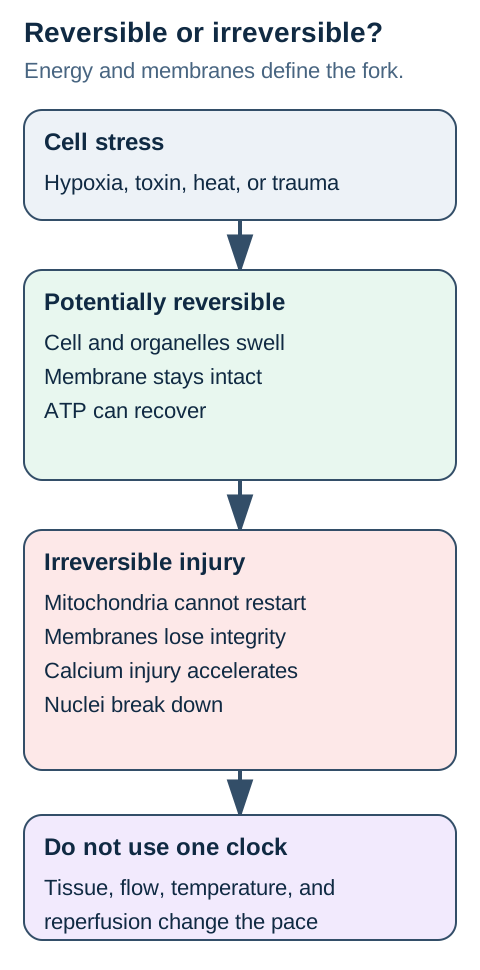
Cell injury is a contest between the intensity of the insult and the cell's remaining reserve. A short or mild insult may disrupt energy use, ion transport, protein synthesis, and lipid handling while membranes and mitochondria remain capable of recovery. A severe or sustained insult crosses into irreversible injury when mitochondria cannot restore oxidative phosphorylation and one or more critical membranes lose integrity. That distinction matters more than a memorized minute count because tissue type, collateral flow, temperature, workload, and intermittent perfusion all change the pace. [1 [1],2 [2],3 [3]]
Reversible injury enlarges cells before it destroys them. Sodium accumulates when the sodium-potassium adenosine triphosphatase slows, water follows, the endoplasmic reticulum dilates, membrane blebs appear, and ribosomes detach. Hepatocytes and other lipid-processing cells may also develop fatty change because energy loss disrupts synthesis, oxidation, and export of lipids. These findings are warning signs, not automatic proof of death. [1 [1],2 [2]]
Irreversible injury is a systems failure, not one isolated molecule. Persistent mitochondrial dysfunction, severe plasma-membrane injury, lysosomal leakage, uncontrolled cytosolic calcium, and nuclear destruction reinforce one another. Pyknosis is nuclear shrinkage and chromatin condensation, karyorrhexis is fragmentation, and karyolysis is fading from deoxyribonuclease activity. The sequence supports necrotic cell death, but it is not a universal stopwatch. [1 [1],2 [2]]
Figure 2. A recoverable branch retains membrane boundaries and energy restoration; the irreversible branch combines mitochondrial nonrecovery, membrane failure, calcium injury, and nuclear destruction.
Do not place a fixed minute at the fork. The border depends on tissue, perfusion, demand, and the severity of injury.
Adenosine triphosphate loss changes transport before it changes identity
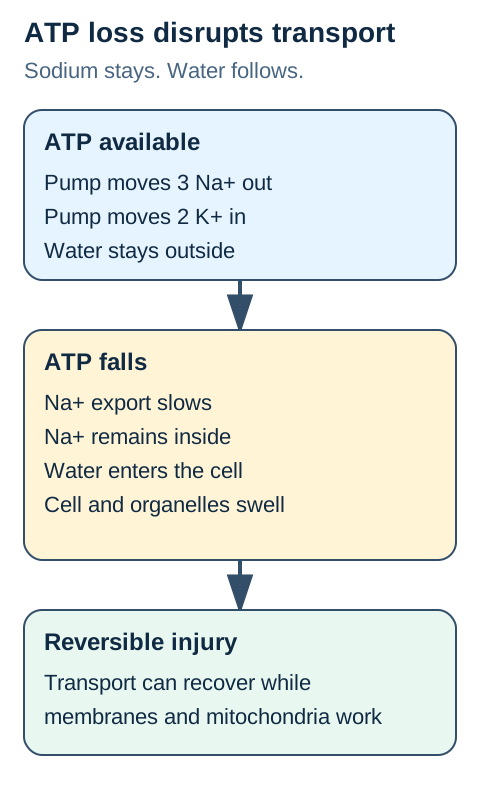
The sodium-potassium pump is the fastest mechanistic bridge from hypoxia to cell swelling. Reduced oxygen delivery slows oxidative phosphorylation, adenosine triphosphate falls, sodium export declines, potassium leaks out, and water enters the cell. The result is hydropic change with swollen cytoplasm and organelles. The cell may recover if energy supply returns before critical membranes and mitochondria are permanently damaged. [1 [1],2 [2]]
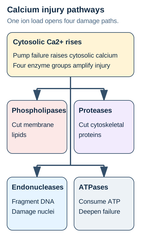
The calcium pump fails in parallel. Sarco-endoplasmic reticulum calcium adenosine triphosphatase normally stores calcium in the endoplasmic reticulum, and plasma-membrane pumps keep cytosolic calcium low. Energy loss and membrane damage permit calcium to accumulate in the cytosol and mitochondria. Calcium then activates phospholipases, proteases, endonucleases, and adenosine triphosphatases, so a pump problem becomes a self-reinforcing injury program. [1 [1],2 [2],6 [6]]
Ribosome detachment explains a different consequence of the same energy crisis. Dilated rough endoplasmic reticulum loses ribosomes, protein synthesis falls, and damaged or misfolded proteins accumulate. That finding does not directly pull water into the cell; it travels beside ion-pump failure as another reversible response until the stress becomes too severe. Matching each organelle to its consequence prevents a common distractor error.
Figure 1. Oxygen loss lowers adenosine triphosphate, slows sodium export, raises intracellular sodium, and draws water into a still-bounded cell.
The visual lock is simple: sodium stays, water follows, and the cell swells while its boundary can still recover.
Try it here · Checkpoint 1 of 3
Make your prediction before reading the choices. A first attempt is just a starting point.
Case 1
Show answer and explanations for case 1
A. Calcium-activated phospholipases destroy membrane lipids and rupture cells (Why this does not fit)
Calcium-dependent phospholipases can amplify severe ischemic injury, but the biopsy lacks the required destructive membrane change.
Reasoning steps for option A
Why could calcium enzymes matter in a threatened infarct?
Sustained severe injury may raise cytosolic calcium and injure membrane lipids.
What does the intact membrane in this early sample oppose?
It weighs against phospholipase-driven rupture as the cause of current swelling.
B. Caspases package fragmented nuclear material into apoptotic bodies (Why this does not fit)
Caspase activation could follow other tissue insults, yet apoptosis usually shrinks and packages isolated cells rather than enlarging an ischemic field.
Reasoning steps for option B
How would a caspase-associated death program package a myocyte?
It would form membrane-bound fragments for phagocytic clearance.
Which feature of the biopsy instead suggests osmotic stress?
Multiple swollen cardiomyocytes remain connected with preserved nuclei and membranes.
C. Low energy impairs sodium export, causing water influx (Best answer)
Energy loss slows the sodium-potassium pump, so sodium and water enter still-intact cells; no death-specific morphology is given.
Reasoning steps for option C
Which initial metabolic deficit follows coronary occlusion?
Oxygen delivery falls and myocytes make less adenosine triphosphate.
Why do intact enlarged cells retain an opportunity to recover?
The weakened sodium gradient draws in water without proving that the membrane has ruptured.
D. Ischemic proteins denature while cell outlines persist as ghost fibers (Why this does not fit)
Protein-denaturation-related ghost fibers explain a developed infarct, not nuclei-intact hydropic cells at the threatened edge.
Reasoning steps for option D
What would retained ghost architecture mean after an infarct?
A dead fiber can keep an outline after its proteins denature.
Which currently observed finding rules out using that morphology here?
The sampled cells are swollen and still retain intact nuclei and membrane boundaries.
E. Ribosome detachment directly forces potassium into the cytosol and draws water inward (Why this does not fit)
Ribosome detachment reduces protein synthesis but does not directly create the sodium-dependent osmotic gradient that explains early swelling.
Reasoning steps for option E
cell-01 alternative 5: Which observation would be required for this choice?
Protein synthesis would fall before structural recovery is assessed.
cell-01 alternative 5: Why does the keyed answer compare better?
The described water entry is governed by ion transport rather than ribosome position.
Takeaway: Early swelling points to energy-dependent ion pump failure; it does not by itself establish whether myocardium is dead.
The point of no return is a coupled membrane-mitochondria failure
Mitochondrial inability to recover is one anchor of irreversible injury. Severe permeability transition collapses the proton gradient, oxidative phosphorylation cannot restart, reactive oxygen species increase, and mitochondrial proteins can enter the cytosol. A second anchor is profound membrane dysfunction: the plasma membrane leaks cellular enzymes, lysosomes release acid hydrolases, and damaged organelles can no longer be compartmentalized. [1 [1],2 [2],6 [6]]
Cytosolic calcium links the anchors. Phospholipases cleave membrane phospholipids, proteases damage cytoskeletal and membrane proteins, endonucleases fragment nuclear material, and adenosine triphosphatases consume the cell's remaining energy. A selective phospholipase inhibitor should reduce lipid cleavage without directly blocking a calcium-activated protease. Applied questions often test that prediction rather than asking for a memorized enzyme list. [1 [1],2 [2]]
Membrane permeability separates a dead core from a threatened border. An extracellular tracer enters cells whose plasma membranes have failed. A neighboring swollen cell that excludes the tracer and later restores sodium transport may still be viable. The same logic applies to serum biomarkers: troponin or aminotransferases require leakage from injured cells, but the clinical interpretation still depends on tissue context and evidence of the underlying disease. [3]
Figure 3. Calcium activates four destructive enzyme groups: phospholipases, proteases, endonucleases, and adenosine triphosphatases.
Each enzyme class predicts a different consequence: lipid loss, cytoskeletal damage, nuclear fragmentation, or deeper energy depletion.
Apoptosis is packaged disposal, not quiet necrosis
Apoptosis shrinks and packages individual cells. Caspases condense chromatin, fragment the nucleus, and form membrane-bound apoptotic bodies that are rapidly phagocytosed. Because intracellular contents are contained, surrounding inflammation is usually limited. Physiologic examples include deletion of self-reactive lymphocytes, hormone-dependent involution, and removal of cells with irreparable deoxyribonucleic acid damage. [1]
Necrosis usually swells, ruptures, leaks, and inflames. The affected field may include many contiguous cells, membranes fail, cytosolic enzymes escape, and damage-associated molecules recruit inflammation. This morphology is not defined by whether the initial trigger was ischemic, toxic, infectious, or traumatic. The same insult can activate several death programs in different cells or at different times. [1]
Regulated necrotic programs do not erase the morphology distinction. Necroptosis, pyroptosis, and ferroptosis are molecularly regulated, yet they can still culminate in membrane failure and inflammatory signaling. For board transfer, first identify what the specimen shows, then use pathway details only when the vignette supplies a receptor, enzyme, iron, lipid-peroxide, or inflammasome signal. [1]
Necrosis patterns report the dominant process
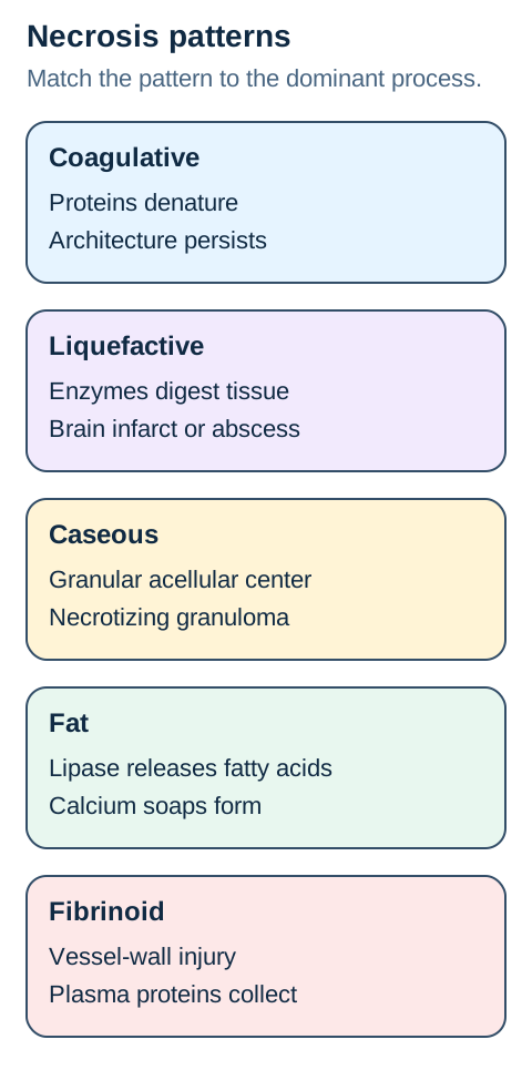
Coagulative necrosis preserves tissue outlines for a time. Protein denaturation slows proteolysis, so eosinophilic anucleate ghost cells remain recognizable. Ischemic infarcts in the heart, kidney, spleen, and most other solid organs use this pattern. The major exception is the brain, where enzymatic digestion dominates after infarction. [2 [2],4 [4],5 [5]]
Liquefactive necrosis digests tissue into viscous material or a cavity. Neutrophil enzymes create the liquid center of a bacterial abscess, while brain infarction evolves toward macrophage-rich digestion and cavitation. The organ does not dictate every outcome: one kidney can contain a coagulative arterial infarct and a separate liquefactive abscess because the mechanisms differ. [2]
Caseous necrosis is the granular center of a necrotizing granuloma. It is classically associated with tuberculosis and some fungal infections, but morphology alone does not name the organism. Acid-fast stains, fungal stains, culture, antigen testing, or molecular assays establish cause. A board item that shows a granuloma plus a detected fungus is testing integration, not the word caseous by itself. [2]
Fat necrosis follows local lipid destruction. In acute pancreatitis, escaped lipase releases fatty acids from triglycerides; fatty acids bind calcium and form chalky soaps, a process called saponification. Breast trauma can also damage adipocytes, producing oil cysts, calcification, and a mass that may mimic carcinoma. Pancreatic fat injury is enzymatic fat necrosis, not simply a synonym for liquefactive necrosis. [2 [2],7 [7],8 [8]]
Fibrinoid necrosis is centered in a vessel wall. Severe immune-mediated vascular injury or extreme hypertension permits plasma proteins and fibrin-like material to accumulate in the wall, producing bright eosinophilic change. Hyaline arteriolosclerosis is more homogeneous chronic wall thickening, while hyperplastic arteriolosclerosis forms concentric layers. Compartment is the fastest discriminator. [2]
Figure 4. Match the dominant process to the pattern: denaturation, digestion, granulomatous debris, lipid-calcium soaps, or vessel-wall injury.
Start with location and mechanism. Organ identity refines the answer but does not replace the mechanism.
Try it here · Checkpoint 2 of 3
Make your prediction before reading the choices. A first attempt is just a starting point.
Case 15
Show answer and explanations for case 15
A. Denatured acinar proteins preserve the outlines of infarcted fat (Why this does not fit)
Denaturation of ischemic acinar proteins might occur in severe pancreatitis, but ghost protein outlines do not explain calcium deposits inside fat.
Reasoning steps for option A
Why could coagulative injury coexist with pancreatic inflammation?
Severe regional ischemia can kill parenchymal cells.
What material must the chosen mechanism account for instead?
The operative report localizes chalky deposits to adipose tissue.
B. Immunoglobulin complexes trap fibrin within peripancreatic vessel walls (Why this does not fit)
Immune vascular injury can produce fibrin-like deposits, but peripancreatic fat nodules and low calcium point to saponification rather than arteriolar walls.
Reasoning steps for option B
Which compartment would immune-complex fibrinoid injury target?
Damage would primarily appear within small blood-vessel walls.
What site and chemistry do the current findings emphasize?
Necrotic fat with white calcium-containing deposits lies around the pancreas.
C. Neutrophil lysosomes turn adipose tissue into a purulent cavity (Why this does not fit)
Neutrophil-rich abscesses can liquefy inflamed tissues, but no pus cavity is described among the chalky fat flecks.
Reasoning steps for option C
Why could pancreatitis make inflammatory digestion plausible?
Severe inflammation attracts immune cells and enzyme release.
What distinguishes this biopsy from a pyogenic abscess?
The deposits are calcium soaps within fat rather than neutrophilic liquid debris.
D. Caspase packaging traps calcium inside intact apoptotic bodies (Why this does not fit)
Chalky peripancreatic deposits arise from fatty-acid binding to calcium, not from sequestration inside apoptotic fragments.
Reasoning steps for option D
cell-15 alternative 4: Which observation would be required for this choice?
The gross deposits are extracellular soaps in injured fat.
cell-15 alternative 4: Why does the keyed answer compare better?
The pancreatic enzyme signal points to lipid hydrolysis.
E. Liberated fatty acids bind calcium to form insoluble soaps (Best answer)
Pancreatic lipase injures nearby fat, and released fatty acids combine with calcium to form chalky soaps that can accompany lowered blood calcium.
Reasoning steps for option E
What enzyme can reach peripancreatic adipocytes in this illness?
First locate the lesion. Is the abnormal material inside parenchymal cells, in a pus-filled cavity, at the center of a granuloma, within adipose tissue, or in a vessel wall? That first localization prevents a bright pink vessel wall from being mislabeled as a solid-organ infarct and prevents chalky peripancreatic deposits from being called dystrophic calcification without recognizing fat saponification.
Next ask whether architecture is preserved or digested. Preserved ghost outlines favor coagulative necrosis. Loss of architecture with fluid debris favors liquefaction. A granular acellular center surrounded by epithelioid macrophages favors caseation. Shadowy adipocytes with basophilic calcium deposits favor fat necrosis. Then use the clinical trigger to confirm the pattern rather than substituting the trigger for the morphology.
Finally connect morphology to the first safe action. A stable traumatic breast oil cyst still requires appropriate imaging and tissue assessment when malignancy remains possible. Acute pancreatitis requires severity assessment, analgesia, moderately aggressive isotonic crystalloid rather than automatic aggressive loading, and early oral feeding as tolerated rather than routine prolonged fasting. Prophylactic antibiotics do not treat sterile pancreatic necrosis. [7 [7],8 [8]]
Laboratory leakage is evidence of injury, not a complete diagnosis. Cardiac troponin above the ninety-ninth percentile defines myocardial injury; a rise or fall indicates an acute process. Myocardial infarction additionally requires evidence of ischemia, such as ischemic symptoms, electrocardiographic changes, imaging evidence, or an identified coronary thrombus. Sepsis, tachyarrhythmia, pulmonary embolism, and renal disease can produce myocardial injury without satisfying the infarction definition. [3]
Reperfusion saves tissue and can injure it
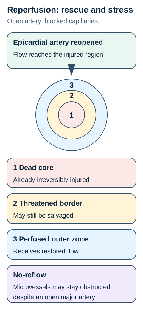
Ischemic death spreads as a wavefront rather than arriving everywhere at one fixed minute. The subendocardium is often injured first because wall stress and perfusion patterns leave it vulnerable. A central core may become irreversibly injured while a surrounding border remains threatened but viable. Collaterals, oxygen demand, temperature, intermittent flow, and the completeness of occlusion alter the map. Prompt reperfusion remains valuable because surviving tissue can still be rescued. [3 [3],4 [4],5 [5],6 [6]]
Reoxygenation can add a second injury to tissue that has already been stressed. Mitochondrial reactive oxygen species, calcium loading, inflammation, complement, endothelial dysfunction, and mitochondrial permeability transition can worsen cell damage. This does not mean reperfusion should be delayed. The correct synthesis is that flow restoration is necessary while reperfusion biology explains why an open artery does not guarantee complete tissue recovery. [6]
No-reflow is a microvascular problem behind a successful epicardial result. Endothelial swelling, capillary plugging, edema, hemorrhage, and microthrombi may impair tissue perfusion even after the large artery is open. Cardiac magnetic resonance imaging may show microvascular obstruction within an infarcted core while the peripheral zone has different viability. Separate epicardial patency from capillary perfusion when interpreting post-intervention findings. [6]
Figure 5. The ischemic field contains a dead core, a threatened border, and a microcirculation that may remain obstructed after large-vessel reopening.
Reperfusion is both rescue and stress: it restores substrate delivery while reactive oxygen species, calcium, and microvascular injury may add damage.
The brain has its own vulnerability map
Neurons depend heavily on continuous oxygen and glucose delivery. Energy failure disrupts ion gradients, glutamate handling, calcium control, and mitochondrial function. Global hypoperfusion can injure watershed zones between major arterial territories, while focal arterial occlusion follows a vascular distribution. The mature infarct usually becomes liquefactive and may leave a cystic cavity. [12 [12],13 [13],14 [14]]
Selective vulnerability means not every neuron responds equally. Hippocampal cornu ammonis 1 (CA1) pyramidal neurons, cerebellar Purkinje cells, and selected cortical neurons are classically sensitive to global ischemia. The pattern is a tendency, not an absolute rule, and severity, duration, age, temperature, and reperfusion alter it. Delayed neuronal death also means a patient can have evolving injury after circulation returns. [12 [12],13 [13]]
Watershed injury explains bilateral border-zone deficits after profound hypotension. A patient with cardiac arrest or severe shock may develop symmetric cortical or deep border-zone lesions even without one persistently blocked artery. In contrast, a middle cerebral artery infarct produces a focal territorial pattern. Use the distribution with the hemodynamic history rather than assuming every ischemic brain lesion has the same mechanism. [13 [13],14 [14]]
The lesion continues after the insult
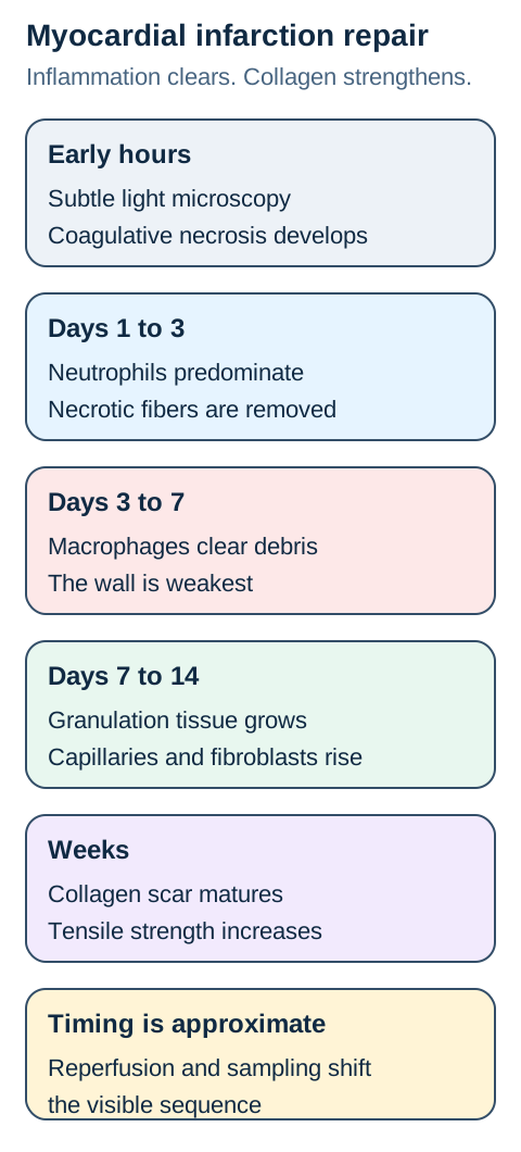
Acute myocardial infarction evolves from necrosis to inflammation to scar. Early light-microscopic findings may be subtle even when ultrastructural injury has begun. Neutrophils predominate over the next several days, macrophages remove dead tissue, granulation tissue grows from the margins, and collagenous scar matures over weeks. Exact timing varies with sampling, reperfusion, infarct size, and survival interval. [4 [4],5 [5]]
Mechanical weakness peaks while macrophages are clearing necrotic myocardium. Free-wall rupture, ventricular septal rupture, or papillary-muscle rupture classically occurs several days after a transmural infarct, before a strong collagen scar has formed. The complication depends on location: papillary-muscle rupture causes acute severe mitral regurgitation, septal rupture creates a new harsh murmur and left-to-right flow, and free-wall rupture can cause tamponade. [4 [4],5 [5]]
Granulation tissue is repair tissue, not a granuloma. It contains proliferating capillaries, fibroblasts, loose extracellular matrix, and inflammatory cells. A granuloma is a macrophage-organized immune structure. Confusing the similar names loses the causal story: granulation tissue fills and strengthens a wound, while a granuloma contains a persistent agent or immune stimulus. [15 [15],16 [16]]
Long-term outcome depends on what tissue can regenerate. Labile epithelia can proliferate if stem cells and matrix are preserved. Permanent cells such as cardiomyocytes have limited regenerative capacity, so a large infarct heals predominantly by fibrosis. In the brain, removed necrotic tissue leaves gliosis around a cavity rather than a collagen scar replacing neurons. [15 [15],16 [16]]
Figure 6. Myocardial injury progresses from subtle early change to neutrophils, macrophage-rich clearance, granulation tissue, and mature scar.
The clinical danger period follows biology: tissue is weakest during active clearance before collagen has matured.
Try it here · Checkpoint 3 of 3
Make your prediction before reading the choices. A first attempt is just a starting point.
Case 26
Show answer and explanations for case 26
A. Macrophage-rich clearance weakens the infarcted posteromedial papillary muscle before a strong collagen scar forms (Best answer)
Several days after infarction, macrophage removal of necrotic tissue can weaken the posteromedial papillary muscle and cause acute severe mitral regurgitation.
Reasoning steps for option A
cell-26 option 1: What case feature tests this alternative?
The inferior infarct and flail leaflet localize the structural failure.
cell-26 option 1: What comparison resolves the choice?
The timing falls within active debris removal before collagen maturation.
B. Neutrophils create a mature collagen scar that shortens the anterior papillary muscle (Why this does not fit)
Neutrophils do not create a mature scar, and mature collagen would strengthen rather than acutely rupture the papillary muscle.
Reasoning steps for option B
cell-26 option 2: What case feature tests this alternative?
Early inflammatory cells clear injury rather than synthesize durable collagen.
cell-26 option 2: What comparison resolves the choice?
Scar contraction is not the cause of a sudden flail leaflet on day 5.
C. Liquefactive necrosis converts the ventricular myocardium into a cystic cavity (Why this does not fit)
Myocardial infarction usually undergoes coagulative necrosis and fibrous repair, not the cavity-forming liquefaction typical of brain infarction.
Reasoning steps for option C
cell-26 option 3: What case feature tests this alternative?
The heart retains ghost architecture before repair.
cell-26 option 3: What comparison resolves the choice?
A cystic ventricular cavity does not explain the specific valvular apparatus failure.
D. Endocardial apoptosis silently removes chordae without inflammatory tissue weakening (Why this does not fit)
The acute post-infarct complication reflects necrotic structural failure, not isolated apoptotic removal of chordae.
Reasoning steps for option D
cell-26 option 4: What case feature tests this alternative?
The regional infarct places the papillary muscle at risk.
cell-26 option 4: What comparison resolves the choice?
A contained cellular program would not usually create abrupt gross rupture.
E. Dystrophic calcification stiffens the mitral leaflet during the first week (Why this does not fit)
Dystrophic calcification is not the expected first-week cause of a flail leaflet after infarction.
Reasoning steps for option E
cell-26 option 5: What case feature tests this alternative?
Mineral deposition is not the dominant event in this healing interval.
cell-26 option 5: What comparison resolves the choice?
The new regurgitation follows support-muscle failure rather than leaflet stiffening.
Takeaway: Connect macrophage-rich infarct clearance to papillary-muscle rupture.
Gangrene is a clinical state, not a fifth microscopic pattern
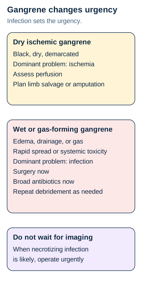
Dry gangrene is ischemic tissue with a dry, sharply demarcated appearance and no dominant infection. The underlying microscopic pattern is usually coagulative necrosis. Management requires perfusion assessment, protection of the tissue, risk-factor treatment, and vascular evaluation for possible revascularization or planned amputation. A clear boundary does not justify abandonment of follow-up. [10 [10],11 [11]]
Wet gangrene adds infection, edema, drainage, and enzymatic digestion. It can spread quickly and requires urgent source control, broad empiric antibiotics, and hemodynamic support as needed. Gas in soft tissue, severe pain, systemic toxicity, bullae, anesthesia, or rapid progression should raise concern for a necrotizing soft-tissue infection. Imaging can help define extent in selected stable patients, but it must not delay surgery when clinical suspicion is high. [9]
Clostridial myonecrosis is an operative emergency. Immediate exploration and debridement plus broad empiric therapy come first. Once clostridial myonecrosis is established, penicillin plus clindamycin is a recommended directed regimen because clindamycin suppresses toxin production while penicillin treats susceptible organisms. Hyperbaric oxygen must not delay debridement and is not a substitute for source control. [9]
Figure 7. Dry ischemic necrosis prompts perfusion planning; wet or gas-forming necrosis prompts immediate infection control and surgery.
The bedside split is infection and spread. Rapid progression overrides the desire for one more test.
Beyond: thresholds that change action
Do not diagnose myocardial infarction from troponin alone. Use the ninety-ninth-percentile upper reference limit for myocardial injury, look for a rise or fall to establish an acute process, and require evidence of ischemia before naming infarction. This distinction prevents sepsis-related, tachyarrhythmia-related, or renal-associated myocardial injury from being mislabeled. [3]
Do not use one ischemic duration as a universal declaration of death. Ultrastructural change may begin within minutes, routine light-microscopic necrosis takes longer to become visible, and the rate of irreversible injury varies across tissue and patient conditions. Treat time as a probability modifier while acting urgently to restore perfusion when indicated. [3 [3],4 [4],5 [5],6 [6]]
Acute pancreatitis management has changed away from reflexive fasting and maximal fluid loading. Begin oral feeding early as tolerated, prefer a low-fat solid diet when appropriate, use isotonic crystalloid with frequent reassessment, and avoid routine prophylactic antibiotics for sterile necrosis. A multicenter randomized trial found more fluid overload with aggressive resuscitation without improved outcomes, supporting a moderated and monitored strategy. [7 [7],8 [8]]
Critical limb-threatening ischemia needs vascular planning rather than a morphology-only label. Ischemic rest pain, nonhealing wounds, or gangrene attributable to peripheral artery disease warrants objective perfusion assessment and multidisciplinary evaluation. Infection, tissue loss, anatomy, procedural risk, and patient goals determine whether revascularization, debridement, or amputation is appropriate. [10 [10],11 [11]]
For documented group A streptococcal necrotizing fasciitis or clostridial myonecrosis, typical adult directed dosing in the Infectious Diseases Society of America table is penicillin G 2 to 4 million units intravenously every 4 to 6 hours plus clindamycin 600 to 900 mg intravenously every 8 hours. Verify against current labeling, organ function, allergies, culture results, and local guidance. Surgery remains the primary treatment. [9]
The safe referral rule is based on consequence. Suspected necrotizing infection, rapidly spreading wet gangrene, acute limb ischemia, new mechanical complication after infarction, or severe pancreatitis with organ failure requires urgent specialty care. The pathologic pattern explains what is happening; the physiologic threat determines how quickly the team must act.
Practice transfer
Answer each case by naming the dominant mechanism before reading the options. Then use location, architecture, membrane integrity, inflammation, time course, and physiologic threat to eliminate near-neighbor answers. A correct label without the causal link is fragile; a causal model transfers to unfamiliar specimens and management questions.
Case 2
Show answer and explanations for case 2
A. The transport pump can recover as energy supply returns (Best answer)
Restored supply can regenerate transport energy, matching normalization of membrane contour and organelles in serial samples.
Reasoning steps for option A
What mechanism could shrink a pale swollen tubule after unclamping?
Recovering oxidative phosphorylation can repower ion export.
How does the follow-up biopsy change a prediction based on initial vacuoles?
Recovered structure supports reversible damage rather than established cortical infarction.
B. Membrane blebs imply an evolving cortical infarct despite restored flow (Why this does not fit)
A wedge-shaped cortical infarct can follow severe artery obstruction, but recovered membranes and function weigh against permanent tissue death in this preparation.
Reasoning steps for option B
Why is cortical scarring possible during prolonged renal artery blockage?
Absent flow can kill a regional wedge of renal parenchyma.
Which observed recovery conflicts with that destination?
Serial samples regain normal contours and do not show persistent dead tissue.
C. Cytosolic protein leakage can persist after the membranes recover (Why this does not fit)
Membrane protein leakage could signal lethal injury elsewhere, but the reported membranes remain intact on serial assessment.
Reasoning steps for option C
What would allow tubular cytosolic proteins to spill outward?
The plasma-membrane barrier would need to be severely disrupted.
What tissue observation favors a reversible event here?
The tubules reestablish a normal boundary after perfusion returns.
D. The transient loss of filtration predicts medullary liquefaction (Why this does not fit)
Liquefactive destruction is associated with brain infarction or abscess, neither of which matches recoverable kidney tubules without a cavity.
Reasoning steps for option D
When does enzyme digestion create a fluid-filled lesion?
In an abscess or evolving cerebral infarct it can erase tissue structure.
What is incompatible with that claim in the unclamped kidney?
Recognizable intact tubules remain and their swollen organelles normalize.
E. Dystrophic calcification proves that the swollen mitochondria had crossed into irreversible injury (Why this does not fit)
Calcification can occur in dead tissue, but complete structural normalization after reperfusion directly supports reversible injury.
Reasoning steps for option E
cell-02 alternative 5: Which observation would be required for this choice?
Mineral deposition would not disappear with prompt restoration of perfusion.
cell-02 alternative 5: Why does the keyed answer compare better?
Observed return of architecture is stronger evidence than a hypothetical late deposit.
Takeaway: Swollen organelles with restored architecture support reversible injury, unlike membrane loss or necrosis.
A. Less membrane-lipid cleavage with similarly less cytoskeletal protein cleavage (Why this does not fit)
The inhibitor directly suppresses lipid digestion, but the experiment specifically leaves the protease activity that cuts cytoskeletal proteins unchanged.
Reasoning steps for option A
Why might protecting membranes secondarily appear to protect the scaffold?
A more stable cell boundary could potentially reduce overall stress downstream.
What stated selective intervention limits that inference here?
Proteases stay active, so cytoskeletal protein breakdown is not directly blocked.
B. Persistent membrane-lipid cleavage with reduced cytoskeletal protein cleavage (Why this does not fit)
This reversal would fit a selective protease inhibitor more closely, not an agent that suppresses phospholipases alone.
Reasoning steps for option B
What would need to be inhibited for the scaffold to be protected?
Calcium-activated proteases that cleave structural proteins.
Which enzyme is actually inhibited in group A?
Phospholipases, whose direct substrate is membrane lipid.
C. Persistent membrane-lipid cleavage with persistent cytoskeletal protein cleavage (Why this does not fit)
Structural protein digestion can persist, but lipid damage should decline when the membrane-targeted phospholipase is selectively inhibited.
Reasoning steps for option C
Why is continued structural injury a credible part of this prediction?
Protease activity does not change after the intervention.
Which separate result fails to incorporate the pharmacologic perturbation?
It forecasts no change in lipid cleavage despite blocking the enzymes that attack phospholipids.
D. Less membrane-lipid cleavage with persistent cytoskeletal protein cleavage (Best answer)
Which measured product should fall after the specified inhibitor?
Phospholipid cleavage products should decrease because the targeted lipase class is suppressed.
Which injury should persist in group A despite improved lipid integrity?
Active proteases still degrade cytoskeletal proteins while adenosine triphosphate remains low.
E. Less endonuclease activity with unchanged membrane-lipid cleavage (Why this does not fit)
The inhibitor targets phospholipases, so lipid cleavage should fall; it does not directly block calcium-activated endonucleases.
Reasoning steps for option E
cell-05 alternative 5: Which observation would be required for this choice?
The named drug target is the enzyme that cuts phospholipids.
cell-05 alternative 5: Why does the keyed answer compare better?
Nuclear nuclease activity lies outside the selective target.
Takeaway: Selective phospholipase inhibition predicts less lipid damage but persistent calcium-activated protease injury when proteases are not inhibited.
A. Pyknosis, karyorrhexis and karyolysis reflect nuclear injury (Best answer)
A shrunken dark nucleus can condense, fragment and finally fade, representing related nuclear morphologies in severely damaged cells rather than distinct diseases.
Reasoning steps for option A
Which microscopic description belongs to pyknosis?
A nucleus contracts into a dense, dark focus.
What distinguishes the other two regions in this infarct?
One shows broken nuclear pieces and the other loss of nuclear staining.
B. Small dense nuclei and absent nuclei both reflect metabolic adaptation (Why this does not fit)
Reversible chromatin clumping can occur with stress, but fragmentation and nuclear loss after infarction exceed transient swollen-cell changes.
Reasoning steps for option B
How could nuclear appearance change during an earlier reversible insult?
Chromatin might clump without complete nuclear destruction.
Why is the third region not merely hydropic change?
Its nuclei have faded away and tissue is already infarcted.
C. Condensation and fragmentation instead indicate quiet apoptotic deletion (Why this does not fit)
Apoptosis also involves chromatin changes, but scattered packaged bodies and quiet clearance are not described in these infarct zones.
Reasoning steps for option C
Why could condensation recall an apoptotic cell?
Individual apoptotic cells may display condensed chromatin.
Which broader finding argues against making every zone apoptosis?
The sampled territory is a contiguous arterial infarct with nuclear fragmentation and dissolution.
D. The nuclear pattern alone permits precise dating of the arterial occlusion (Why this does not fit)
Nuclear progression reflects degree and evolution of injury, but it cannot date each ischemic region to an exact minute.
Reasoning steps for option D
Why might older and younger nuclear changes coexist?
Perfusion deficits and collateral flow can vary across the infarct.
Which clinical claim would exceed these morphological observations?
Assigning a universal precise occlusion duration from the nuclei alone.
E. The nuclear sequence proves apoptosis because surrounding inflammation is not described (Why this does not fit)
Pyknosis, karyorrhexis, and karyolysis occur in lethal injury and do not by themselves establish apoptosis or a lack of inflammation.
Reasoning steps for option E
cell-06 alternative 5: Which observation would be required for this choice?
A missing statement about inflammation cannot override infarct morphology.
cell-06 alternative 5: Why does the keyed answer compare better?
Fading nuclei in a territorial infarct supports necrotic destruction.
Takeaway: Condensation, fragmentation and dissolution are characteristic nuclear changes in dying cells; their timing is not a universal clock.
A. Untreated: intact apoptotic bodies; inhibited: a greater number of similarly packaged bodies (Why this does not fit)
Untreated receptor signaling can package bodies, but caspase inhibition should dampen rather than amplify this caspase-dependent morphology.
Reasoning steps for option A
Which part of this prediction matches the untreated culture?
Membrane-bounded fragments can form downstream of a death receptor.
What action of the inhibitor disproves increased identical fragments?
It blocks the caspase cascade required for this ordered cleavage process.
B. Untreated: extensive cellular leakage; inhibited: fewer membrane-bound fragments (Why this does not fit)
Inhibition may diminish characteristic fragments, but the intact boundaries specified in untreated wells argue against extensive leakage as their main pattern.
Reasoning steps for option B
What makes cytosolic leakage a plausible toxicity concern?
Some severely injured tumor cells can rupture in other death pathways.
What observation opposes that interpretation for untreated cells?
Their contents remain enclosed in fragments ready for phagocytosis.
C. Untreated: widespread neutrophil-rich lysis; inhibited: the same lysis without nuclear change (Why this does not fit)
A culture of packaged isolated cells does not have the ruptured tissue or recruited neutrophils required for necrotic inflammation, and inhibition changes programmed fragmentation.
Reasoning steps for option C
What sample would support inflammatory lysis instead?
Ruptured clusters could spill contents and recruit inflammatory cells in perfused tissue.
Which findings here require different untreated and inhibited responses?
The untreated cells are membrane-bounded, while direct caspase inhibition interrupts their fragmentation.
D. Untreated cells swell and leak, while caspase-inhibited cells form more apoptotic bodies (Why this does not fit)
The observed membrane-bound fragments and limited leakage already identify apoptosis, and caspase inhibition should reduce rather than increase that packaging.
Reasoning steps for option D
cell-09 alternative 4: Which observation would be required for this choice?
The untreated morphology is contained rather than lytic.
cell-09 alternative 4: Why does the keyed answer compare better?
Blocking executioner enzymes reduces the characteristic fragments.
E. Untreated: many intact apoptotic bodies with little leakage; inhibited: fewer such bodies (Best answer)
An active receptor-caspase pathway packages cells quietly; direct caspase inhibition reduces that characteristic programmed fragmentation without proving another death pathway absent.
Reasoning steps for option E
Why does ligand exposure yield little extracellular cell content in untreated wells?
The shrinking cells partition their material into membrane-bound bodies that phagocytes can clear.
What characteristic morphology should decline when caspases are inhibited?
Fewer cells should complete ordered apoptotic fragmentation after death-receptor stimulation.
Takeaway: An extrinsic death-receptor signal engages caspases to package apoptotic bodies with little leakage; inhibition reduces that characteristic morphology.
What releases soluble cytosolic enzyme from the first specimen?
The disrupted cardiomyocyte membranes no longer retain cellular contents.
Why is the second specimen expected to show less leakage?
Membrane-bound apoptotic fragments are quietly ingested rather than spilling contents.
E. Both fluids contain similar enzyme concentrations because caspases perforate plasma membranes (Why this does not fit)
Caspase-mediated apoptosis usually preserves membrane containment until fragments are cleared, so it causes less extracellular enzyme release than necrosis.
Reasoning steps for option E
cell-10 alternative 5: Which observation would be required for this choice?
Apoptotic packaging limits exposure of cytosolic contents.
cell-10 alternative 5: Why does the keyed answer compare better?
Infarcted fibers have lost the barrier that normally retains enzymes.
Takeaway: Membrane rupture makes necrotic myocardium leak cytosolic enzymes and recruit inflammation; packaged apoptotic cells leak less.
A. Coagulation preserves neuronal cell outlines around the cavity (Why this does not fit)
Coagulative necrosis can retain outlines in a kidney after vascular blockage, but a brain cavity with absent tissue structure contradicts preserved neuronal ghosts.
Reasoning steps for option A
What does a coagulated renal wedge often retain initially?
Dead tubular outlines can persist while nuclei are lost.
Why is the cerebral lesion not described that way?
The original brain architecture has disappeared into a fluid-filled space.
B. Enzymatic digestion favors liquefactive necrosis in the brain (Best answer)
After a cerebral infarct, enzymatic digestion removes local architecture and can leave a fluid cavity unlike an ordinary ischemic kidney infarct.
Reasoning steps for option B
What happened to the normal parenchymal pattern at the stroke site?
The earlier neuronal and supporting tissue framework has been destroyed.
How does this organ change the usual solid-organ expectation?
Cerebral infarcts typically liquefy while ischemic kidney tissue usually coagulates.
C. Caseous granulomas surround ischemic neurons throughout the lesion (Why this does not fit)
Some brain infections provoke granulomas with necrotic centers, but the history is an arterial occlusion with cavity evolution rather than granulomatous inflammation.
Reasoning steps for option C
What finding would support a caseating brain infection?
A macrophage-rimmed acellular center and relevant pathogen evidence would help.
Which provided lesion history supports an ischemic alternative?
A named cerebral artery infarct preceded progressive liquefaction.
D. Fat saponification converts neuronal membranes to calcium soaps (Why this does not fit)
The brain contains lipids, yet typical cerebral infarct cavitation is enzymatic tissue digestion rather than pancreatic-type calcium soap formation.
Reasoning steps for option D
Why might the brain's lipid content suggest a fat-related idea?
A. Ischemic denaturation preserves recognizable parenchymal ghosts (Why this does not fit)
Coagulative preservation can occur in ischemic solid organs, but this lesion contains pus and has lost tissue outlines.
Reasoning steps for option A
Why might focal tissue loss suggest an ischemic infarct?
An arterial blockage can also produce a discrete nonviable region.
How does the aspirate change the mechanism?
Neutrophilic pus points to digestion, not retained ghosts.
B. Epithelioid macrophages form a granuloma around dry acellular debris (Why this does not fit)
Granulomatous infections can have necrotic centers, but a dense neutrophilic aspirate favors acute pyogenic abscess over epithelioid macrophage organization.
Reasoning steps for option B
What would make caseous necrosis reasonable?
A granuloma surrounding granular acellular debris would be characteristic.
Which cell type actually dominates the obtained sample?
Neutrophils are abundant in the aspirated pus.
C. Neutrophil-derived enzymes digest tissue into liquid debris (Best answer)
Neutrophils discharge tissue-digesting enzymes into a pyogenic abscess, turning the involved soft tissue into the pus-filled cavity observed.
Reasoning steps for option C
What does aspiration reveal about the inflammatory infiltrate?
Thick pus includes many neutrophils rather than an organized granulomatous rim.
What happens to adjacent thigh soft tissue under their enzymatic action?
It is broken down until a soft liquid center forms.
D. Lipase-mediated calcium soaps accumulate in necrotic adipocytes (Why this does not fit)
Fat necrosis can have chalky calcium soaps, but the central material here is purulent soft tissue rather than injured adipocytes.
Reasoning steps for option D
What substrate do lipases require for saponification?
Damaged adipose triglycerides must release fatty acids.
What is instead documented in the thigh lesion?
A cavity of neutrophils and digested soft tissue.
E. Apoptotic bodies are removed without recruiting inflammatory cells (Why this does not fit)
A neutrophil-rich pus-filled cavity with complete tissue digestion is not the contained, minimally inflammatory pattern of apoptosis.
Reasoning steps for option E
cell-13 alternative 5: Which observation would be required for this choice?
The abundant neutrophils indicate an inflammatory lytic process.
cell-13 alternative 5: Why does the keyed answer compare better?
Pus requires enzymatic tissue breakdown rather than isolated packaging.
Takeaway: Pyogenic abscesses show liquefactive digestion because neutrophil enzymes break down tissue.
A. Traumatic fat necrosis with local lipid release (Best answer)
Prior impact, an oil cyst and foamy macrophages around injured adipocytes support traumatic fat necrosis while biopsy excludes carcinoma in sampled tissue.
Reasoning steps for option A
Why does an oil cyst fit prior blunt breast trauma?
Damaged adipocytes release lipid and stimulate a local macrophage response.
Why still examine a tissue specimen rather than reassure from trauma alone?
Fat necrosis can resemble a breast malignancy clinically or radiographically.
B. Enzymatic fat necrosis from pancreatic lipase reaching breast tissue (Why this does not fit)
Pancreatic lipase causes fat necrosis near an inflamed pancreas, but this isolated breast nodule followed local impact with no pancreatic illness.
Reasoning steps for option B
Why is enzyme-mediated fat necrosis an initially plausible label?
Both causes can damage adipocytes and release stored lipid.
What selects a different cause for this patient's breast lesion?
A handlebar hit immediately preceded the nodule and imaging shows an oil cyst.
C. Fibrinoid arteriolar injury from immune-complex vasculitis (Why this does not fit)
Vasculitis can damage breast vessels, but the biopsied focus centers on adipocytes and macrophages rather than pink injured arteriole walls.
Reasoning steps for option C
What pathology would indicate fibrinoid injury?
Small-vessel walls would contain fibrin-like eosinophilic material.
Which cellular compartment instead explains the oil cyst?
Traumatized fat cells released lipid into the breast tissue.
D. Caseating granulomatous infection of the mammary parenchyma (Why this does not fit)
Granulomatous infection can produce a palpable lesion, but this core shows oil-related macrophages without a caseating macrophage-rimmed center.
Reasoning steps for option D
Why should clinicians not ignore infection in a breast mass?
Some inflammatory infections form nodules that mimic tumors.
What specific tissue evidence favors the traumatic alternative?
The biopsied lesion consists of damaged adipocytes and foamy lipid-clearing macrophages.
E. Coagulative necrosis from an arterial embolus to the breast (Why this does not fit)
The trauma, oil cyst, foamy macrophages, and damaged adipocytes favor traumatic fat necrosis rather than a territorial arterial infarct.
Reasoning steps for option E
cell-16 alternative 5: Which observation would be required for this choice?
The lesion follows local adipocyte disruption after blunt injury.
cell-16 alternative 5: Why does the keyed answer compare better?
An oil cyst records released lipid rather than preserved parenchymal ghosts.
Takeaway: Breast trauma can produce fat necrosis and oil cysts that clinically mimic a tumor; pathology should still rule out malignancy.
A. Renal epithelial ischemia preserves the outlines of necrotic tubules (Why this does not fit)
Renal cortical ischemia can yield tubular ghost outlines, but the pink material specifically occupies arteriole walls with neighboring glomeruli retained.
Reasoning steps for option A
What pattern would tubular coagulative necrosis highlight?
Anucleate tubular epithelial outlines in an ischemic parenchymal territory.
Which anatomical location disproves that primary diagnosis here?
The bright homogeneous material replaces vascular walls.
B. Severe vascular injury permits plasma proteins to accumulate in arteriolar walls (Best answer)
Severe immune-mediated vascular injury permits plasma proteins and fibrin-like material to accumulate where the biopsy shows pink wall replacement.
Reasoning steps for option B
What immune-injured structure is abnormal in the specimen?
The walls of small renal arterioles rather than surrounding tubules.
Why do they become homogeneous and eosinophilic?
Severe wall damage allows plasma proteins to enter and replace normal structure.
C. Neutrophils digest interstitial kidney tissue into fluid debris (Why this does not fit)
A bacterial renal abscess would collect neutrophils in a liquefying interstitial focus, not selectively make arteriolar walls uniformly pink.
Reasoning steps for option C
Why could renal infection cause acute kidney injury?
A focal suppurative lesion can damage nearby functioning tissue.
What feature lacks the architecture of an abscess?
No purulent cavity or neutrophil-rich center is reported; damage is wall-centered.
D. Macrophages surround granular necrosis inside glomerular granulomas (Why this does not fit)
Granulomas can contain necrotic debris and macrophages, yet no glomerular granuloma is present and the lesion is confined to arterioles.
Reasoning steps for option D
What morphology would establish caseation?
Granular cell-free material surrounded by organized macrophages.
Which compartment currently contains the pathology?
The walls of small arteries display the eosinophilic replacement.
E. Hyaline arteriolosclerosis from chronic low-grade wall thickening (Why this does not fit)
Hyaline change is smoother chronic thickening; destructive immune-mediated injury with fibrin-like wall material favors fibrinoid necrosis.
Reasoning steps for option E
cell-17 alternative 5: Which observation would be required for this choice?
The biopsy describes wall disruption rather than bland chronic thickening.
cell-17 alternative 5: Why does the keyed answer compare better?
The autoimmune setting supports severe inflammatory vascular injury.
Takeaway: Fibrinoid necrosis localizes to injured vessel walls and can accompany severe hypertension or immune-mediated vasculitis.
A. Dry gangrene; start empiric antibiotics without evaluating distal perfusion (Why this does not fit)
The classification fits, but no infection is described and neglecting documented poor arterial flow misses a key opportunity to protect adjacent viable tissue.
Reasoning steps for option A
Why might antibiotics be considered for a dead toe?
Devitalized tissue can later become secondarily infected.
Which current data make perfusion evaluation the more immediate focused step?
There is no redness or discharge, but Doppler demonstrates severe arterial insufficiency.
B. Wet gangrene; perform infectious source control without assessing arterial supply (Why this does not fit)
New moisture or spreading infection would change urgency, but the toe is dry and stable while perfusion is demonstrably compromised.
Reasoning steps for option B
When would infected wet gangrene merit urgent source control?
Moist gangrenous tissue accompanied by swelling, purulence or systemic toxicity.
What two current findings select another assessment?
No infection signs are present, and Doppler shows low distal arterial flow.
C. Dry gangrene; allow separation without follow-up because the boundary is clear (Why this does not fit)
Dry demarcation does not resolve the severe arterial deficit or establish that adjacent living tissues have adequate blood supply.
Reasoning steps for option C
Why could a sharp black boundary encourage observation?
It appears localized and lacks signs of spreading infection.
What outstanding problem still requires evaluation?
Reduced distal perfusion threatens tissue and may be amenable to a vascular plan.
D. Dry gangrene; assess arterial supply and potential revascularization with a vascular team (Best answer)
A clearly demarcated noninfected ischemic toe is dry gangrene; impaired distal flow warrants vascular evaluation rather than automatic antibiotic or watchful waiting.
Reasoning steps for option D
Which observations classify the lesion as dry ischemic gangrene?
Black mummified tissue and an absence of moist infectious or systemic findings.
Why is a vascular assessment needed even without infection?
Markedly reduced flow affects remaining tissue viability and possible revascularization.
E. Wet gangrene; schedule an outpatient biopsy before checking perfusion (Why this does not fit)
The tissue is dry and noninfected with absent pulses, so urgent perfusion assessment is more relevant than labeling it wet or delaying vascular evaluation.
Reasoning steps for option E
cell-19 alternative 5: Which observation would be required for this choice?
No drainage, edema, fever, or spreading infection is present.
cell-19 alternative 5: Why does the keyed answer compare better?
Absent pulses identify the arterial problem that guides planning.
Takeaway: A dry demarcated ischemic toe without infection warrants vascular perfusion assessment and management planning, not automatic infection treatment or neglect.
A. Reperfusion converts the dry toe to a moist swollen lesion (Why this does not fit)
Reopening blood supply can help viable adjacent tissue, but cannot itself produce fever and purulent drainage or resurrect already black necrotic tissue.
Reasoning steps for option A
Why might an improved perfusion state look different?
A viable margin could become better oxygenated and less painful.
Which new findings argue against perfusion alone explaining the conversion?
High temperature and drainage suggest a new infectious complication.
B. A new granulomatous reaction produces the malodorous drainage (Why this does not fit)
A necrotizing granuloma can destroy tissue, but a fast change with pus and spreading redness is unlike a macrophage-organized center.
Reasoning steps for option B
What morphology would caseating infection require?
A structured macrophage rim around granular acellular debris.
What syndrome does the rapid purulent progression instead suggest?
Acute bacterial infection atop an ischemic foot.
C. Extensive apoptosis in the margin produces the purulent exudate (Why this does not fit)
Apoptosis can remove isolated cells quietly; it does not account for grossly infected tissue with exudate and systemic fever.
Reasoning steps for option C
Why is apoptosis normally less inflammatory?
Fragments remain membrane-wrapped for orderly phagocytosis.
What evidence here points to exposed inflammatory tissue rather than quiet clearance?
The wound drains pus and erythema extends beyond the previous boundary.
D. Dystrophic calcification transforms the dry tissue into a moist infected lesion (Why this does not fit)
Calcification may occur in dead tissue but does not explain foul drainage, fever, edema, and rapid spread.
Reasoning steps for option D
cell-20 alternative 4: Which observation would be required for this choice?
Systemic illness and malodor point to infection.
cell-20 alternative 4: Why does the keyed answer compare better?
Moist tissue digestion requires microbial and inflammatory enzymes.
E. Superimposed infection adds liquefactive digestion to ischemic necrosis (Best answer)
New purulence, fever and spreading redness on an ischemic foot mean infection now superimposes enzymatic tissue digestion on prior dry necrosis.
Reasoning steps for option E
What changed from the formerly stable dry lesion?
The foot became swollen, moist, malodorous and systemically inflammatory.
Why does this alter the urgent clinical plan?
Spreading infected necrotic tissue requires evaluation for source control and antibiotics.
Takeaway: Wet gangrene adds infection and tissue digestion to ischemia; evaluate urgently for spreading infection and source control.
A. Urgent surgical exploration and debridement with broad empiric antibiotics (Best answer)
Gas in muscle plus rapidly escalating pain and shock raises concern for necrotizing myonecrosis that demands immediate debridement and broad therapy before final culture.
Reasoning steps for option A
Which anatomic finding makes this more than a skin-surface wound?
Gas tracks through deep thigh muscle on imaging.
What interventions cannot safely wait for the pathogen's final name?
Operative removal of devitalized tissue and empiric antimicrobial treatment.
B. Start broad antibiotics and monitor tissue progression before exploring (Why this does not fit)
Empiric antibiotics are essential, but observation alone leaves devitalized muscle and toxin-producing infection in place.
Reasoning steps for option B
Why might an antibiotic-first strategy seem convenient?
Drug administration can begin at the bedside without operating room preparation.
What feature makes immediate exploration indispensable?
Crepitus, deep muscle gas and instability imply ongoing tissue destruction requiring source control.
C. Use hyperbaric oxygen alongside antibiotics before definitive muscle exploration (Why this does not fit)
Hyperbaric oxygen has been studied as an adjunct, but guidelines caution against using it to postpone immediate surgery for gas gangrene.
Reasoning steps for option C
What appealing rationale might support oxygen therapy?
Anaerobic organisms can be sensitive to high-oxygen environments.
Why is it not the next definitive action here?
The patient needs urgent debridement and stabilization rather than delay for a chamber.
D. Obtain deep aspirate and await species identification before operation (Why this does not fit)
Aspirate cultures can guide later narrowing, but awaiting species confirmation before surgery risks progression in a critically ill patient.
Reasoning steps for option D
Why obtain samples during surgical exploration?
Deep material can establish the organism and tailor treatment.
Why should testing proceed alongside rather than before source control?
Rapidly progressive muscle necrosis and shock demand action immediately.
E. Begin anticoagulation and defer surgery until perfusion imaging is complete (Why this does not fit)
Shock, crepitus, and intramuscular gas after contamination demand immediate source control, not delay for vascular imaging.
Reasoning steps for option E
cell-21 alternative 5: Which observation would be required for this choice?
The dominant threat is toxin-producing tissue infection.
cell-21 alternative 5: Why does the keyed answer compare better?
Definitive treatment requires removal of devitalized infected muscle.
Takeaway: Suspected gas gangrene or necrotizing myonecrosis requires immediate surgery and empiric antibiotics; do not delay source control for tests.
A. An open epicardial artery implies complete recovery of tissue-level perfusion (Why this does not fit)
An open epicardial artery improves proximal supply, yet it does not guarantee uniform delivery through damaged capillary beds.
Reasoning steps for option A
Why could a clinician expect improving flow after the procedure?
The proximal occlusion has been removed.
What measured regional mismatch disproves complete tissue perfusion?
The core remains poorly perfused despite restored large-vessel patency.
B. The core findings favor isolated reversible edema without vascular damage (Why this does not fit)
Edema may be present after infarction, but the imaging explicitly identifies low perfusion of the core relative to a better-perfused rim.
Reasoning steps for option B
Why can postischemic swelling affect a cardiac image?
Injury can increase tissue water and change magnetic resonance signal.
Which provided variable requires addressing microvascular flow?
Regional perfusion differs between core and rim even while the artery is open.
C. The perfused margin cannot contain viable myocardium after infarction (Why this does not fit)
Core necrosis does not imply every cell in the rim has died; the better-perfused periphery may remain salvageable.
Reasoning steps for option C
Which region appears most damaged from the scan?
The infarct core has persistent poor perfusion.
Why can the peripheral zone respond differently to reperfusion?
Its lesser injury and improved microvascular delivery may support survival.
D. Microvascular obstruction can persist despite an open artery; the rim may remain viable (Best answer)
A patent upstream artery and poorly perfused core can coexist if capillaries are blocked or damaged; the better-perfused rim may still benefit from timely flow.
Reasoning steps for option D
What does the angiogram actually establish?
The culprit large artery is open after intervention.
Which distinct vascular level explains the magnetic resonance finding?
Microcirculation inside the infarct core may still be obstructed.
E. The dark core proves complete capillary reperfusion throughout the infarct (Why this does not fit)
A nonperfused core despite an open artery is evidence against complete capillary recovery and supports microvascular obstruction.
Reasoning steps for option E
cell-24 alternative 5: Which observation would be required for this choice?
Dark tissue on perfusion imaging is not receiving normal microvascular flow.
cell-24 alternative 5: Why does the keyed answer compare better?
Large-vessel success and tissue-level perfusion are separate observations.
Takeaway: Assess the microcirculation and infarct distribution rather than equating an open artery with complete capillary recovery.
A. Both renal lesions remain coagulated because solid-organ identity controls every injury (Why this does not fit)
The embolic infarct can coagulate, but purulent neutrophil digestion can liquefy tissue even within the same kidney.
Reasoning steps for option A
Why is kidney infarction often structurally preserved?
Protein denaturation maintains anucleate tubular ghosts for a time.
What independent insult changes the new lesion's histology?
An abscess adds neutrophils and their tissue-digesting enzymes.
B. Both lesions liquefy because pus diffuses throughout the previously ischemic kidney (Why this does not fit)
The aspirated abscess is a distinct focus and does not imply that the whole prior infarct has lost its retained outlines.
Reasoning steps for option B
Why might nearby pus make widespread digestion sound plausible?
Neutrophil enzymes can destroy tissue locally.
Which localization prevents applying that to the entire kidney?
The pus comes from a discrete new lesion separate from the previously described infarct.
C. The abscess becomes caseous because infection follows the earlier ischemic insult (Why this does not fit)
Caseation needs a necrotizing granuloma, not simply the sequence of a vascular embolus followed by neutrophilic pus.
Reasoning steps for option C
What inflammatory structure would favor caseous necrosis?
An epithelioid macrophage rim would encircle granular acellular debris.
What current aspirate instead identifies a pyogenic focus?
Thick neutrophilic material supports enzyme-driven liquefaction.
D. The infarct becomes fibrinoid because every renal necrosis pattern is centered in vessels (Why this does not fit)
The wedge-shaped cortical lesion is parenchymal coagulative necrosis; fibrinoid change specifically involves the vessel wall.
Reasoning steps for option D
cell-25 alternative 4: Which observation would be required for this choice?
Ghost tubules localize the lesion to renal parenchyma.
cell-25 alternative 4: Why does the keyed answer compare better?
A separate abscess proves the same organ can display another mechanism.
E. Renal infarct keeps ghost tubules; the separate abscess liquefies (Best answer)
Vascular ischemia denatures proteins and preserves renal tubular ghosts; neutrophilic enzymes digest the separate infected focus into liquid debris.
Reasoning steps for option E
What happens after renal artery embolism?
Ischemic tubules can preserve dead cell outlines temporarily.
What does the aspirated neutrophilic pus predict?
Enzymes digest the new infected focus into liquid debris.
Takeaway: The same kidney can show coagulative arterial infarction and liquefactive abscess because the mechanism, not organ identity alone, determines morphology.
A. A single middle cerebral artery embolus selectively destroys every vulnerable region (Why this does not fit)
One focal embolus does not explain bilateral watershed lesions plus hippocampal and cerebellar involvement.
Reasoning steps for option A
cell-27 option 1: What case feature tests this alternative?
The abnormalities cross one arterial territory.
cell-27 option 1: What comparison resolves the choice?
The history supplies systemic circulatory failure rather than a focal blockage.
B. Global substrate failure injures border zones and neurons with high metabolic and excitotoxic susceptibility (Best answer)
Global hypoperfusion preferentially injures watershed regions and selectively vulnerable neurons such as cornu ammonis 1 (CA1) pyramidal cells and Purkinje cells.
Reasoning steps for option B
cell-27 option 2: What case feature tests this alternative?
Low systemic flow first threatens the least-perfused arterial borders.
cell-27 option 2: What comparison resolves the choice?
Regional energy demand and glutamate-calcium biology shape neuronal susceptibility.
C. Coagulative necrosis preserves neural architecture because the brain is a solid organ (Why this does not fit)
Brain infarction is the major solid-organ exception and generally evolves toward liquefactive digestion.
Reasoning steps for option C
cell-27 option 3: What case feature tests this alternative?
Neural tissue does not retain ghost architecture like myocardium.
cell-27 option 3: What comparison resolves the choice?
The mature lesion tends toward macrophage-rich tissue removal.
D. Fibrinoid necrosis of cerebral arterioles directly creates symmetric watershed bands (Why this does not fit)
Fibrinoid change is a vessel-wall lesion and does not by itself explain the global perfusion pattern and selective neuronal loss.
Reasoning steps for option D
cell-27 option 4: What case feature tests this alternative?
The defining distribution follows low-flow borders.
cell-27 option 4: What comparison resolves the choice?
The named neurons are vulnerable even without diffuse wall-centered fibrinoid injury.
E. Caspase inhibition in Purkinje cells causes inflammatory membrane rupture throughout the cortex (Why this does not fit)
The vignette does not provide caspase inhibition, and global ischemia engages multiple injury mechanisms rather than one engineered pathway.
Reasoning steps for option E
cell-27 option 5: What case feature tests this alternative?
No treatment altering caspases was given.
cell-27 option 5: What comparison resolves the choice?
The anatomic pattern is more informative than an unsupported molecular intervention.
Takeaway: Use distribution and cell type to recognize selective vulnerability in global ischemia.
A. Type 1 myocardial infarction from plaque rupture (Why this does not fit)
Type 1 infarction requires an acute coronary atherothrombotic event with evidence of ischemia, which is not provided.
Reasoning steps for option A
cell-32 option 1: What case feature tests this alternative?
No ischemic syndrome or coronary thrombosis is described.
cell-32 option 1: What comparison resolves the choice?
Dynamic troponin alone does not establish plaque rupture.
B. Acute myocardial injury without established myocardial infarction (Best answer)
A troponin rise and fall above the ninety-ninth percentile establishes acute myocardial injury, but infarction requires additional evidence of ischemia.
Reasoning steps for option B
cell-32 option 2: What case feature tests this alternative?
The biomarker pattern is acute rather than chronic.
cell-32 option 2: What comparison resolves the choice?
The required ischemic symptoms, tracing changes, imaging findings, or thrombus are absent.
C. Chronic myocardial injury because sepsis cannot cause an acute troponin change (Why this does not fit)
A rise and fall defines an acute process, and critical illness can cause acute myocardial injury.
Reasoning steps for option C
cell-32 option 3: What case feature tests this alternative?
Serial change argues against a stable chronic baseline.
cell-32 option 3: What comparison resolves the choice?
Sepsis can stress myocardium without proving coronary infarction.
D. Myocardial infarction solely because troponin exceeds the reference limit (Why this does not fit)
The reference-limit threshold defines myocardial injury, not infarction in the absence of ischemic evidence.
Reasoning steps for option D
cell-32 option 4: What case feature tests this alternative?
The biomarker establishes cell injury but not its cause.
cell-32 option 4: What comparison resolves the choice?
The universal definition requires a second ischemic component.
E. Coagulative necrosis confirmed by the laboratory value alone (Why this does not fit)
Troponin leakage does not by itself establish a microscopic necrosis pattern or the clinical diagnosis of infarction.
Reasoning steps for option E
cell-32 option 5: What case feature tests this alternative?
A serum marker cannot directly display retained tissue architecture.
cell-32 option 5: What comparison resolves the choice?
Morphology and clinical etiology require separate evidence.
Takeaway: Separate acute myocardial injury from myocardial infarction.