Squamous lung carcinoma: tissue, calcium and local anatomy
Level 1 and Step 1 connect eosinophilic keratin-rich cytoplasm, keratin pearls, intercellular bridges, stromal invasion and PTHrP physiology.
One lesson, three clinical depths. A central lung mass, high calcium and pink tumor cells are clues, not a complete diagnosis. At Level 1 and Step 1, explain why keratin-rich cytoplasm is eosinophilic, how keratin pearls and intercellular bridges support squamous differentiation, how stromal breach establishes invasion and how PTHrP raises calcium. At Level 2 and Step 2 CK, combine morphology, imaging, biochemistry and sampling to classify and stage the disease. At Level 3 and Step 3, manage dangerous complications, coordinate definitive therapy and monitor the patient over time. The Beyond section adds tissue stewardship, referral thresholds and multidisciplinary pitfalls.
Use the same evidence at three depths
Level 1 and Step 1. Start with cell identity and mechanism. Pink, abundant cytoplasm reflects eosin binding to protein-rich cytoplasm, especially keratin. Concentric keratin within malignant epithelial nests forms a keratin pearl. Desmosome-linked intercellular bridges reinforce the squamous pattern. None of those findings alone proves stromal invasion.
Level 2 and Step 2 CK. Ask what the tissue establishes, what the sample cannot establish and which additional test answers the remaining question. A central location, smoking exposure, cavitation or hypercalcemia can change probability, but morphology, an appropriate immunostain panel and staging studies drive the diagnosis and treatment plan.
Level 3 and Step 3. Separate immediate threats from cancer control. Symptomatic hypercalcemia, airway compromise, superior vena cava obstruction, focal neurologic deficits and treatment toxicity require parallel assessment while the multidisciplinary team addresses stage and fitness.
Beyond the boards. Preserve tissue, compare prior tumors when origin is uncertain, document the biochemical mediator instead of assuming it, and verify any drug regimen against current labeling and institutional protocols.
A bronchial mass explains airflow, not cell type
Airway pattern. Squamous cell carcinoma is a malignant epithelial tumor showing squamous differentiation. Tobacco exposure is an important risk factor. Many pulmonary squamous tumors involve central bronchi, but peripheral tumors occur. Small cell carcinoma can also be central and smoking-associated. Start with the location to understand symptoms; use tissue to classify the tumor. Neither smoking history nor geography can replace a biopsy. [1][2][5]
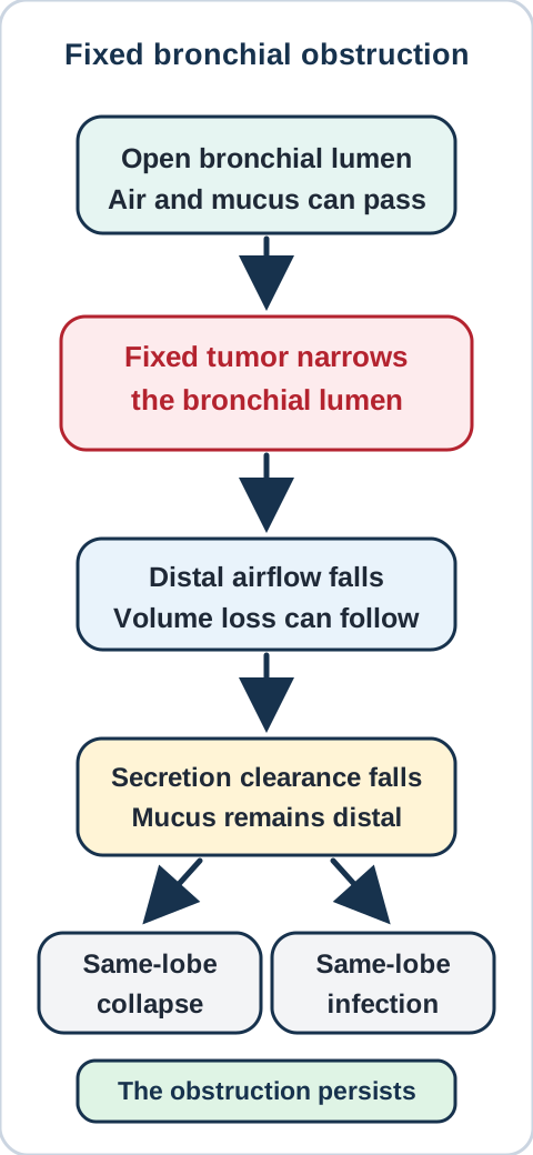
Airway pattern. A tumor projecting into a bronchus narrows a fixed part of the airway. This can produce a localized, single-pitched wheeze rather than widespread variable wheezing. Distal ventilation and secretion clearance decline. The same downstream lobe may repeatedly become infected or collapse. Surface bleeding can cause hemoptysis. Necrosis within a tumor can produce cavitation, but infection and other tumors can also cavitate. A cavity is a structural observation, not a histologic diagnosis. [2]
Connect a fixed bronchial lesion to focal wheeze and recurrent same-lobe opacity. [2] Bone Wizardry; original construction.
Airway pattern. Trace the air path in the drawing. Now imagine that antibiotics clear the infection but leave the obstruction unchanged. Which problem should remain?
Check the airway prediction
Airway pattern. The fixed narrowing remains, so secretion retention and recurrent opacity in the same distribution can recur. Improvement of fever does not establish that the underlying obstruction has disappeared.
Airway pattern. Use persistent symptoms to frame the next clinical question. Repeated same-lobe pneumonia, a focal wheeze, unexplained hemoptysis or weight loss warrants evaluation for an obstructing lesion. Clinical assessment, appropriate imaging and tissue sampling address different parts of that question. After cancer is confirmed, its extent and the patient's functional condition matter for treatment planning; the name of a tumor alone does not select an operation or drug regimen. [2]
Try it here · Checkpoint 1 of 3
Make your prediction before reading the choices. A first attempt is just a starting point.
Case 6
Show answer and explanations for case 6
A. Intermittent widespread bronchial smooth-muscle contraction (Why this does not fit)
A widespread reversible airway process can cause variable wheezing in several regions. A fixed bronchial lesion with persistent volume loss in one lobe is more anatomically specific. Match the distribution and persistence of symptoms to the obstruction.
Reasoning steps for option A
What distribution is typical of a generalized reversible airway process?
A widespread reversible airway process can cause variable wheezing in several regions.
In case lung-scc-06, which stated detail most directly tests option A?
A fixed bronchial lesion with persistent volume loss in one lobe is more anatomically specific.
After resolving case lung-scc-06, what transferable rule distinguishes option A?
Match the distribution and persistence of symptoms to the obstruction.
B. Repeated embolic obstruction of pulmonary arterial branches (Why this does not fit)
An embolus obstructs pulmonary blood flow. The demonstrated lesion obstructs an airway and explains distal secretion retention in the recurring distribution. Distinguish loss of perfusion from loss of airway clearance.
Reasoning steps for option B
What primary structure is affected by a pulmonary embolus?
An embolus obstructs pulmonary blood flow.
In case lung-scc-06, which stated detail most directly tests option B?
The demonstrated lesion obstructs an airway and explains distal secretion retention in the recurring distribution.
After resolving case lung-scc-06, what transferable rule distinguishes option B?
Distinguish loss of perfusion from loss of airway clearance.
C. Diffuse hydrostatic flooding of alveolar spaces (Why this does not fit)
Hydrostatic edema can produce pulmonary opacities through increased vascular pressure. The fixed focal wheeze and repeatedly affected downstream lobe instead track the bronchial lesion. Do not use a diffuse mechanism when a focal structural cause explains the pattern.
Reasoning steps for option C
What pattern can high pulmonary venous pressure produce?
Hydrostatic edema can produce pulmonary opacities through increased vascular pressure.
In case lung-scc-06, which stated detail most directly tests option C?
The fixed focal wheeze and repeatedly affected downstream lobe instead track the bronchial lesion.
After resolving case lung-scc-06, what transferable rule distinguishes option C?
Do not use a diffuse mechanism when a focal structural cause explains the pattern.
D. Persistent impairment of distal ventilation and secretion clearance (Best answer)
Air entry and secretion clearance decline downstream from the obstruction. Antibiotics can treat infection without correcting the fixed lesion that leaves the same lobe vulnerable. Recurrent infection in one distribution warrants assessment of the underlying airway.
Reasoning steps for option D
What changes downstream from a fixed bronchial narrowing?
Air entry and secretion clearance decline downstream from the obstruction.
In case lung-scc-06, which stated detail most directly tests option D?
Antibiotics can treat infection without correcting the fixed lesion that leaves the same lobe vulnerable.
After resolving case lung-scc-06, what transferable rule distinguishes option D?
Recurrent infection in one distribution warrants assessment of the underlying airway.
Takeaway: Resolution of fever does not prove resolution of a persistent bronchial obstruction.
What makes the pink material useful? In hematoxylin and eosin staining, eosinophilic cytoplasm is pink-staining cytoplasm. It does not mean that the tumor is made of eosinophils. Keratinizing squamous carcinoma can contain polygonal malignant cells with conspicuous cytoplasm, abnormal nuclei and concentric accumulations of keratin within tumor nests. These accumulations are keratin pearls. Intercellular bridges reflect prominent cell contacts associated with desmosomes, which link intermediate filaments to mechanical cell-cell attachments. [16] Read these features in their malignant epithelial context, not as isolated proof from one pink structure. [1][3]
Pink is a cytoplasmic clue, not a diagnosis. Abundant eosinophilic cytoplasm points toward a protein-rich, keratinizing phenotype. By contrast, a basaloid population is often described as smaller and more blue or basophilic, with less visible cytoplasm. Use that color-and-cytoplasm contrast to generate the differential, then let architecture, atypia, keratinization, immunophenotype and tissue boundaries decide it. A small biopsy may miss a keratin pearl, so absence of a pearl does not exclude squamous differentiation.
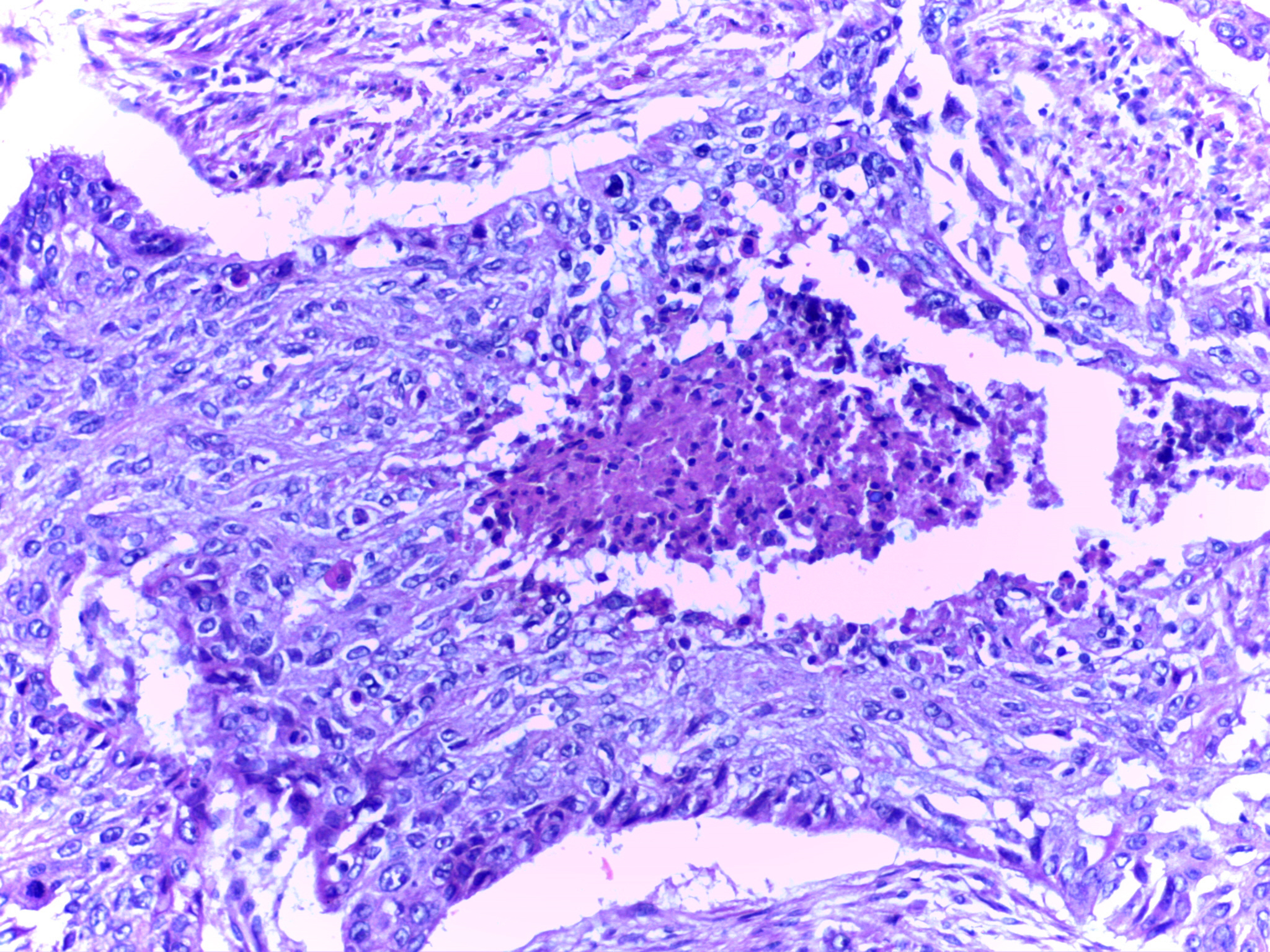
Inspect the solid pink keratinized material within the tumor nests. This photograph shows pulmonary squamous carcinoma. This field alone does not establish stage, primary origin or a calcium mechanism. [3]Department of Pathology, Calicut Medical College; CC BY-SA 4.0. No alterations.
Squamous morphology. In the photograph, follow the curved arrangement of cells around the pale pink keratinized center. Compare that solid accumulation with an open glandular lumen. The source identifies pulmonary squamous carcinoma; this single field does not establish the patient's stage, calcium mechanism or primary site by itself.
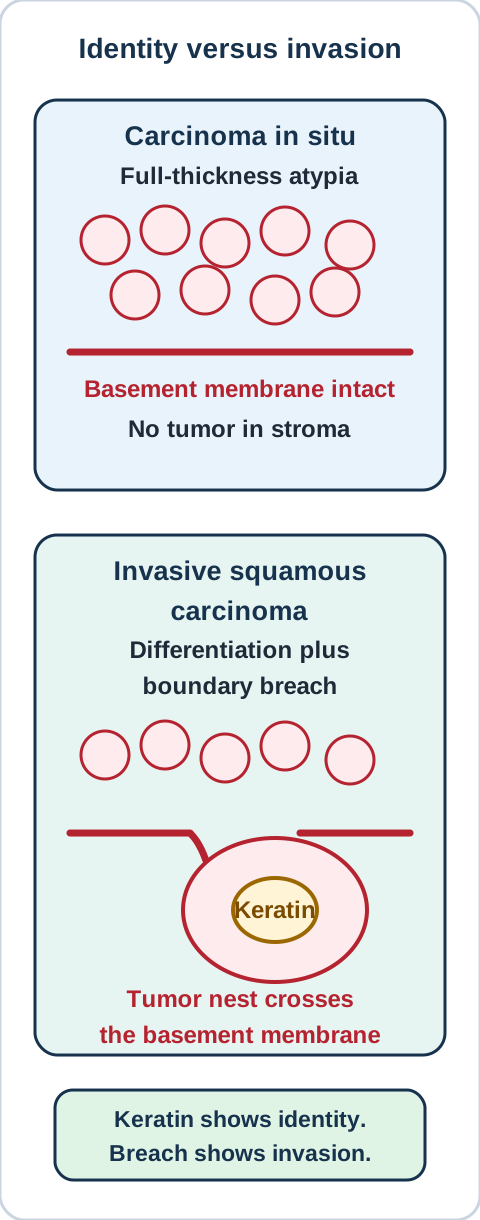
Squamous morphology. The normal large-airway lining is predominantly pseudostratified ciliated columnar epithelium, not stratified squamous epithelium. Chronic injury can favor squamous metaplasia, an adaptive change in epithelial differentiation. Dysplasia adds disordered maturation and cytologic atypia. Full-thickness atypia confined above the basement membrane is carcinoma in situ; invasion requires extension into the underlying tissue. These are useful distinctions, not a compulsory sequence in which every metaplastic patch becomes cancer. Persistent dysplasia identifies increased risk, while individual lesions can regress or remain noninvasive. [15]
Separate squamous differentiation from proof of stromal invasion. [1][15] Bone Wizardry; original construction.
Squamous morphology. Point to the finding that changes the lower panel from an identity question to an invasion question.
Check the boundary distinction
Squamous morphology. The tumor nest extends below the basement membrane into stroma. Keratin production identifies differentiation, whereas its relationship to the surrounding tissue establishes invasion. A superficial sample may show differentiation without resolving the deepest extent.
Squamous morphology. Not every sample contains pearls or obvious bridges. In a poorly differentiated non-small cell carcinoma, a focused immunostain panel can help: substantial nuclear p40 staining supports squamous differentiation, while TTF1 can support pulmonary adenocarcinoma in the appropriate context. p40 is more specific for squamous differentiation than p63. Weak focal p40 or p63 positivity alone should not force a squamous diagnosis.
Keratinizing areas can be p40-negative; unequivocal keratinizing morphology should not be discarded because that component does not stain. A limited, nondiagnostic sample may remain non-small cell carcinoma, not otherwise specified, rather than being labeled large cell carcinoma from insufficient tissue. [1]
Squamous morphology. There is another boundary: differentiation versus origin. A p40-positive squamous tumor in the lung can be primary or metastatic. Prior skin, head and neck, cervical or other squamous tumors require clinical comparison, review of prior morphology and appropriate additional testing. No single routine immunostain proves pulmonary origin in every squamous tumor. For transfer, ask what information is missing when a lung biopsy and an earlier head and neck tumor both keratinize. [1]
A circulating signal can change bone and kidney function
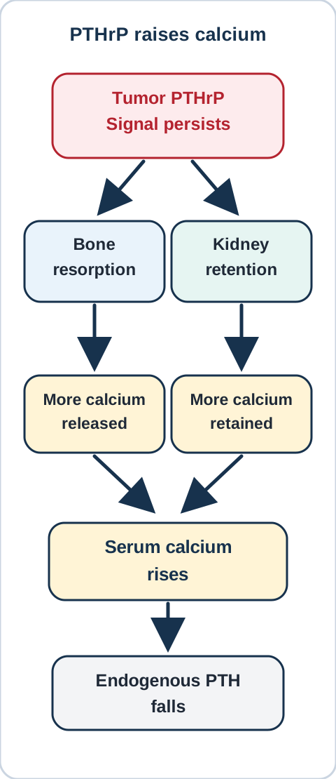
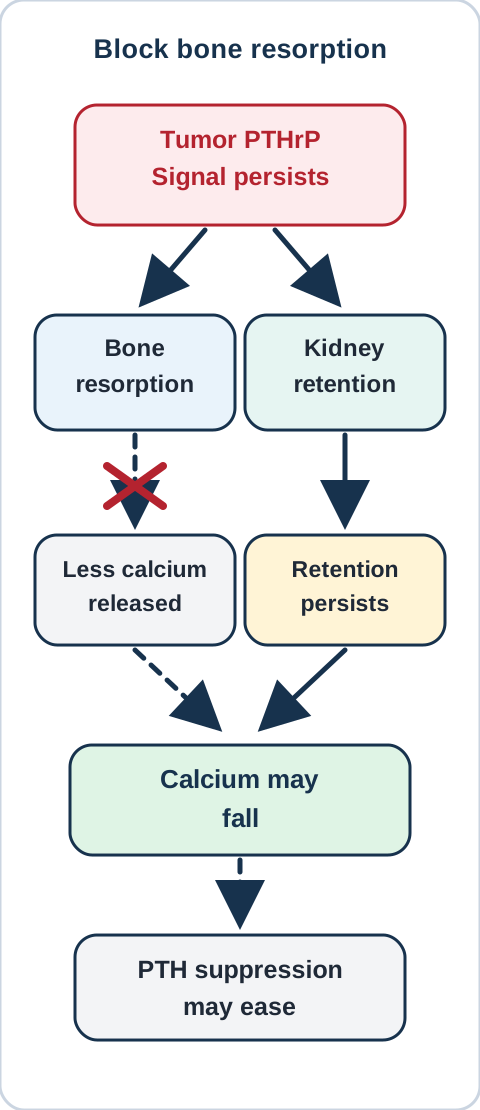
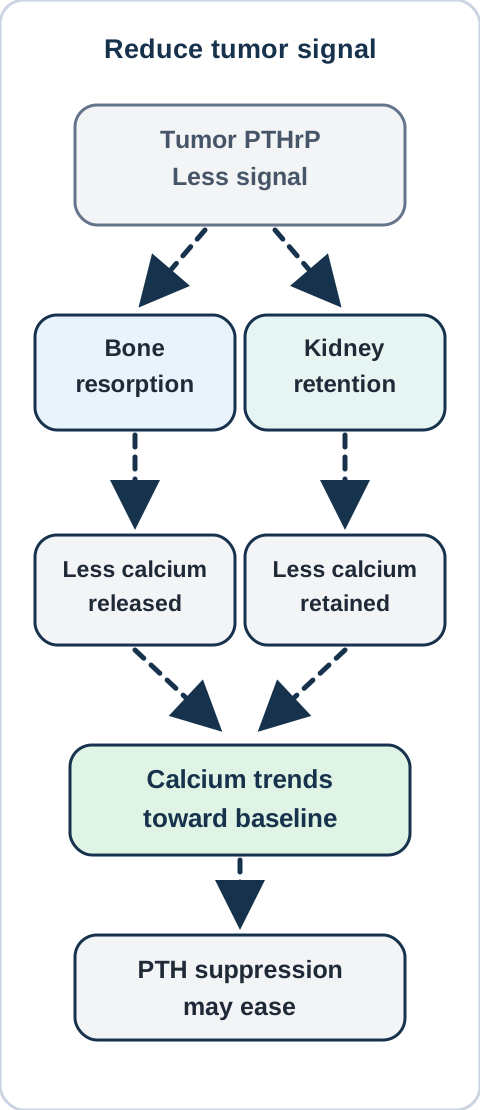
Why can calcium rise while native PTH falls? Some squamous carcinomas produce parathyroid hormone-related peptide, or PTHrP. Its PTH-like receptor effects promote calcium release through osteoclast-mediated bone resorption and increase renal calcium reabsorption. Signaling through osteoblast-lineage cells favors RANKL-dependent osteoclast activity; the simplified diagram groups these intermediate steps into its bone pathway. PTHrP also reduces renal phosphate reabsorption. Thus high calcium, phosphate loss and suppressed endogenous PTH can occur together. The parathyroid glands are responding appropriately to high calcium, while the tumor-derived stimulus persists. [8][11]
Predict which pathways change when bone resorption is inhibited versus when tumor PTHrP falls; immediate comparison without animation. [8][10][11] Bone Wizardry; original construction.
Calcium physiology. PTH-like renal signaling can also increase urinary cyclic AMP despite low endogenous PTH. This downstream result does not identify which peptide stimulated the receptor or establish the tumor histology. [17]
Calcium physiology. Bone metastases are not required for this humoral effect. A distant bone can respond to a circulating signal even when no tumor deposit is found there. Conversely, malignancy-associated hypercalcemia can arise through osteolysis or excess calcitriol, and causes unrelated to the cancer can coexist. Suppressed PTH establishes a PTH-independent pattern; it does not automatically establish PTHrP as the cause. Measure and interpret the relevant biochemical evidence rather than assigning every high calcium value to the lung lesion. [4][8]
Calcium physiology. In typical PTHrP-mediated hypercalcemia, calcitriol is low or within the reference range rather than showing the sustained rise expected from an excess-calcitriol mechanism. This is a pattern, not an absolute exclusion criterion. Primary hyperparathyroidism is different: PTH is not appropriately suppressed for the calcium concentration. A PTH value inside the laboratory's reference interval can still be inappropriate when calcium is clearly high. Read the pair together. [8][10]
Calcium physiology. Choose one intervention below after predicting what stays active. These are qualitative comparisons with other calcium determinants held constant, not dose-response simulations.
Inhibit bone resorption and compare the pathways
Predict which pathways change when bone resorption is inhibited versus when tumor PTHrP falls; immediate comparison without animation. [8][10][11] Bone Wizardry; original construction.
Calcium physiology. The bone contribution falls, but the tumor signal and renal response can persist. A lower calcium concentration therefore does not by itself prove that the cancer has responded.
Reduce tumor PTHrP and compare the pathways
Predict which pathways change when bone resorption is inhibited versus when tumor PTHrP falls; immediate comparison without animation. [8][10][11] Bone Wizardry; original construction.
Calcium physiology. Both PTHrP-dependent pathways become less driven. With other determinants unchanged, calcium tends toward baseline and suppression of endogenous PTH eases. Reopen the first comparison to distinguish control of a downstream effect from control of its source.
Calcium physiology. The important results remain the same without opening either comparison: an antiresorptive can lower calcium despite persistently high PTHrP, whereas effective reduction of the tumor signal can reduce both bone and renal stimulation. Actual responses require serial measurements because kidney function, treatment and other calcium mechanisms may also change. In a prospective study, calcium-lowering bisphosphonate treatment did not necessarily lower PTHrP. [10]
Calcium physiology. Hypercalcemia can cause constipation, nausea, lethargy, confusion and thirst. Impaired renal concentrating ability produces polyuria and volume depletion, which can further limit calcium clearance. It is not simply calcium competitively blocking an ADH receptor: experimental evidence includes altered collecting-duct water-channel abundance. Chronic calcium excess can contribute to stones or renal calcium deposition, but these are not required findings. [8][14]
Calcium physiology. Severe or symptomatic hypercalcemia needs urgent clinical assessment rather than outpatient diagnostic labeling alone. Management includes monitored correction of volume depletion with cardiac and renal constraints considered, plus calcium-lowering treatment. The Endocrine Society recommends an intravenous bisphosphonate or denosumab for hypercalcemia of malignancy and conditionally suggests adding calcitonin initially in severe cases. Calcitonin is a short-term measure because its effect wanes. Treating the malignancy remains important for preventing recurrence; this lesson is not an individualized dosing protocol. [4]
Calcium physiology. Transfer the model: after calcium falls with an antiresorptive, PTHrP remains high. Which observation would actually help assess tumor control? Follow the tumor and its circulating signal rather than assuming that improved calcium alone proves regression.
Try it here · Checkpoint 2 of 3
Make your prediction before reading the choices. A first attempt is just a starting point.
Case 10
Show answer and explanations for case 10
A. The tumor source of PTHrP has been suppressed (Why this does not fit)
A fall in PTHrP would support a reduction in that circulating signal. PTHrP remains high and nearly unchanged despite the calcium decrease. A downstream biochemical response does not establish suppression of the tumor product.
Reasoning steps for option A
What would provide evidence that the circulating tumor signal decreased?
A fall in PTHrP would support a reduction in that circulating signal.
In case lung-scc-10, which stated detail most directly tests option A?
PTHrP remains high and nearly unchanged despite the calcium decrease.
After resolving case lung-scc-10, what transferable rule distinguishes option A?
A downstream biochemical response does not establish suppression of the tumor product.
B. A downstream calcium-raising pathway has been reduced (Best answer)
An antiresorptive reduces the bone-resorption contribution to serum calcium. The calcium decrease despite persistent PTHrP is consistent with an effect downstream from the hormone source. Distinguish control of calcium from control of the malignancy.
Reasoning steps for option B
What does an antiresorptive primarily change?
An antiresorptive reduces the bone-resorption contribution to serum calcium.
In case lung-scc-10, which stated detail most directly tests option B?
The calcium decrease despite persistent PTHrP is consistent with an effect downstream from the hormone source.
After resolving case lung-scc-10, what transferable rule distinguishes option B?
Distinguish control of calcium from control of the malignancy.
C. The original hypercalcemia was PTH-dependent (Why this does not fit)
PTH dependence requires inappropriately nonsuppressed endogenous PTH during hypercalcemia. A response to an antiresorptive does not change the documented PTHrP-mediated setting into a native PTH disorder. Treatment response cannot replace the initial hormone assessment.
Reasoning steps for option C
What prior hormone pattern would establish PTH dependence?
PTH dependence requires inappropriately nonsuppressed endogenous PTH during hypercalcemia.
In case lung-scc-10, which stated detail most directly tests option C?
A response to an antiresorptive does not change the documented PTHrP-mediated setting into a native PTH disorder.
After resolving case lung-scc-10, what transferable rule distinguishes option C?
Treatment response cannot replace the initial hormone assessment.
D. The renal effect of PTHrP has necessarily ceased (Why this does not fit)
An antiresorptive does not necessarily eliminate renal responses to circulating PTHrP. The circulating signal persists, so its renal effects can remain active. Track each affected organ separately when predicting a drug response.
Reasoning steps for option D
Does inhibiting bone resorption directly eliminate PTHrP signaling in the kidney?
An antiresorptive does not necessarily eliminate renal responses to circulating PTHrP.
In case lung-scc-10, which stated detail most directly tests option D?
The circulating signal persists, so its renal effects can remain active.
After resolving case lung-scc-10, what transferable rule distinguishes option D?
Track each affected organ separately when predicting a drug response.
Takeaway: Calcium can improve after antiresorptive treatment while the tumor-derived PTHrP signal persists.
Let the distribution identify the threatened structure
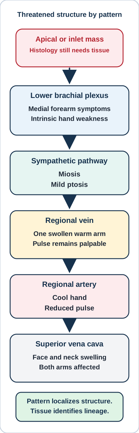
Does an apical syndrome identify squamous histology? No. A superior sulcus tumor can invade the thoracic inlet and produce a Pancoast presentation. This is an anatomic description, not a synonym for squamous carcinoma. Other tumor types can produce the same syndrome. Tissue identification and assessment of local extent still matter. [2][7]
Separate nerve localization, venous outflow and arterial inflow without equating a superior sulcus syndrome with one histology. [2][7] Bone Wizardry; original construction.
Anatomic localization. Lower brachial plexus involvement, often involving C8 and T1 contributions, can cause shoulder pain radiating into the medial arm or forearm with intrinsic hand weakness. Medial forearm sensory loss is not fully explained by an isolated ulnar lesion at the elbow. Involvement of nearby sympathetic pathways can produce ipsilateral Horner findings: miosis and mild ptosis, with sweating changes depending on the site of interruption. These neighboring deficits together help localize a lesion near the apex. They do not establish tumor lineage. [2][7]
Anatomic localization. On the map, follow the nerve path and then the vessel paths. Predict how a swollen warm arm with a preserved pulse differs from a cold hand with a reduced pulse.
Compare venous outflow with arterial inflow
Anatomic localization. Venous obstruction raises upstream venous pressure, favoring swelling while arterial inflow may remain intact. Arterial compromise reduces delivery, favoring a cool hand and diminished pulse. The findings localize different problems even when the same regional tumor could cause either.
Anatomic localization. Subclavian venous obstruction can predominantly affect one arm. Obstruction of the superior vena cava affects drainage from the head and both upper limbs, potentially causing facial swelling, distended neck or chest veins and bilateral arm swelling. Severe airway symptoms, stridor or neurologic compromise require urgent assessment. Do not substitute a routine muscle diagnosis for a new combination of vascular and neurologic abnormalities. [2]
Anatomic localization. Persistent hoarseness requires another anatomic question. The recurrent laryngeal nerves are branches of the vagus that supply most intrinsic laryngeal muscles. The left nerve returns beneath the aortic arch; the right returns beneath the right subclavian artery. A hilar or mediastinal lesion can therefore impair vocal fold function without producing a complete apical syndrome. The phrenic nerve, arising mainly from C3 through C5, serves the diaphragm, so hemidiaphragm dysfunction and vocal fold paralysis should not be assigned to the same nerve. [2]
Anatomic localization. Finally, do not let a correct calcium mechanism explain every new symptom. Lung cancer can spread to the brain and bone. New seizures, a focal deficit or persistent neurologic change after calcium correction warrants investigation for a structural or other neurologic cause. Focal bone pain also requires assessment rather than automatic attribution to circulating PTHrP. The transfer question is always: does the distribution fit the proposed mechanism? [2]
Compare mechanisms without assigning exclusive labels
What should outweigh an expected association? Direct evidence about the tissue or physiology. A person who smokes can have adenocarcinoma. A central lesion can have neuroendocrine differentiation. A patient with known squamous carcinoma can also have a medication-related electrolyte disorder. Keep the observations separate long enough to test whether one explanation accounts for them. [1][2][5]
Tissue observations answer a different question from symptoms
Observed pattern
Useful interpretation
Observed patternMalignant epithelial nests with keratinization and bridges
Useful interpretationSquamous differentiation; assess invasion, origin and extent separately.
Observed patternSmall cells with scant cytoplasm, nuclear molding and neuroendocrine morphology
Useful interpretationConsider small cell carcinoma; correlate morphology with an appropriate stain panel.
Observed patternMalignant glands or mucin-producing epithelial cells
Useful interpretationGlandular differentiation supports adenocarcinoma; a peripheral site is not required.
Observed patternIron-coated fibers or pleural plaques
Useful interpretationExposure-related evidence, not a diagnosis of a malignant cell population.
Evidence hierarchy. Crush artifact may obscure small cell morphology, but crush artifact alone is not diagnostic. Chromogranin and synaptophysin support neuroendocrine differentiation when the morphology fits. Positivity for one marker does not by itself establish small cell carcinoma. Well-differentiated carcinoid tumors are also neuroendocrine. In selected patients, serotonin-related flushing, diarrhea and bronchospasm suggest a different physiological syndrome from humoral hypercalcemia. Symptoms and stains should be interpreted together rather than used as mutually exclusive tumor labels. [1][5]
Evidence hierarchy. Small cell carcinoma is associated with several systemic syndromes. In SIADH, retained water produces hypotonic hyponatremia with urine that remains inappropriately concentrated after the relevant clinical exclusions. Ectopic ACTH can drive cortisol excess, hypertension, hypokalemia and metabolic alkalosis. Lambert-Eaton myasthenic syndrome affects presynaptic calcium-dependent acetylcholine release; proximal weakness, reduced reflexes, autonomic symptoms and improvement after brief contraction support that mechanism. P/Q-type voltage-gated calcium-channel antibodies are established biomarkers, whereas isolated N-type positivity is not equivalent evidence. None of these associations substitutes for histology. [5][12]
Evidence hierarchy. Adenocarcinoma can form glands, produce mucin or grow along preexisting alveolar septa in a lepidic pattern. A small biopsy showing lepidic growth does not assess the entire lesion well enough to guarantee absence of invasion elsewhere. Likewise, digital clubbing with painful periosteal new bone formation suggests hypertrophic osteoarthropathy, not a particular histologic subtype or proof of bone metastases. This differs from osteolytic destruction and from a circulating calcium-raising signal. [1][2]
Evidence hierarchy. Asbestos exposure increases risk for lung cancer and mesothelioma. Smoking and asbestos together substantially increase lung-cancer risk; smoking does not have the same established association with mesothelioma. Pleural plaques are benign exposure-related lesions, whereas suspicious diffuse pleural thickening requires evaluation for a pleural malignancy. A Prussian blue-positive coating identifies iron around a fiber, not malignant squamous cells. Many characteristic asbestos bodies have an asbestos core, but ferruginous bodies can form around other fibers too. Neither the coating nor an exposure history establishes mesothelioma. [6][9][13]
Evidence hierarchy. Try three substitutions: glands in a central mass, pleural plaques beside a keratinizing bronchial tumor, and low sodium in a patient with known squamous carcinoma. Which observation identifies differentiation, which identifies exposure, and which needs a separate physiological workup?
Check the three distinctions
Evidence hierarchy. Glands support glandular differentiation despite central location. Plaques can coexist with a bronchial carcinoma without changing its histology. Low sodium requires evaluation of tonicity, volume status, medications and other causes rather than relabeling the tumor from an association.
Evidence hierarchy. Read tissue for differentiation and invasion. Read paired laboratory results for physiology. Read the distribution of deficits for anatomy. Then combine the conclusions with imaging, prior tumors and clinical course.
Try it here · Checkpoint 3 of 3
Make your prediction before reading the choices. A first attempt is just a starting point.
Case 24
Show answer and explanations for case 24
A. Keratinized tumor material and iron-coated fibers (Best answer)
Prussian blue establishes iron in the coating, not the chemical identity of the fiber core. The concentric material belongs to keratinizing tumor nests, whereas the elongated bodies are a separate fiber-associated finding. Distinguish tumor differentiation, coating composition and core identity.
Reasoning steps for option A
What does the coating stain directly establish?
Prussian blue establishes iron in the coating, not the chemical identity of the fiber core.
In case lung-scc-24, which stated detail most directly tests option A?
The concentric material belongs to keratinizing tumor nests, whereas the elongated bodies are a separate fiber-associated finding.
After resolving case lung-scc-24, what transferable rule distinguishes option A?
Distinguish tumor differentiation, coating composition and core identity.
B. Keratinized tumor material and proven asbestos fibers (Why this does not fit)
Mineral identity needs evidence about the core rather than only an iron-positive coating. The cores are explicitly uncharacterized, so the stain does not prove they are asbestos. Many asbestos bodies are ferruginous, but not every ferruginous body establishes asbestos.
Reasoning steps for option B
When can a fiber be confidently assigned a mineral identity?
Mineral identity needs evidence about the core rather than only an iron-positive coating.
In case lung-scc-24, which stated detail most directly tests option B?
The cores are explicitly uncharacterized, so the stain does not prove they are asbestos.
After resolving case lung-scc-24, what transferable rule distinguishes option B?
Many asbestos bodies are ferruginous, but not every ferruginous body establishes asbestos.
C. Calcified glandular material and iron-coated fibers (Why this does not fit)
Glandular architecture with calcified material would support that description. The supplied concentric material is continuous with keratinizing malignant epithelial cells. Use cellular context instead of equating every concentric structure with calcium deposition.
Reasoning steps for option C
What would support calcified glandular material rather than keratin?
Glandular architecture with calcified material would support that description.
In case lung-scc-24, which stated detail most directly tests option C?
The supplied concentric material is continuous with keratinizing malignant epithelial cells.
After resolving case lung-scc-24, what transferable rule distinguishes option C?
Use cellular context instead of equating every concentric structure with calcium deposition.
D. Calcified glandular material and proven asbestos fibers (Why this does not fit)
That interpretation requires evidence of glandular calcification and identification of an asbestos core. The observed material is keratin-associated and only the coating of the separate fibers has been identified. Do not promote a morphological resemblance or an iron stain beyond what it establishes.
Reasoning steps for option D
What independent evidence is missing for each component?
That interpretation requires evidence of glandular calcification and identification of an asbestos core.
In case lung-scc-24, which stated detail most directly tests option D?
The observed material is keratin-associated and only the coating of the separate fibers has been identified.
After resolving case lung-scc-24, what transferable rule distinguishes option D?
Do not promote a morphological resemblance or an iron stain beyond what it establishes.
Takeaway: Prussian blue identifies iron; it does not by itself identify the core of a ferruginous body.
Sample the safest high-yield target. A central endobronchial lesion may be approached by bronchoscopy, while nodal disease may be sampled with endobronchial ultrasound and a peripheral lesion may require image-guided biopsy. The exact route depends on anatomy, procedural risk and which specimen can answer both diagnosis and stage. [1][2]
Preserve tissue for the unresolved questions. Unequivocal keratinizing morphology can establish squamous differentiation. Poorly differentiated non-small cell carcinoma may need a focused panel, commonly p40 and TTF1, rather than a broad stain battery that exhausts the block. A shared squamous phenotype does not establish pulmonary origin when a prior squamous tumor exists. [1]
Stage before selecting definitive therapy. Chest and upper-abdominal imaging, nodal assessment and evaluation for distant disease answer different questions from the biopsy. Brain imaging is particularly important when neurologic symptoms are present or when the planned stage-specific pathway requires it. Functional status and cardiopulmonary reserve are part of operability, not afterthoughts. [2]
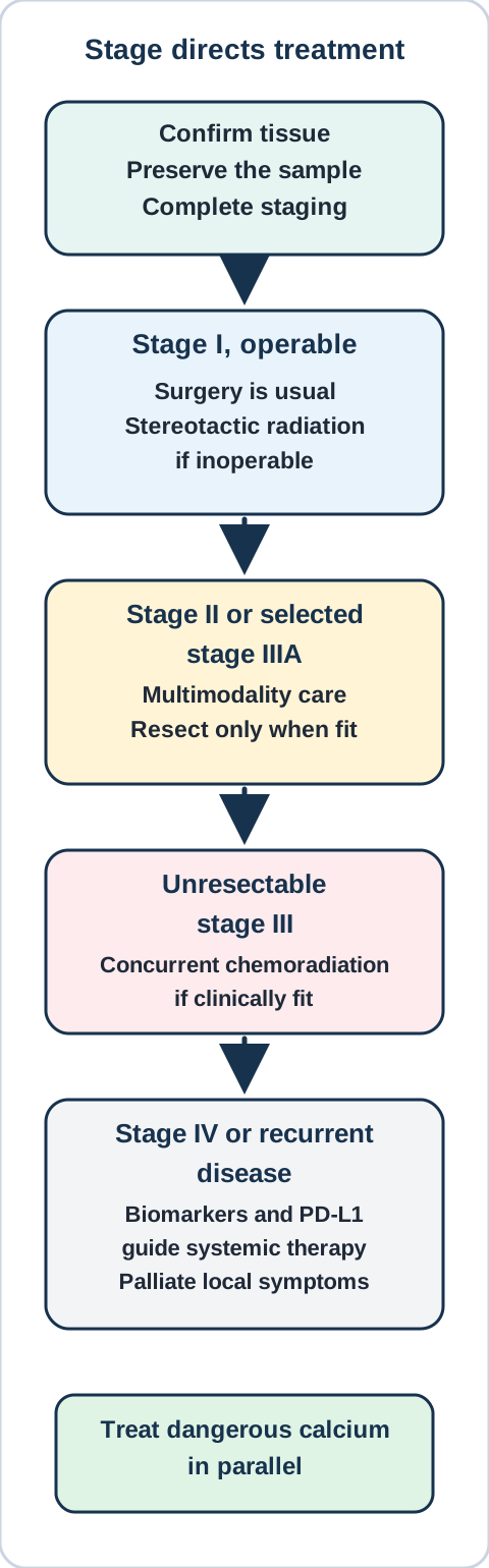
Use stage and fitness to choose the treatment pathway while urgent metabolic complications are treated in parallel. This is not a dosing protocol. [2][4] Bone Wizardry; original construction.
Staging check. A biopsy proves keratinizing squamous carcinoma, but mediastinal nodes have not been assessed. Which missing information can change both the stage and the treatment pathway?
Check the staging distinction
Nodal status matters. Mediastinal nodal involvement can move a patient from an operation-centered pathway to a multimodality or nonsurgical pathway. Histology alone cannot answer that question.
Choose therapy from stage, fitness and tumor features
Early operable disease. Surgery is the usual definitive approach for operable stage I non-small cell lung cancer. Stereotactic body radiation is an important definitive option when a patient with localized disease is medically inoperable or declines surgery after informed discussion. [2]
Selected stage II and IIIA disease. These patients need multidisciplinary planning because surgery, systemic therapy and radiation may be combined in different sequences. Resectability, nodal burden, pulmonary reserve and perioperative risk must be resolved before promising an operation. [2]
Unresectable stage III disease. Clinically fit patients are commonly evaluated for definitive concurrent chemoradiation followed by guideline-directed consolidation when eligible. Frailty, organ function and treatment tolerance can require a different plan. Verify the current regimen against the active guideline, drug labeling and local protocol. [2]
Metastatic or recurrent disease. Systemic treatment depends on performance status, comorbidity, PD-L1 expression, molecular features, prior therapy and patient goals. Squamous morphology narrows some probabilities but does not remove the need for current biomarker-directed decision making. Symptom-directed radiation, airway procedures and palliative care can be integrated early rather than reserved for the final days. [2]
Do not confuse calcium control with tumor control. Hydration when clinically appropriate, an antiresorptive and short-term calcitonin can lower calcium while the tumor-derived signal persists. Reassess the malignancy separately with symptoms, imaging and the relevant biochemical marker. [4][10]
Beyond the boards: decisions that prevent avoidable harm
Tissue stewardship is a treatment decision. Before ordering another stain, ask whether morphology already answers the lineage question and whether the remaining tissue is needed for molecular testing. When a prior head and neck, skin, cervical or other squamous tumor exists, retrieve the old slides and compare morphology rather than assuming a new pulmonary primary. [1]
Severe hypercalcemia is an emergency pathway. The Endocrine Society defines severe hypercalcemia of malignancy as calcium above 14 mg/dL. In severe disease it conditionally suggests calcitonin with an intravenous bisphosphonate or denosumab, and limits calcitonin to 48 to 72 hours because tachyphylaxis develops. Confirm the corrected or ionized calcium, assess kidney and cardiac constraints, and verify treatment against current labeling and local protocols. [4]
Typical adult treatment details. Verify every regimen against current labeling and local protocols. For severe hypercalcemia of malignancy, begin individualized isotonic intravenous fluid replacement when clinically appropriate, then add rapid and durable calcium-lowering therapy. A labeled calcitonin salmon starting dose is 4 IU/kg subcutaneously or intramuscularly every 12 hours; limit use to 48 to 72 hours because tachyphylaxis reduces benefit. A labeled zoledronic acid regimen is 4 mg intravenously over at least 15 minutes after adequate hydration, with serum creatinine assessed before treatment and no retreatment sooner than 7 days. [18][19]
When bisphosphonate therapy is refractory. The labeled denosumab regimen for hypercalcemia of malignancy refractory to bisphosphonate therapy is 120 mg subcutaneously every 4 weeks, with additional 120 mg doses on days 8 and 15 of the first month. Monitor for treatment-emergent hypocalcemia and assess calcium, phosphate and magnesium as clinically indicated. Complete an oral examination and preventive dentistry when feasible because ongoing antiresorptive therapy can cause osteonecrosis of the jaw. [20]
Surveillance after curative-intent therapy. A guideline-based schedule is chest CT every 6 months for the first 2 years, then annually to detect recurrence or a new primary. Routine PET-CT is not recommended for surveillance. New focal neurologic deficits, focal bone pain, worsening airway symptoms or recurrent hypercalcemia require symptom-directed evaluation rather than waiting for the next routine scan. [21]
Escalate by threatened organ. Stridor, progressive superior vena cava symptoms, spinal or focal neurologic deficits, a cold pulseless limb and severe symptomatic hypercalcemia require urgent assessment. Engage pulmonology or interventional radiology for airway and sampling questions, thoracic surgery and radiation oncology for local control, medical oncology for systemic therapy, pathology for tissue comparison and endocrinology or nephrology for refractory calcium disorders.
Build the longitudinal plan before discharge. Reconcile medications and supplements, document smoking-cessation support, explain symptoms that require emergency care, define who will review pending pathology and biomarkers, and schedule assessment of treatment response and toxicity. Follow the tumor, the complication and the patient's function as separate outcomes.
Apply what you have learned
Practice method. Work from the observations in each case. Choose one answer before reviewing why each alternative fits or fails.
Case 1
Show answer and explanations for case 1
A. Squamous differentiation; invasion not demonstrated (Why this does not fit)
Keratinization can establish squamous differentiation without by itself proving invasion. Here, serial sections also demonstrate epithelial nests extending into stroma. Assess the tissue boundary separately from cell differentiation.
Reasoning steps for option A
What can keratinization establish without depth information?
Keratinization can establish squamous differentiation without by itself proving invasion.
In case lung-scc-01, which stated detail most directly tests option A?
Here, serial sections also demonstrate epithelial nests extending into stroma.
After resolving case lung-scc-01, what transferable rule distinguishes option A?
Assess the tissue boundary separately from cell differentiation.
B. Squamous differentiation; stromal invasion demonstrated (Best answer)
Keratinized epithelial nests support squamous differentiation. Extension through the basement membrane into desmoplastic stroma demonstrates invasion. Use morphology for identity and tissue relationships for invasion.
Reasoning steps for option B
What do concentric keratinized nests indicate?
Keratinized epithelial nests support squamous differentiation.
In case lung-scc-01, which stated detail most directly tests option B?
Extension through the basement membrane into desmoplastic stroma demonstrates invasion.
After resolving case lung-scc-01, what transferable rule distinguishes option B?
Use morphology for identity and tissue relationships for invasion.
C. Glandular differentiation; stromal invasion demonstrated (Why this does not fit)
Glandular differentiation is supported by malignant gland formation or mucin production. The described solid keratinized nests support squamous rather than glandular differentiation. Recognize the material and architecture before classifying the carcinoma.
Reasoning steps for option C
What would support glandular differentiation?
Glandular differentiation is supported by malignant gland formation or mucin production.
In case lung-scc-01, which stated detail most directly tests option C?
The described solid keratinized nests support squamous rather than glandular differentiation.
After resolving case lung-scc-01, what transferable rule distinguishes option C?
Recognize the material and architecture before classifying the carcinoma.
D. Glandular differentiation; invasion not demonstrated (Why this does not fit)
Glandular architecture confined above an intact boundary would support that interpretation. This sample instead contains keratinized nests invading desmoplastic stroma. Neither differentiation nor invasion should be inferred from location alone.
Reasoning steps for option D
What two findings would make this interpretation appropriate?
Glandular architecture confined above an intact boundary would support that interpretation.
In case lung-scc-01, which stated detail most directly tests option D?
This sample instead contains keratinized nests invading desmoplastic stroma.
After resolving case lung-scc-01, what transferable rule distinguishes option D?
Neither differentiation nor invasion should be inferred from location alone.
Takeaway: Keratinization and stromal invasion answer two separate diagnostic questions.
A. Squamous carcinoma; p63 establishes the subtype (Why this does not fit)
p63 can be expressed in tumors other than squamous carcinoma. Diffuse p63 with only weak focal p40 and no keratinization does not securely establish this subtype. Do not treat p63 and p40 as interchangeable evidence.
Reasoning steps for option A
How specific is p63 for squamous differentiation?
p63 can be expressed in tumors other than squamous carcinoma.
In case lung-scc-02, which stated detail most directly tests option A?
Diffuse p63 with only weak focal p40 and no keratinization does not securely establish this subtype.
After resolving case lung-scc-02, what transferable rule distinguishes option A?
Do not treat p63 and p40 as interchangeable evidence.
B. Adenocarcinoma; absent keratin establishes the subtype (Why this does not fit)
A poorly differentiated tumor may lack visible keratin without being glandular. There are neither glands nor supporting TTF1 findings in this limited sample. Absence of one lineage feature does not establish the competing lineage.
Reasoning steps for option B
Does absence of keratinization prove glandular differentiation?
A poorly differentiated tumor may lack visible keratin without being glandular.
In case lung-scc-02, which stated detail most directly tests option B?
There are neither glands nor supporting TTF1 findings in this limited sample.
After resolving case lung-scc-02, what transferable rule distinguishes option B?
Absence of one lineage feature does not establish the competing lineage.
C. Large cell carcinoma; the biopsy excludes other subtypes (Why this does not fit)
A large cell designation requires adequate exclusion of other differentiation in appropriately sampled tumor. A small indeterminate biopsy cannot exclude unsampled differentiated components. Use a limited-sample category rather than a resection-level conclusion.
Reasoning steps for option C
What sampling does a definitive large cell designation require?
A large cell designation requires adequate exclusion of other differentiation in appropriately sampled tumor.
In case lung-scc-02, which stated detail most directly tests option C?
A small indeterminate biopsy cannot exclude unsampled differentiated components.
After resolving case lung-scc-02, what transferable rule distinguishes option C?
Use a limited-sample category rather than a resection-level conclusion.
D. Non-small cell carcinoma, NOS; subtype remains unresolved (Best answer)
Weak focal p40 does not establish squamous differentiation. The absence of diagnostic morphology or supportive lineage staining leaves this small sample unclassified within non-small cell carcinoma. Preserve diagnostic uncertainty when the sample does not resolve it.
Reasoning steps for option D
What does weak focal p40 establish in this setting?
Weak focal p40 does not establish squamous differentiation.
In case lung-scc-02, which stated detail most directly tests option D?
The absence of diagnostic morphology or supportive lineage staining leaves this small sample unclassified within non-small cell carcinoma.
After resolving case lung-scc-02, what transferable rule distinguishes option D?
Preserve diagnostic uncertainty when the sample does not resolve it.
Takeaway: Weak focal p40 or p63 alone should not force a squamous diagnosis on limited tissue.
A. Glandular carcinoma; PTH-independent hypercalcemia (Best answer)
Malignant mucin-producing glands support glandular carcinoma. Suppressed PTH during confirmed hypercalcemia identifies a PTH-independent pattern without specifying its cause. Classify the tissue and investigate the calcium mechanism separately.
Reasoning steps for option A
Which findings identify glandular differentiation?
Malignant mucin-producing glands support glandular carcinoma.
In case lung-scc-03, which stated detail most directly tests option A?
Suppressed PTH during confirmed hypercalcemia identifies a PTH-independent pattern without specifying its cause.
After resolving case lung-scc-03, what transferable rule distinguishes option A?
Classify the tissue and investigate the calcium mechanism separately.
B. Squamous carcinoma; PTH-independent hypercalcemia (Why this does not fit)
A central cavitary mass with hypercalcemia can raise suspicion for squamous carcinoma. The biopsy instead directly demonstrates glandular differentiation. An association cannot override diagnostic morphology.
Reasoning steps for option B
Why might squamous carcinoma initially be considered?
A central cavitary mass with hypercalcemia can raise suspicion for squamous carcinoma.
In case lung-scc-03, which stated detail most directly tests option B?
The biopsy instead directly demonstrates glandular differentiation.
After resolving case lung-scc-03, what transferable rule distinguishes option B?
An association cannot override diagnostic morphology.
C. Glandular carcinoma; PTH-dependent hypercalcemia (Why this does not fit)
PTH-dependent hypercalcemia has an inappropriately nonsuppressed PTH concentration. This patient has suppressed PTH despite high calcium. Read PTH relative to the simultaneous calcium level.
Reasoning steps for option C
What PTH response supports a PTH-dependent process?
PTH-dependent hypercalcemia has an inappropriately nonsuppressed PTH concentration.
In case lung-scc-03, which stated detail most directly tests option C?
This patient has suppressed PTH despite high calcium.
After resolving case lung-scc-03, what transferable rule distinguishes option C?
Read PTH relative to the simultaneous calcium level.
D. Squamous carcinoma; PTH-dependent hypercalcemia (Why this does not fit)
Squamous morphology and inappropriately nonsuppressed PTH would support those separate conclusions. This biopsy shows glands and the measured PTH is suppressed. Do not let smoking history replace either tissue or biochemical evidence.
Reasoning steps for option D
What evidence would support both parts of this interpretation?
Squamous morphology and inappropriately nonsuppressed PTH would support those separate conclusions.
In case lung-scc-03, which stated detail most directly tests option D?
This biopsy shows glands and the measured PTH is suppressed.
After resolving case lung-scc-03, what transferable rule distinguishes option D?
Do not let smoking history replace either tissue or biochemical evidence.
Takeaway: A central tumor with high calcium need not be squamous; low PTH does not identify PTHrP without further evidence.
A. Compare central versus peripheral position on CT (Why this does not fit)
Location can change the clinical probability of a primary lung tumor. It cannot distinguish pulmonary origin from metastasis when both tumors share squamous differentiation. Use geography as context rather than a definitive origin marker.
Reasoning steps for option A
How much does location establish about a squamous tumor origin?
Location can change the clinical probability of a primary lung tumor.
In case lung-scc-04, which stated detail most directly tests option A?
It cannot distinguish pulmonary origin from metastasis when both tumors share squamous differentiation.
After resolving case lung-scc-04, what transferable rule distinguishes option A?
Use geography as context rather than a definitive origin marker.
B. Compare the intensity of p40 staining between specimens (Why this does not fit)
Strong p40 supports squamous differentiation. Both lesions share that differentiation, so their p40 intensity does not establish which site was primary. A lineage marker is not automatically an organ-of-origin marker.
Reasoning steps for option B
What does strong p40 primarily identify?
Strong p40 supports squamous differentiation.
In case lung-scc-04, which stated detail most directly tests option B?
Both lesions share that differentiation, so their p40 intensity does not establish which site was primary.
After resolving case lung-scc-04, what transferable rule distinguishes option B?
A lineage marker is not automatically an organ-of-origin marker.
C. Compare prior morphology and context with targeted ancillary testing (Best answer)
Morphologic comparison can identify similarities and differences not resolved by a shared lineage stain. The prior oropharyngeal tumor makes metastatic disease a real alternative requiring clinicopathologic correlation and appropriate additional testing. Separate the diagnosis of squamous differentiation from determination of its primary site.
Reasoning steps for option C
Why review the original tumor directly?
Morphologic comparison can identify similarities and differences not resolved by a shared lineage stain.
In case lung-scc-04, which stated detail most directly tests option C?
The prior oropharyngeal tumor makes metastatic disease a real alternative requiring clinicopathologic correlation and appropriate additional testing.
After resolving case lung-scc-04, what transferable rule distinguishes option C?
Separate the diagnosis of squamous differentiation from determination of its primary site.
D. Compare the current calcium and PTHrP concentrations (Why this does not fit)
That pair can help assess a humoral calcium mechanism. It would not distinguish the origin of two keratinizing squamous tumors. Match the investigation to the unresolved diagnostic question.
Reasoning steps for option D
What question does the calcium-PTHrP pair address?
That pair can help assess a humoral calcium mechanism.
In case lung-scc-04, which stated detail most directly tests option D?
It would not distinguish the origin of two keratinizing squamous tumors.
After resolving case lung-scc-04, what transferable rule distinguishes option D?
Match the investigation to the unresolved diagnostic question.
Takeaway: Squamous markers do not by themselves distinguish a primary lung tumor from a squamous metastasis.
A. Sealing the paracellular diffusion pathway (Why this does not fit)
Tight junctions restrict passage between adjacent cells. Dense plaques attached to keratin filaments describe desmosomal contacts rather than the apical sealing apparatus. Do not infer sealing function from any visible epithelial contact.
Reasoning steps for option A
Which junction forms a paracellular seal?
Tight junctions restrict passage between adjacent cells.
In case lung-scc-05, which stated detail most directly tests option A?
Dense plaques attached to keratin filaments describe desmosomal contacts rather than the apical sealing apparatus.
After resolving case lung-scc-05, what transferable rule distinguishes option A?
Do not infer sealing function from any visible epithelial contact.
B. Distributing tensile force between neighboring cells (Best answer)
Desmosomes anchor intermediate filaments to cell-cell attachment sites. The keratin-linked plaques explain how adjacent squamous cells can share mechanical stress. Visible bridges reflect adhesion, not gland formation or hormone production.
Reasoning steps for option B
How do desmosomes connect to the cytoskeleton?
Desmosomes anchor intermediate filaments to cell-cell attachment sites.
In case lung-scc-05, which stated detail most directly tests option B?
The keratin-linked plaques explain how adjacent squamous cells can share mechanical stress.
After resolving case lung-scc-05, what transferable rule distinguishes option B?
Visible bridges reflect adhesion, not gland formation or hormone production.
C. Transmitting small ions directly between cells (Why this does not fit)
Gap junction channels allow small ions and molecules to pass between cells. The keratin-linked dense plaques are mechanical attachments, not communicating channels. Distinguish mechanical anchoring from electrical or metabolic coupling.
Reasoning steps for option C
Which junction permits direct intercellular ion passage?
Gap junction channels allow small ions and molecules to pass between cells.
In case lung-scc-05, which stated detail most directly tests option C?
The keratin-linked dense plaques are mechanical attachments, not communicating channels.
After resolving case lung-scc-05, what transferable rule distinguishes option C?
Distinguish mechanical anchoring from electrical or metabolic coupling.
D. Anchoring basal cells to the extracellular matrix (Why this does not fit)
Hemidesmosomal contacts anchor epithelial cells to the underlying matrix. These contacts join two neighboring tumor cells rather than a cell to the basement membrane. First identify the two structures joined by a junction.
Reasoning steps for option D
Which attachment connects an epithelial cell to its substrate?
Hemidesmosomal contacts anchor epithelial cells to the underlying matrix.
In case lung-scc-05, which stated detail most directly tests option D?
These contacts join two neighboring tumor cells rather than a cell to the basement membrane.
After resolving case lung-scc-05, what transferable rule distinguishes option D?
First identify the two structures joined by a junction.
Takeaway: Desmosome-associated bridges transmit mechanical force through intermediate-filament networks.
A. Circulating PTHrP stimulates bone resorption and renal calcium retention (Best answer)
A circulating signal can alter bone and kidney function at sites without tumor deposits. High PTHrP with suppressed PTH supports that humoral explanation despite negative skeletal imaging. Do not require bone metastases for humoral hypercalcemia.
Reasoning steps for option A
Must a bone contain tumor to respond to PTHrP?
A circulating signal can alter bone and kidney function at sites without tumor deposits.
In case lung-scc-07, which stated detail most directly tests option A?
High PTHrP with suppressed PTH supports that humoral explanation despite negative skeletal imaging.
After resolving case lung-scc-07, what transferable rule distinguishes option A?
Do not require bone metastases for humoral hypercalcemia.
B. Autonomous parathyroid PTH stimulates bone resorption and renal calcium retention (Why this does not fit)
A PTH-dependent cause has inappropriately nonsuppressed endogenous PTH. The measured PTH is suppressed while PTHrP is high. Identify which hormone is actually providing the stimulus.
Reasoning steps for option B
What PTH pattern would support an autonomous parathyroid source?
A PTH-dependent cause has inappropriately nonsuppressed endogenous PTH.
In case lung-scc-07, which stated detail most directly tests option B?
The measured PTH is suppressed while PTHrP is high.
After resolving case lung-scc-07, what transferable rule distinguishes option B?
Identify which hormone is actually providing the stimulus.
C. Local skeletal tumor deposits release calcium through destructive invasion (Why this does not fit)
Osteolytic tumor involvement can contribute to malignancy-associated hypercalcemia. A measured circulating PTHrP signal provides an explanation that does not depend on finding such deposits. Distinguish a humoral mechanism from local destruction rather than treating them as synonymous.
Reasoning steps for option C
Can direct skeletal invasion produce hypercalcemia?
Osteolytic tumor involvement can contribute to malignancy-associated hypercalcemia.
In case lung-scc-07, which stated detail most directly tests option C?
A measured circulating PTHrP signal provides an explanation that does not depend on finding such deposits.
After resolving case lung-scc-07, what transferable rule distinguishes option C?
Distinguish a humoral mechanism from local destruction rather than treating them as synonymous.
D. Excess calcitriol increases calcium absorption across the intestinal epithelium (Why this does not fit)
Excess calcitriol can increase intestinal calcium absorption. The supplied positive mediator is PTHrP, not a high calcitriol result. Choose the mechanism supported by the biochemical evidence without assigning all malignancy-related calcium excess one cause.
Reasoning steps for option D
How can calcitriol raise calcium?
Excess calcitriol can increase intestinal calcium absorption.
In case lung-scc-07, which stated detail most directly tests option D?
The supplied positive mediator is PTHrP, not a high calcitriol result.
After resolving case lung-scc-07, what transferable rule distinguishes option D?
Choose the mechanism supported by the biochemical evidence without assigning all malignancy-related calcium excess one cause.
Takeaway: A circulating tumor product can affect distant organs without metastatic deposits in them.
A. Appropriately suppressed; favor a PTH-independent process (Why this does not fit)
Normal feedback should suppress endogenous PTH during hypercalcemia. Values in the middle of the reference interval are not appropriately suppressed at calcium near 13 mg/dL. Interpret the hormone against the simultaneous physiological stimulus.
Reasoning steps for option A
What response is appropriate when calcium is clearly high?
Normal feedback should suppress endogenous PTH during hypercalcemia.
In case lung-scc-08, which stated detail most directly tests option A?
Values in the middle of the reference interval are not appropriately suppressed at calcium near 13 mg/dL.
After resolving case lung-scc-08, what transferable rule distinguishes option A?
Interpret the hormone against the simultaneous physiological stimulus.
B. Physiologically irrelevant; favor the lung mass as the cause (Why this does not fit)
The mass changes clinical probability but does not determine the calcium mechanism. Repeated nonsuppressed PTH requires explanation even in a patient with a tumor. Evaluate a possible coexisting endocrine cause instead of anchoring on cancer.
Reasoning steps for option B
Does finding a lung mass make paired hormone testing irrelevant?
The mass changes clinical probability but does not determine the calcium mechanism.
In case lung-scc-08, which stated detail most directly tests option B?
Repeated nonsuppressed PTH requires explanation even in a patient with a tumor.
After resolving case lung-scc-08, what transferable rule distinguishes option B?
Evaluate a possible coexisting endocrine cause instead of anchoring on cancer.
C. Inappropriately nonsuppressed; investigate PTH-dependent causes (Best answer)
Yes, a reference-range PTH can be inappropriate when calcium is high. Both paired measurements show failure of the expected suppression. Investigate PTH-dependent causes, including primary hyperparathyroidism, without declaring a specific cause from this pair alone.
Reasoning steps for option C
Can a reference-range PTH be abnormal for the calcium level?
Yes, a reference-range PTH can be inappropriate when calcium is high.
In case lung-scc-08, which stated detail most directly tests option C?
Both paired measurements show failure of the expected suppression.
After resolving case lung-scc-08, what transferable rule distinguishes option C?
Investigate PTH-dependent causes, including primary hyperparathyroidism, without declaring a specific cause from this pair alone.
D. Compensatorily increased; favor renal secondary hyperparathyroidism (Why this does not fit)
Chronic kidney disease can impair mineral regulation and stimulate PTH secretion. Normal kidney function and high calcium do not support that proposed compensatory setting. A normal-range hormone is not automatically an appropriate compensatory response.
Reasoning steps for option D
What setting usually supports renal secondary hyperparathyroidism?
Chronic kidney disease can impair mineral regulation and stimulate PTH secretion.
In case lung-scc-08, which stated detail most directly tests option D?
Normal kidney function and high calcium do not support that proposed compensatory setting.
After resolving case lung-scc-08, what transferable rule distinguishes option D?
A normal-range hormone is not automatically an appropriate compensatory response.
Takeaway: A laboratory-normal PTH can be physiologically inappropriate during hypercalcemia.
A. Autonomous endogenous PTH secretion with renal calcium retention (Why this does not fit)
Autonomous PTH secretion produces an inappropriately nonsuppressed PTH result. Her intact PTH is suppressed during hypercalcemia. A confirmed feedback response argues against a PTH-dependent source.
Reasoning steps for option A
What hormone pattern supports an endogenous PTH source?
Autonomous PTH secretion produces an inappropriately nonsuppressed PTH result.
In case lung-scc-09, which stated detail most directly tests option A?
Her intact PTH is suppressed during hypercalcemia.
After resolving case lung-scc-09, what transferable rule distinguishes option A?
A confirmed feedback response argues against a PTH-dependent source.
B. PTHrP excess with renal phosphate loss and calcium retention (Why this does not fit)
A high PTHrP result would support a PTHrP-mediated explanation. PTHrP is not increased while the active vitamin D metabolite is markedly high. Investigate the measured abnormal mediator rather than assigning the expected association.
Reasoning steps for option B
What would directly support this humoral mechanism?
A high PTHrP result would support a PTHrP-mediated explanation.
In case lung-scc-09, which stated detail most directly tests option B?
PTHrP is not increased while the active vitamin D metabolite is markedly high.
After resolving case lung-scc-09, what transferable rule distinguishes option B?
Investigate the measured abnormal mediator rather than assigning the expected association.
C. High vitamin D substrate exposure with increased intestinal calcium entry (Why this does not fit)
Excess vitamin D intake often produces a high 25-hydroxyvitamin D concentration. The substrate concentration is normal and supplement exposure is absent. Distinguish excess substrate from inappropriate production of the active metabolite.
Reasoning steps for option C
What pattern would support excess vitamin D substrate intake?
Excess vitamin D intake often produces a high 25-hydroxyvitamin D concentration.
In case lung-scc-09, which stated detail most directly tests option C?
The substrate concentration is normal and supplement exposure is absent.
After resolving case lung-scc-09, what transferable rule distinguishes option C?
Distinguish excess substrate from inappropriate production of the active metabolite.
D. Unregulated calcitriol production with increased intestinal calcium entry (Best answer)
Calcitriol is the active vitamin D metabolite and is markedly high in this patient. Its increase despite suppressed PTH supports investigation of PTH-independent calcitriol production. Identify the responsible process without inferring a new tumor histology from the biochemical pattern alone.
Reasoning steps for option D
Which measured molecule is directly abnormal here?
Calcitriol is the active vitamin D metabolite and is markedly high in this patient.
In case lung-scc-09, which stated detail most directly tests option D?
Its increase despite suppressed PTH supports investigation of PTH-independent calcitriol production.
After resolving case lung-scc-09, what transferable rule distinguishes option D?
Identify the responsible process without inferring a new tumor histology from the biochemical pattern alone.
Takeaway: Low PTH identifies a branch of the differential; the measured mediator determines the next mechanism to investigate.
A. Increased proximal phosphate reabsorption despite low PTH (Why this does not fit)
Greater tubular reabsorption would conserve phosphate. The kidney is instead continuing to excrete a substantial fraction despite hypophosphatemia. Interpret excretion relative to the need for conservation.
Reasoning steps for option A
What would increased phosphate reabsorption do to urinary loss?
Greater tubular reabsorption would conserve phosphate.
In case lung-scc-11, which stated detail most directly tests option A?
The kidney is instead continuing to excrete a substantial fraction despite hypophosphatemia.
After resolving case lung-scc-11, what transferable rule distinguishes option A?
Interpret excretion relative to the need for conservation.
B. Reduced phosphate filtration from a fall in glomerular function (Why this does not fit)
Reduced filtration can limit phosphate elimination and contribute to retention. Kidney function is normal and phosphate is low with continuing renal loss. Do not substitute reduced filtration for a demonstrated reabsorption problem.
Reasoning steps for option B
What happens to phosphate handling when filtration is markedly impaired?
Reduced filtration can limit phosphate elimination and contribute to retention.
In case lung-scc-11, which stated detail most directly tests option B?
Kidney function is normal and phosphate is low with continuing renal loss.
After resolving case lung-scc-11, what transferable rule distinguishes option B?
Do not substitute reduced filtration for a demonstrated reabsorption problem.
C. Reduced proximal phosphate reabsorption from PTH-like signaling (Best answer)
PTH-like signaling reduces proximal tubular phosphate reabsorption. High PTHrP explains continuing phosphate loss even while endogenous PTH is suppressed. Suppression of native PTH does not mean every PTH-receptor stimulus has ceased.
Reasoning steps for option C
How does PTH-like signaling affect proximal phosphate transport?
In case lung-scc-11, which stated detail most directly tests option C?
High PTHrP explains continuing phosphate loss even while endogenous PTH is suppressed.
After resolving case lung-scc-11, what transferable rule distinguishes option C?
Suppression of native PTH does not mean every PTH-receptor stimulus has ceased.
D. Reduced intestinal phosphate absorption with renal conservation (Why this does not fit)
It should conserve phosphate by reducing urinary loss. Continued fractional phosphate excretion of 18% despite low plasma phosphate indicates a renal contribution. Use urinary handling to distinguish renal from extrarenal phosphate deficits.
Reasoning steps for option D
How should an intact kidney respond to an isolated intestinal phosphate deficit?
It should conserve phosphate by reducing urinary loss.
In case lung-scc-11, which stated detail most directly tests option D?
Continued fractional phosphate excretion of 18% despite low plasma phosphate indicates a renal contribution.
After resolving case lung-scc-11, what transferable rule distinguishes option D?
Use urinary handling to distinguish renal from extrarenal phosphate deficits.
A. Reduced renal responsiveness to vasopressin during hypercalcemia (Best answer)
It supports impaired renal responsiveness rather than an isolated shortage of vasopressin. Hypercalcemia provides an acquired explanation for the concentrating defect in this patient. Separate a renal water-conservation defect from excess solute excretion.
Reasoning steps for option A
What does persistently dilute urine after desmopressin suggest?
It supports impaired renal responsiveness rather than an isolated shortage of vasopressin.
In case lung-scc-12, which stated detail most directly tests option A?
Hypercalcemia provides an acquired explanation for the concentrating defect in this patient.
After resolving case lung-scc-12, what transferable rule distinguishes option A?
Separate a renal water-conservation defect from excess solute excretion.
B. Reduced hypothalamic secretion of vasopressin as the sole defect (Why this does not fit)
Urine generally becomes substantially more concentrated when the missing hormone is replaced. Little response during monitored desmopressin testing favors a renal defect instead. Use the response to replacement to distinguish central from renal mechanisms.
Reasoning steps for option B
What response is expected with an isolated central vasopressin deficit?
Urine generally becomes substantially more concentrated when the missing hormone is replaced.
In case lung-scc-12, which stated detail most directly tests option B?
Little response during monitored desmopressin testing favors a renal defect instead.
After resolving case lung-scc-12, what transferable rule distinguishes option B?
Use the response to replacement to distinguish central from renal mechanisms.
C. Increased renal glucose delivery causing an osmotic diuresis (Why this does not fit)
High filtered glucose with glycosuria supports a glucose-driven solute diuresis. Glucose is normal and absent from urine, which remains very dilute. Verify the proposed urinary solute before attributing polyuria to it.
Reasoning steps for option C
What evidence would support glucose-driven osmotic diuresis?
High filtered glucose with glycosuria supports a glucose-driven solute diuresis.
In case lung-scc-12, which stated detail most directly tests option C?
Glucose is normal and absent from urine, which remains very dilute.
After resolving case lung-scc-12, what transferable rule distinguishes option C?
Verify the proposed urinary solute before attributing polyuria to it.
D. Increased water intake suppressing an otherwise normal concentrating system (Why this does not fit)
Excess water intake tends to lower plasma tonicity. This patient has high plasma osmolality despite dilute urine, indicating failed water conservation. Interpret urine concentration together with plasma tonicity.
Reasoning steps for option D
How would primary excess water intake usually affect plasma tonicity?
Excess water intake tends to lower plasma tonicity.
In case lung-scc-12, which stated detail most directly tests option D?
This patient has high plasma osmolality despite dilute urine, indicating failed water conservation.
After resolving case lung-scc-12, what transferable rule distinguishes option D?
Interpret urine concentration together with plasma tonicity.
Takeaway: Hypercalcemia can cause a renal concentrating defect; it is not simply competitive blockade of an ADH receptor.
A. An antiresorptive without an initial rapid-acting adjunct (Why this does not fit)
An antiresorptive addresses the sustained contribution from bone resorption. Severe symptomatic hypercalcemia also favors an initial rapid-acting adjunct while that effect develops. Do not confuse an appropriate durable treatment with the complete initial plan for a severe presentation.
Reasoning steps for option A
What useful effect does an antiresorptive provide?
An antiresorptive addresses the sustained contribution from bone resorption.
In case lung-scc-13, which stated detail most directly tests option A?
Severe symptomatic hypercalcemia also favors an initial rapid-acting adjunct while that effect develops.
After resolving case lung-scc-13, what transferable rule distinguishes option A?
Do not confuse an appropriate durable treatment with the complete initial plan for a severe presentation.
B. Calcitonin alone until the tumor is treated (Why this does not fit)
Calcitonin can act quickly but its effect is short-lived and wanes with continued use. Severe PTHrP-mediated hypercalcemia needs a more sustained calcium-lowering strategy as well. Use a temporary adjunct without mistaking it for durable control.
Reasoning steps for option B
What is the limitation of relying on calcitonin alone?
Calcitonin can act quickly but its effect is short-lived and wanes with continued use.
In case lung-scc-13, which stated detail most directly tests option B?
Severe PTHrP-mediated hypercalcemia needs a more sustained calcium-lowering strategy as well.
After resolving case lung-scc-13, what transferable rule distinguishes option B?
Use a temporary adjunct without mistaking it for durable control.
C. A glucocorticoid while deferring antiresorptive treatment (Why this does not fit)
Glucocorticoids are particularly relevant to excess-calcitriol mechanisms. This patient has high PTHrP without an increased calcitriol result. Choose the treatment strategy for the measured mechanism rather than for cancer in general.
Reasoning steps for option C
Which hypercalcemia mechanism particularly supports glucocorticoid treatment?
Glucocorticoids are particularly relevant to excess-calcitriol mechanisms.
In case lung-scc-13, which stated detail most directly tests option C?
This patient has high PTHrP without an increased calcitriol result.
After resolving case lung-scc-13, what transferable rule distinguishes option C?
Choose the treatment strategy for the measured mechanism rather than for cancer in general.
D. Calcitonin together with an antiresorptive (Best answer)
Calcitonin can provide a rapid temporary effect while an antiresorptive provides more sustained control. Confusion and calcium of 15.4 mg/dL with high PTHrP support this initial combination alongside monitored supportive care. Urgent calcium control and cancer-directed treatment address different time scales.
Reasoning steps for option D
Why combine agents in a severe initial presentation?
Calcitonin can provide a rapid temporary effect while an antiresorptive provides more sustained control.
In case lung-scc-13, which stated detail most directly tests option D?
Confusion and calcium of 15.4 mg/dL with high PTHrP support this initial combination alongside monitored supportive care.
After resolving case lung-scc-13, what transferable rule distinguishes option D?
Urgent calcium control and cancer-directed treatment address different time scales.
Takeaway: Severe symptomatic hypercalcemia requires urgent coordinated treatment, not a wait for oncologic response.
A. Upper brachial plexus and cervical sympathetic pathway (Why this does not fit)
Upper plexus involvement more strongly affects shoulder and elbow functions. Medial forearm sensory loss and intrinsic hand weakness favor lower rather than upper plexus involvement. Match the limb distribution before adding the nearby autonomic structure.
Reasoning steps for option A
Which limb functions are most associated with the upper plexus?
Upper plexus involvement more strongly affects shoulder and elbow functions.
In case lung-scc-14, which stated detail most directly tests option A?
Medial forearm sensory loss and intrinsic hand weakness favor lower rather than upper plexus involvement.
After resolving case lung-scc-14, what transferable rule distinguishes option A?
Match the limb distribution before adding the nearby autonomic structure.
B. Lower brachial plexus and cervical sympathetic pathway (Best answer)
Lower plexus involvement fits the C8 and T1-predominant limb abnormalities. Ipsilateral miosis and mild ptosis additionally localize sympathetic interruption near the apex. Combined somatic and autonomic deficits can identify neighboring structures.
Reasoning steps for option B
Which plexus region fits medial forearm and intrinsic hand findings?
Lower plexus involvement fits the C8 and T1-predominant limb abnormalities.
In case lung-scc-14, which stated detail most directly tests option B?
Ipsilateral miosis and mild ptosis additionally localize sympathetic interruption near the apex.
After resolving case lung-scc-14, what transferable rule distinguishes option B?
Combined somatic and autonomic deficits can identify neighboring structures.
C. Ulnar nerve at the elbow and phrenic nerve (Why this does not fit)
An ulnar lesion can impair finger abduction and sensation in part of the hand. It does not explain medial forearm sensory loss or the ocular sympathetic findings. Do not reduce a broader distribution to one distal nerve.
Reasoning steps for option C
Can an isolated ulnar lesion explain the entire sensory distribution?
An ulnar lesion can impair finger abduction and sensation in part of the hand.
In case lung-scc-14, which stated detail most directly tests option C?
It does not explain medial forearm sensory loss or the ocular sympathetic findings.
After resolving case lung-scc-14, what transferable rule distinguishes option C?
Do not reduce a broader distribution to one distal nerve.
D. Lower brachial plexus and recurrent laryngeal nerve (Why this does not fit)
Recurrent laryngeal injury can impair vocal fold function. It does not account for the miosis and ptosis accompanying the lower plexus findings. Identify the autonomic pathway rather than assigning every nearby nerve the same deficit.
Reasoning steps for option D
What function would recurrent laryngeal injury affect?
Recurrent laryngeal injury can impair vocal fold function.
In case lung-scc-14, which stated detail most directly tests option D?
It does not account for the miosis and ptosis accompanying the lower plexus findings.
After resolving case lung-scc-14, what transferable rule distinguishes option D?
Identify the autonomic pathway rather than assigning every nearby nerve the same deficit.
Takeaway: Lower plexus findings plus ipsilateral Horner findings localize a thoracic inlet process.
A. Glandular carcinoma with superior sulcus involvement (Best answer)
Malignant mucin-producing glands support glandular differentiation. The apical mass with hand and sympathetic deficits establishes a superior sulcus presentation without making the tumor squamous. Local syndrome and histologic subtype must be assigned independently.
Reasoning steps for option A
What does gland formation identify?
Malignant mucin-producing glands support glandular differentiation.
In case lung-scc-15, which stated detail most directly tests option A?
The apical mass with hand and sympathetic deficits establishes a superior sulcus presentation without making the tumor squamous.
After resolving case lung-scc-15, what transferable rule distinguishes option A?
Local syndrome and histologic subtype must be assigned independently.
B. Squamous carcinoma with superior sulcus involvement (Why this does not fit)
Squamous carcinoma can produce a superior sulcus presentation. The actual biopsy demonstrates glandular rather than squamous differentiation. An anatomic syndrome is not an exclusive histologic label.
Reasoning steps for option B
Why might squamous carcinoma be considered in an apical mass?
Squamous carcinoma can produce a superior sulcus presentation.
In case lung-scc-15, which stated detail most directly tests option B?
The actual biopsy demonstrates glandular rather than squamous differentiation.
After resolving case lung-scc-15, what transferable rule distinguishes option B?
An anatomic syndrome is not an exclusive histologic label.
C. Glandular carcinoma with a paraneoplastic transmission defect (Why this does not fit)
A transmission disorder more often produces a characteristic weakness pattern without a focal sensory distribution. Painful focal hand deficits and ipsilateral Horner findings beside an invasive apical mass favor direct local involvement. Use distribution to separate structural injury from a remote systemic syndrome.
Reasoning steps for option C
What pattern would favor a neuromuscular transmission disorder?
A transmission disorder more often produces a characteristic weakness pattern without a focal sensory distribution.
In case lung-scc-15, which stated detail most directly tests option C?
Painful focal hand deficits and ipsilateral Horner findings beside an invasive apical mass favor direct local involvement.
After resolving case lung-scc-15, what transferable rule distinguishes option C?
Use distribution to separate structural injury from a remote systemic syndrome.
D. Squamous carcinoma with a paraneoplastic transmission defect (Why this does not fit)
Squamous differentiation and a transmission-disorder pattern would be needed to support that combination. The tissue is glandular and the focal findings match direct thoracic inlet involvement. Do not let a familiar tumor association displace two contrary observations.
Reasoning steps for option D
What evidence would be needed for both proposed components?
Squamous differentiation and a transmission-disorder pattern would be needed to support that combination.
In case lung-scc-15, which stated detail most directly tests option D?
The tissue is glandular and the focal findings match direct thoracic inlet involvement.
After resolving case lung-scc-15, what transferable rule distinguishes option D?
Do not let a familiar tumor association displace two contrary observations.
Takeaway: A Pancoast presentation describes local anatomy; the biopsy can establish a non-squamous tumor.
A. Reduced arterial pressure distal to the left subclavian artery (Why this does not fit)
Arterial compromise can cause a cool hand and diminished pulse. The warm hand with preserved symmetric pulses instead supports maintained arterial inflow. Separate failure of arterial delivery from impaired venous drainage.
Reasoning steps for option A
What distal findings suggest impaired arterial delivery?
Arterial compromise can cause a cool hand and diminished pulse.
In case lung-scc-16, which stated detail most directly tests option A?
The warm hand with preserved symmetric pulses instead supports maintained arterial inflow.
After resolving case lung-scc-16, what transferable rule distinguishes option A?
Separate failure of arterial delivery from impaired venous drainage.
B. Increased venous pressure upstream from the superior vena cava (Why this does not fit)
The head, neck and both upper limbs drain through the superior vena cava. Isolated left arm swelling without face, neck or right arm findings favors a more localized outflow problem. Use the full drainage territory to distinguish central from unilateral venous obstruction.
Reasoning steps for option B
Which territories drain through the superior vena cava?
The head, neck and both upper limbs drain through the superior vena cava.
In case lung-scc-16, which stated detail most directly tests option B?
Isolated left arm swelling without face, neck or right arm findings favors a more localized outflow problem.
After resolving case lung-scc-16, what transferable rule distinguishes option B?
Use the full drainage territory to distinguish central from unilateral venous obstruction.
C. Increased venous pressure upstream from the left subclavian vein (Best answer)
Venous obstruction can cause arm swelling while the hand remains warm and well perfused. A regional left-sided outflow problem fits the unilateral swelling beside the apical mass. A normal pulse does not exclude a clinically important venous obstruction.
Reasoning steps for option C
What can venous obstruction produce despite normal arterial inflow?
Venous obstruction can cause arm swelling while the hand remains warm and well perfused.
In case lung-scc-16, which stated detail most directly tests option C?
A regional left-sided outflow problem fits the unilateral swelling beside the apical mass.
After resolving case lung-scc-16, what transferable rule distinguishes option C?
A normal pulse does not exclude a clinically important venous obstruction.
D. Increased pulmonary venous pressure from left ventricular dysfunction (Why this does not fit)
Increased left-sided filling pressure principally affects the pulmonary venous circulation. It does not explain isolated swelling of the arm beside a regional mass as well as local venous obstruction. Choose a pressure abnormality that matches the observed drainage territory.
Reasoning steps for option D
What compartment is directly affected by increased left-sided filling pressure?
Increased left-sided filling pressure principally affects the pulmonary venous circulation.
In case lung-scc-16, which stated detail most directly tests option D?
It does not explain isolated swelling of the arm beside a regional mass as well as local venous obstruction.
After resolving case lung-scc-16, what transferable rule distinguishes option D?
Choose a pressure abnormality that matches the observed drainage territory.
Takeaway: A swollen warm arm with preserved pulses suggests impaired venous outflow rather than reduced arterial inflow.
A. The left phrenic nerve descending anterior to the lung root (Why this does not fit)
The phrenic nerve provides motor supply to the diaphragm. Preserved diaphragm excursion and isolated vocal fold paralysis do not match that deficit. Do not equate every thoracic motor nerve with laryngeal function.
Reasoning steps for option A
What function would left phrenic dysfunction primarily impair?
The phrenic nerve provides motor supply to the diaphragm.
In case lung-scc-17, which stated detail most directly tests option A?
Preserved diaphragm excursion and isolated vocal fold paralysis do not match that deficit.
After resolving case lung-scc-17, what transferable rule distinguishes option A?
Do not equate every thoracic motor nerve with laryngeal function.
B. The left recurrent laryngeal nerve returning beneath the aortic arch (Best answer)
Its recurrent course passes beneath the aortic arch before ascending to the larynx. This course matches both the left vocal fold deficit and the nodal location. Combine the function of the injured nerve with its actual thoracic course.
Reasoning steps for option B
Why is the left recurrent laryngeal nerve exposed to aortic-window disease?
Its recurrent course passes beneath the aortic arch before ascending to the larynx.
In case lung-scc-17, which stated detail most directly tests option B?
This course matches both the left vocal fold deficit and the nodal location.
After resolving case lung-scc-17, what transferable rule distinguishes option B?
Combine the function of the injured nerve with its actual thoracic course.
C. The right recurrent laryngeal nerve returning beneath the right subclavian artery (Why this does not fit)
It returns beneath the right subclavian artery rather than beneath the aortic arch. That course and side do not match the left vocal fold deficit beside the aortic window. The two recurrent laryngeal nerves have different thoracic relationships.
Reasoning steps for option C
Where does the right recurrent laryngeal nerve turn?
It returns beneath the right subclavian artery rather than beneath the aortic arch.
In case lung-scc-17, which stated detail most directly tests option C?
That course and side do not match the left vocal fold deficit beside the aortic window.
After resolving case lung-scc-17, what transferable rule distinguishes option C?
The two recurrent laryngeal nerves have different thoracic relationships.
D. The left sympathetic trunk descending along the vertebral column (Why this does not fit)
Cervical sympathetic interruption can cause ipsilateral Horner findings. The observed deficit is vocal fold paralysis, not an ocular sympathetic deficit. Match the missing function before attributing a symptom to an adjacent pathway.
Reasoning steps for option D
What ocular pattern can cervical sympathetic interruption produce?
Cervical sympathetic interruption can cause ipsilateral Horner findings.
In case lung-scc-17, which stated detail most directly tests option D?
The observed deficit is vocal fold paralysis, not an ocular sympathetic deficit.
After resolving case lung-scc-17, what transferable rule distinguishes option D?
Match the missing function before attributing a symptom to an adjacent pathway.
Takeaway: Left vocal fold paralysis can localize to the recurrent laryngeal course beneath the aortic arch.
A. Subclavian vein obstruction; routine outpatient sampling (Why this does not fit)
A unilateral subclavian lesion chiefly impairs drainage from one arm. Face and bilateral arm swelling exceed that territory, and stridor makes routine outpatient timing inappropriate. Both anatomic extent and physiological severity matter.
Reasoning steps for option A
What distribution can an isolated subclavian obstruction produce?
A unilateral subclavian lesion chiefly impairs drainage from one arm.
In case lung-scc-18, which stated detail most directly tests option A?
Face and bilateral arm swelling exceed that territory, and stridor makes routine outpatient timing inappropriate.
After resolving case lung-scc-18, what transferable rule distinguishes option A?
Both anatomic extent and physiological severity matter.
B. Superior vena cava obstruction; routine outpatient sampling (Why this does not fit)
Superior vena cava obstruction fits the upper-body drainage pattern. New stridor and positional breathing difficulty require urgent assessment rather than routine outpatient sampling. Urgency follows airway or neurologic compromise, not the syndrome label alone.
Reasoning steps for option B
Does identifying the correct venous level settle the timing of care?
Superior vena cava obstruction fits the upper-body drainage pattern.
In case lung-scc-18, which stated detail most directly tests option B?
New stridor and positional breathing difficulty require urgent assessment rather than routine outpatient sampling.
After resolving case lung-scc-18, what transferable rule distinguishes option B?
Urgency follows airway or neurologic compromise, not the syndrome label alone.
C. Subclavian vein obstruction; urgent airway assessment (Why this does not fit)
Urgent airway assessment is appropriate for new stridor. A unilateral subclavian obstruction does not account for the full head-and-both-arms distribution. Keep the urgent response while correcting the anatomic localization.
Reasoning steps for option C
Which part of this pairing matches the severe symptom?
Urgent airway assessment is appropriate for new stridor.
In case lung-scc-18, which stated detail most directly tests option C?
A unilateral subclavian obstruction does not account for the full head-and-both-arms distribution.
After resolving case lung-scc-18, what transferable rule distinguishes option C?
Keep the urgent response while correcting the anatomic localization.
D. Superior vena cava obstruction; urgent airway assessment (Best answer)
Superior vena cava obstruction fits the face and bilateral upper-limb findings. Stridor indicates possible airway compromise requiring urgent clinical assessment. Do not delay assessment of a threatened airway while pursuing routine tissue workup.
Reasoning steps for option D
What combines the venous distribution and current danger?
Superior vena cava obstruction fits the face and bilateral upper-limb findings.
In case lung-scc-18, which stated detail most directly tests option D?
Stridor indicates possible airway compromise requiring urgent clinical assessment.
After resolving case lung-scc-18, what transferable rule distinguishes option D?
Do not delay assessment of a threatened airway while pursuing routine tissue workup.
Takeaway: Upper-body venous congestion with stridor is not a routine outpatient localization exercise.
A. Repeat PTHrP measurement to explain the focal weakness (Why this does not fit)
It assesses a circulating calcium-related tumor signal. Normal calcium with a new focal seizure and face-arm deficit requires a neurologic explanation beyond the resolved metabolic disturbance. A systemic mediator test does not localize a focal brain deficit.
Reasoning steps for option A
What does PTHrP measurement assess?
It assesses a circulating calcium-related tumor signal.
In case lung-scc-19, which stated detail most directly tests option A?
Normal calcium with a new focal seizure and face-arm deficit requires a neurologic explanation beyond the resolved metabolic disturbance.
After resolving case lung-scc-19, what transferable rule distinguishes option A?
A systemic mediator test does not localize a focal brain deficit.
B. Limb nerve-conduction studies to assess presynaptic transmission (Why this does not fit)
A neuromuscular transmission disorder can cause characteristic limb weakness without a focal cortical seizure. The seizure and face-arm distribution instead require evaluation of the central nervous system. Use the seizure and deficit distribution to localize before selecting a test.
Reasoning steps for option B
What distribution would favor a peripheral transmission disorder?
A neuromuscular transmission disorder can cause characteristic limb weakness without a focal cortical seizure.
In case lung-scc-19, which stated detail most directly tests option B?
The seizure and face-arm distribution instead require evaluation of the central nervous system.
After resolving case lung-scc-19, what transferable rule distinguishes option B?
Use the seizure and deficit distribution to localize before selecting a test.
C. Brain imaging to assess an intracranial structural process (Best answer)
A focal seizure with persistent face-arm weakness localizes concern to the brain. The deficit appears after the diffuse confusion and hypercalcemia have resolved. Investigate structural and other neurologic causes rather than automatically labeling every symptom metabolic or metastatic.
Reasoning steps for option C
Why does the new pattern call for brain assessment?
A focal seizure with persistent face-arm weakness localizes concern to the brain.
In case lung-scc-19, which stated detail most directly tests option C?
The deficit appears after the diffuse confusion and hypercalcemia have resolved.
After resolving case lung-scc-19, what transferable rule distinguishes option C?
Investigate structural and other neurologic causes rather than automatically labeling every symptom metabolic or metastatic.
D. Cervical spine imaging to assess a lower plexus root lesion (Why this does not fit)
A lower cervical process can affect arm or hand function. It does not explain a focal seizure and facial weakness. Require one anatomical explanation to account for the full distribution.
Reasoning steps for option D
What deficit can a lower cervical lesion produce?
A lower cervical process can affect arm or hand function.
In case lung-scc-19, which stated detail most directly tests option D?
It does not explain a focal seizure and facial weakness.
After resolving case lung-scc-19, what transferable rule distinguishes option D?
Require one anatomical explanation to account for the full distribution.
Takeaway: New focal neurologic findings need their own evaluation even when a genuine metabolic complication has improved.
A. Reduced free-water excretion despite hypotonic plasma (Best answer)
Appropriate regulation favors excretion of dilute urine. Urine remains concentrated despite hypotonic plasma after the stated exclusions, supporting inappropriate antidiuresis. Interpret sodium through water balance and tonicity rather than tumor location alone.
Reasoning steps for option A
What should the kidney do with water when plasma is hypotonic?
Appropriate regulation favors excretion of dilute urine.
In case lung-scc-20, which stated detail most directly tests option A?
Urine remains concentrated despite hypotonic plasma after the stated exclusions, supporting inappropriate antidiuresis.
After resolving case lung-scc-20, what transferable rule distinguishes option A?
Interpret sodium through water balance and tonicity rather than tumor location alone.
B. Primary renal salt loss causing extracellular volume depletion (Why this does not fit)
Clinically important primary salt loss commonly produces volume depletion. The supplied assessment is euvolemic and the urine remains inappropriately concentrated in a setting compatible with antidiuresis. Distinguish primary sodium depletion from excess water relative to solute.
Reasoning steps for option B
What physical state would support primary salt loss?
Clinically important primary salt loss commonly produces volume depletion.
In case lung-scc-20, which stated detail most directly tests option B?
The supplied assessment is euvolemic and the urine remains inappropriately concentrated in a setting compatible with antidiuresis.
After resolving case lung-scc-20, what transferable rule distinguishes option B?
Distinguish primary sodium depletion from excess water relative to solute.
C. Hyperglycemia drawing intracellular water into extracellular fluid (Why this does not fit)
Marked hyperglycemia can shift water into extracellular fluid and lower sodium. Glucose is normal and measured plasma osmolality is low. Check measured tonicity before assigning a transcellular water shift.
Reasoning steps for option C
How can high glucose lower measured sodium?
Marked hyperglycemia can shift water into extracellular fluid and lower sodium.
In case lung-scc-20, which stated detail most directly tests option C?
Glucose is normal and measured plasma osmolality is low.
After resolving case lung-scc-20, what transferable rule distinguishes option C?
Check measured tonicity before assigning a transcellular water shift.
D. Excess water intake with appropriate maximal urinary dilution (Why this does not fit)
An intact regulatory response usually produces very dilute urine during excess water intake. Urine osmolality of 510 is not an appropriate dilution response to plasma osmolality of 258. Compare urine behavior with plasma tonicity before choosing a water-intake explanation.
Reasoning steps for option D
What urinary response would favor primary excess water intake?
An intact regulatory response usually produces very dilute urine during excess water intake.
In case lung-scc-20, which stated detail most directly tests option D?
Urine osmolality of 510 is not an appropriate dilution response to plasma osmolality of 258.
After resolving case lung-scc-20, what transferable rule distinguishes option D?
Compare urine behavior with plasma tonicity before choosing a water-intake explanation.
Takeaway: Hypotonic hyponatremia with inappropriate urine concentration requires physiological and clinical exclusions, not association alone.
A. Loss of postsynaptic nicotinic acetylcholine receptors (Why this does not fit)
Myasthenia gravis impairs postsynaptic responsiveness and commonly causes fatigable weakness. Autonomic symptoms, reduced reflexes and marked postexercise facilitation favor a presynaptic syndrome here. Compare autonomic and reflex findings rather than weakness alone.
Reasoning steps for option A
What disorder is associated with postsynaptic receptor loss?
Myasthenia gravis impairs postsynaptic responsiveness and commonly causes fatigable weakness.
In case lung-scc-21, which stated detail most directly tests option A?
Autonomic symptoms, reduced reflexes and marked postexercise facilitation favor a presynaptic syndrome here.
After resolving case lung-scc-21, what transferable rule distinguishes option A?
Compare autonomic and reflex findings rather than weakness alone.
B. Proteolytic disruption of presynaptic vesicle-fusion proteins (Why this does not fit)
Botulism can impair acetylcholine release by disrupting presynaptic fusion proteins. The gradual proximal syndrome with exercise facilitation and small cell carcinoma favors autoimmune Lambert-Eaton syndrome rather than an acute toxin syndrome. Presynaptic dysfunction has several causes, so include time course and context.
Reasoning steps for option B
Which syndrome can result from toxin-mediated vesicle-fusion disruption?
Botulism can impair acetylcholine release by disrupting presynaptic fusion proteins.
In case lung-scc-21, which stated detail most directly tests option B?
The gradual proximal syndrome with exercise facilitation and small cell carcinoma favors autoimmune Lambert-Eaton syndrome rather than an acute toxin syndrome.
After resolving case lung-scc-21, what transferable rule distinguishes option B?
Presynaptic dysfunction has several causes, so include time course and context.
C. Demyelination with slowed conduction along peripheral motor axons (Why this does not fit)
Demyelination can slow conduction and produce conduction block. A large postexercise amplitude increment with autonomic symptoms points instead to impaired transmitter release. Distinguish axonal conduction from synaptic release.
Reasoning steps for option C
What electrophysiologic effect would demyelination favor?
Demyelination can slow conduction and produce conduction block.
In case lung-scc-21, which stated detail most directly tests option C?
A large postexercise amplitude increment with autonomic symptoms points instead to impaired transmitter release.
After resolving case lung-scc-21, what transferable rule distinguishes option C?
Distinguish axonal conduction from synaptic release.
D. Reduced presynaptic calcium-dependent acetylcholine release (Best answer)
Accumulating presynaptic calcium during activity can transiently improve acetylcholine release. The proximal weakness, autonomic symptoms, reduced reflexes and increment support this presynaptic mechanism. P/Q-type calcium-channel autoimmunity is the established association, not a postsynaptic receptor defect.
Reasoning steps for option D
Why can brief activity improve transmission in Lambert-Eaton syndrome?
Accumulating presynaptic calcium during activity can transiently improve acetylcholine release.
In case lung-scc-21, which stated detail most directly tests option D?
The proximal weakness, autonomic symptoms, reduced reflexes and increment support this presynaptic mechanism.
After resolving case lung-scc-21, what transferable rule distinguishes option D?
P/Q-type calcium-channel autoimmunity is the established association, not a postsynaptic receptor defect.
Takeaway: Facilitation, autonomic symptoms and reduced reflexes support a presynaptic release disorder.
A. Potassium rises; bicarbonate rises (Why this does not fit)
Reduced mineralocorticoid activity reduces the drive for renal hydrogen loss. That favors improvement rather than worsening of the metabolic alkalosis. Correct the potassium and acid-base predictions together.
Reasoning steps for option A
What happens to renal hydrogen loss when mineralocorticoid activity falls?
Reduced mineralocorticoid activity reduces the drive for renal hydrogen loss.
In case lung-scc-22, which stated detail most directly tests option A?
That favors improvement rather than worsening of the metabolic alkalosis.
After resolving case lung-scc-22, what transferable rule distinguishes option A?
Correct the potassium and acid-base predictions together.
B. Potassium rises; bicarbonate falls (Best answer)
Excess mineralocorticoid activity promotes potassium and hydrogen loss. Reducing that activity permits potassium to recover and the high bicarbonate concentration to fall toward normal. Use the shared renal mechanism to predict both laboratory changes.
Reasoning steps for option B
What two renal losses are promoted by excess mineralocorticoid activity?
Excess mineralocorticoid activity promotes potassium and hydrogen loss.
In case lung-scc-22, which stated detail most directly tests option B?
Reducing that activity permits potassium to recover and the high bicarbonate concentration to fall toward normal.
After resolving case lung-scc-22, what transferable rule distinguishes option B?
Use the shared renal mechanism to predict both laboratory changes.
C. Potassium falls; bicarbonate falls (Why this does not fit)
It should reduce renal potassium wasting. A further potassium decrease is not the expected direction with other determinants unchanged. Do not infer every electrolyte direction from the bicarbonate change alone.
Reasoning steps for option C
How should reduced mineralocorticoid activity affect potassium wasting?
It should reduce renal potassium wasting.
In case lung-scc-22, which stated detail most directly tests option C?
A further potassium decrease is not the expected direction with other determinants unchanged.
After resolving case lung-scc-22, what transferable rule distinguishes option C?
Do not infer every electrolyte direction from the bicarbonate change alone.
D. Potassium falls; bicarbonate rises (Why this does not fit)
Increasing mineralocorticoid activity would favor further potassium loss and alkalosis. The supplied change is reduced cortisol excess, which points in the opposite direction. Anchor a prediction to the stated direction of the physiological change.
Reasoning steps for option D
What hormonal direction would favor worsening of both abnormalities?
Increasing mineralocorticoid activity would favor further potassium loss and alkalosis.
In case lung-scc-22, which stated detail most directly tests option D?
The supplied change is reduced cortisol excess, which points in the opposite direction.
After resolving case lung-scc-22, what transferable rule distinguishes option D?
Anchor a prediction to the stated direction of the physiological change.
Takeaway: Reducing ectopic ACTH-driven cortisol excess can improve both hypokalemia and metabolic alkalosis.
A. Pleural mesothelioma with endobronchial extension (Why this does not fit)
Malignant pleural tissue, interpreted with appropriate morphology and testing, is needed to establish mesothelioma. The sampled pleural process is benign fibrosis and the bronchial lesion shows squamous differentiation. Do not diagnose mesothelioma from exposure and plaques alone.
Reasoning steps for option A
What evidence would establish malignant pleural disease?
Malignant pleural tissue, interpreted with appropriate morphology and testing, is needed to establish mesothelioma.
In case lung-scc-23, which stated detail most directly tests option A?
The sampled pleural process is benign fibrosis and the bronchial lesion shows squamous differentiation.
After resolving case lung-scc-23, what transferable rule distinguishes option A?
Do not diagnose mesothelioma from exposure and plaques alone.
B. Pleural plaque transformation into bronchial carcinoma (Why this does not fit)
Pleural plaques are exposure-related fibrous lesions, not a squamous epithelial precursor. The new invasive tumor is a separate bronchial epithelial process. Shared exposure context does not establish direct transformation between unrelated tissues.
Reasoning steps for option B
Are benign pleural plaques a precursor to a squamous bronchial tumor?
Pleural plaques are exposure-related fibrous lesions, not a squamous epithelial precursor.
In case lung-scc-23, which stated detail most directly tests option B?
The new invasive tumor is a separate bronchial epithelial process.
After resolving case lung-scc-23, what transferable rule distinguishes option B?
Shared exposure context does not establish direct transformation between unrelated tissues.
C. Bronchial squamous carcinoma with benign pleural plaques (Best answer)
Invasive keratinizing epithelial nests establish a squamous carcinoma in the sampled bronchial lesion. Stable plaques with benign collagenous histology can coexist without redefining that tumor as mesothelioma. Classify each tissue process before considering their shared risk context.
Reasoning steps for option C
What establishes the identity of the bronchial lesion?
Invasive keratinizing epithelial nests establish a squamous carcinoma in the sampled bronchial lesion.
In case lung-scc-23, which stated detail most directly tests option C?
Stable plaques with benign collagenous histology can coexist without redefining that tumor as mesothelioma.
After resolving case lung-scc-23, what transferable rule distinguishes option C?
Classify each tissue process before considering their shared risk context.
D. Benign bronchial metaplasia with malignant pleural disease (Why this does not fit)
Extension of malignant epithelial nests into stroma establishes invasion. The bronchial sample is invasive while the supplied pleural sample contains benign fibrous tissue. Do not interchange exposure-related fibrosis with invasive epithelial cancer.
Reasoning steps for option D
What finding distinguishes the bronchial sample from uncomplicated metaplasia?
Extension of malignant epithelial nests into stroma establishes invasion.
In case lung-scc-23, which stated detail most directly tests option D?
The bronchial sample is invasive while the supplied pleural sample contains benign fibrous tissue.
After resolving case lung-scc-23, what transferable rule distinguishes option D?
Do not interchange exposure-related fibrosis with invasive epithelial cancer.
Takeaway: Exposure-related plaques and a bronchial squamous carcinoma can coexist as distinct tissue processes.
A. Direct osteolytic invasion by skeletal tumor deposits (Why this does not fit)
Focal destructive lesions can support direct skeletal tumor involvement. The supplied films instead show symmetric periosteal formation without focal destruction. Distinguish an observed bone pattern from an assumption that any cancer-related pain is metastatic.
Reasoning steps for option A
What pattern would favor local destructive skeletal metastases?
Focal destructive lesions can support direct skeletal tumor involvement.
In case lung-scc-25, which stated detail most directly tests option A?
The supplied films instead show symmetric periosteal formation without focal destruction.
After resolving case lung-scc-25, what transferable rule distinguishes option A?
Distinguish an observed bone pattern from an assumption that any cancer-related pain is metastatic.
B. Paraneoplastic periosteal bone formation (Best answer)
Periosteal new bone formation with clubbing supports hypertrophic osteoarthropathy. Symmetry and absence of focal destruction fit that pattern in this patient. The syndrome can occur with different thoracic tumors and does not reclassify the biopsy.
Reasoning steps for option B
What pattern combines periostosis and digital clubbing?
Periosteal new bone formation with clubbing supports hypertrophic osteoarthropathy.
In case lung-scc-25, which stated detail most directly tests option B?
Symmetry and absence of focal destruction fit that pattern in this patient.
After resolving case lung-scc-25, what transferable rule distinguishes option B?
The syndrome can occur with different thoracic tumors and does not reclassify the biopsy.
C. PTHrP-driven bone resorption causing hypercalcemia (Why this does not fit)
PTHrP can increase bone resorption and contribute to high serum calcium. Normal calcium and periosteal new bone formation do not support that explanation for these findings. Do not equate periosteal formation with the calcium-releasing resorption pathway.
Reasoning steps for option C
What principal biochemical effect would support this proposed process?
PTHrP can increase bone resorption and contribute to high serum calcium.
In case lung-scc-25, which stated detail most directly tests option C?
Normal calcium and periosteal new bone formation do not support that explanation for these findings.
After resolving case lung-scc-25, what transferable rule distinguishes option C?
Do not equate periosteal formation with the calcium-releasing resorption pathway.
D. Primary inflammatory erosion of joint surfaces (Why this does not fit)
Joint-centered erosions would support destruction of articular surfaces. The supplied abnormality is symmetric new bone along shafts with preserved joint spaces. Localize the abnormal tissue before selecting a musculoskeletal mechanism.
Reasoning steps for option D
What radiographic change would support an erosive joint process?
Joint-centered erosions would support destruction of articular surfaces.
In case lung-scc-25, which stated detail most directly tests option D?
The supplied abnormality is symmetric new bone along shafts with preserved joint spaces.
After resolving case lung-scc-25, what transferable rule distinguishes option D?
Localize the abnormal tissue before selecting a musculoskeletal mechanism.
Takeaway: Clubbing with symmetric periostosis supports hypertrophic osteoarthropathy, not automatic bone metastasis or a new tumor subtype.
A. Classify the complete lesion as adenocarcinoma in situ (Why this does not fit)
The lesion must be adequately assessed to exclude an invasive component. A small core from one region cannot evaluate the unsampled solid component. A noninvasive growth pattern in one sample does not establish the entire lesion as noninvasive.
Reasoning steps for option A
What must be established for a whole-lesion noninvasive classification?
The lesion must be adequately assessed to exclude an invasive component.
In case lung-scc-26, which stated detail most directly tests option A?
A small core from one region cannot evaluate the unsampled solid component.
After resolving case lung-scc-26, what transferable rule distinguishes option A?
A noninvasive growth pattern in one sample does not establish the entire lesion as noninvasive.
B. Classify the complete lesion as minimally invasive carcinoma (Why this does not fit)
It requires assessment of the invasive component and the whole-lesion criteria. No invasive component has been measured and the solid region is unsampled. Do not assign a precise invasion category from incomplete tissue coverage.
Reasoning steps for option B
What does a minimally invasive designation require?
It requires assessment of the invasive component and the whole-lesion criteria.
In case lung-scc-26, which stated detail most directly tests option B?
No invasive component has been measured and the solid region is unsampled.
After resolving case lung-scc-26, what transferable rule distinguishes option B?
Do not assign a precise invasion category from incomplete tissue coverage.
C. Classify the solid focus as invasive squamous carcinoma (Why this does not fit)
A solid imaging component can raise concern for invasion but does not establish its cell lineage. The sampled tissue shows lepidic glandular growth and no squamous evidence is supplied for the solid focus. Do not convert imaging density into a histologic subtype.
Reasoning steps for option C
Does a solid CT component identify squamous differentiation?
A solid imaging component can raise concern for invasion but does not establish its cell lineage.
In case lung-scc-26, which stated detail most directly tests option C?
The sampled tissue shows lepidic glandular growth and no squamous evidence is supplied for the solid focus.
After resolving case lung-scc-26, what transferable rule distinguishes option C?
Do not convert imaging density into a histologic subtype.
D. Assess unsampled tissue before assigning invasive extent (Best answer)
The presence and extent of invasion elsewhere in the lesion remain unresolved. The solid component was not sampled by the core showing lepidic growth. Separate a sampled pattern from a whole-tumor classification.
Reasoning steps for option D
What remains unresolved despite the observed growth pattern?
The presence and extent of invasion elsewhere in the lesion remain unresolved.
In case lung-scc-26, which stated detail most directly tests option D?
The solid component was not sampled by the core showing lepidic growth.
After resolving case lung-scc-26, what transferable rule distinguishes option D?
Separate a sampled pattern from a whole-tumor classification.
Takeaway: Lepidic growth in a small biopsy does not establish that the complete lesion lacks invasion.
A. Concentric keratin within malignant epithelial nests (Best answer)
Extensive strong p40 supports squamous differentiation in this setting. Keratin within malignant epithelial nests would provide concordant morphologic evidence. Absence of pearls in a limited sample does not exclude squamous differentiation.
Reasoning steps for option A
Which differentiation is supported by extensive strong p40?
Extensive strong p40 supports squamous differentiation in this setting.
In case lung-scc-27, which stated detail most directly tests option A?
Keratin within malignant epithelial nests would provide concordant morphologic evidence.
After resolving case lung-scc-27, what transferable rule distinguishes option A?
Absence of pearls in a limited sample does not exclude squamous differentiation.
B. Mucin within malignant glandular lumina (Why this does not fit)
Malignant glands containing mucin support glandular differentiation. That would not corroborate the squamous differentiation favored by this strong p40 pattern. Use additional morphology to test concordance rather than ignore discordance.
Reasoning steps for option B
What differentiation does malignant gland formation support?
Malignant glands containing mucin support glandular differentiation.
In case lung-scc-27, which stated detail most directly tests option B?
That would not corroborate the squamous differentiation favored by this strong p40 pattern.
After resolving case lung-scc-27, what transferable rule distinguishes option B?
Use additional morphology to test concordance rather than ignore discordance.
C. Nuclear molding in cells with scant cytoplasm (Why this does not fit)
That pattern can support small cell neuroendocrine morphology. The sample lacks neuroendocrine morphology and has a strong squamous-associated stain pattern. A new discordant pattern would require reassessment, not be called corroboration.
Reasoning steps for option C
What tumor pattern commonly includes nuclear molding with scant cytoplasm?
That pattern can support small cell neuroendocrine morphology.
In case lung-scc-27, which stated detail most directly tests option C?
The sample lacks neuroendocrine morphology and has a strong squamous-associated stain pattern.
After resolving case lung-scc-27, what transferable rule distinguishes option C?
A new discordant pattern would require reassessment, not be called corroboration.
D. Uniform organoid nests with finely stippled chromatin (Why this does not fit)
Uniform organoid nests with finely stippled chromatin can suggest a well-differentiated neuroendocrine tumor. That morphology would not corroborate squamous differentiation. Neuroendocrine architecture and squamous keratinization are different tissue patterns.
Reasoning steps for option D
What differentiation does this organoid pattern suggest?
Uniform organoid nests with finely stippled chromatin can suggest a well-differentiated neuroendocrine tumor.
In case lung-scc-27, which stated detail most directly tests option D?
That morphology would not corroborate squamous differentiation.
After resolving case lung-scc-27, what transferable rule distinguishes option D?
Neuroendocrine architecture and squamous keratinization are different tissue patterns.
Takeaway: Strong p40 can support squamous differentiation even when a small sample lacks keratin pearls.
A. Basaloid squamous cell carcinoma (Why this does not fit)
Squamous-associated morphology or immunostaining would support a basaloid squamous carcinoma. Uniform organoid architecture and the neuroendocrine pattern do not supply that evidence. Classify the tissue rather than using bronchial location to force a squamous diagnosis.
Reasoning steps for option A
What evidence would support basaloid squamous differentiation?
Squamous-associated morphology or immunostaining would support a basaloid squamous carcinoma.
In case lung-scc-28, which stated detail most directly tests option A?
Uniform organoid architecture and the neuroendocrine pattern do not supply that evidence.
After resolving case lung-scc-28, what transferable rule distinguishes option A?
Classify the tissue rather than using bronchial location to force a squamous diagnosis.
B. Small cell neuroendocrine carcinoma (Why this does not fit)
Small cell carcinoma is neuroendocrine but typically has high-grade morphology and proliferative activity. The supplied uniform cells, rare mitoses and absent necrosis favor a well-differentiated tumor instead. Neuroendocrine lineage and high-grade classification are separate questions.
Reasoning steps for option B
Does neuroendocrine staining establish a high-grade tumor?
Small cell carcinoma is neuroendocrine but typically has high-grade morphology and proliferative activity.
In case lung-scc-28, which stated detail most directly tests option B?
The supplied uniform cells, rare mitoses and absent necrosis favor a well-differentiated tumor instead.
After resolving case lung-scc-28, what transferable rule distinguishes option B?
Neuroendocrine lineage and high-grade classification are separate questions.
C. Bronchial carcinoid tumor (Best answer)
Uniform organoid cells with rare mitoses and absent necrosis support a well-differentiated neuroendocrine tumor. The positive neuroendocrine stains are concordant with a carcinoid in this bronchial sample. Use proliferation and morphology together; do not subtype all carcinoids from Ki-67 alone.
Reasoning steps for option C
What distinguishes this pattern from a high-grade neuroendocrine carcinoma?
Uniform organoid cells with rare mitoses and absent necrosis support a well-differentiated neuroendocrine tumor.
In case lung-scc-28, which stated detail most directly tests option C?
The positive neuroendocrine stains are concordant with a carcinoid in this bronchial sample.
After resolving case lung-scc-28, what transferable rule distinguishes option C?
Use proliferation and morphology together; do not subtype all carcinoids from Ki-67 alone.
D. Pulmonary adenocarcinoma (Why this does not fit)
Malignant glands or mucin production support adenocarcinoma. The supplied architecture and chromatin instead support neuroendocrine differentiation. A lung epithelial tumor should not be assigned glandular differentiation without supporting evidence.
Reasoning steps for option D
What morphology would favor adenocarcinoma?
Malignant glands or mucin production support adenocarcinoma.
In case lung-scc-28, which stated detail most directly tests option D?
The supplied architecture and chromatin instead support neuroendocrine differentiation.
After resolving case lung-scc-28, what transferable rule distinguishes option D?
A lung epithelial tumor should not be assigned glandular differentiation without supporting evidence.
Takeaway: Chromogranin or synaptophysin positivity does not by itself establish small cell carcinoma.