Separate colorectal screening from diagnosis, interpret tumor extent and biology, and choose stage-specific treatment and surveillance without misusing CEA.
A normal screening result does not explain new iron deficiency, and a cancer diagnosis does not yet tell you how far it has spread. Which question is being answered now: screening, diagnosis, staging, treatment, or surveillance? Follow the patient from that first distinction to a plan based on anatomy and tumor biology.
Is this screening or investigation?
Colorectal cancer usually means an adenocarcinoma arising from the gland-forming lining of the colon or rectum. Finding disease before symptoms and investigating a current symptom are different tasks. By the end of this lesson, you should be able to explain why a test was chosen, what it can establish, and what remains unresolved after its result. [3][9]
Start with a comparison. An asymptomatic 47-year-old without risk modifiers can choose a screening program. A 47-year-old with persistent blood mixed with stool and unexplained weight loss needs diagnostic evaluation. The second person does not need to become older, anemic, or stool-test-positive before the symptoms matter. [1][9]
The USPSTF 2021 framework recommends screening asymptomatic, average-risk adults from 45 through 75. At 76 through 85, weigh health, previous screening and preferences; stop screening after 85. These are screening boundaries, not prohibitions against evaluating a new symptom. Prior cancer, adenomatous polyps, colonic inflammatory bowel disease, and hereditary risk require separate plans. [1]
Examples of complete screening strategies in that framework
Test
Usual interval after a negative result
TestFIT or high-sensitivity guaiac testing
Usual interval after a negative resultEvery year
TestStool DNA-FIT
Usual interval after a negative resultEvery 1 to 3 years
TestColonoscopy
Usual interval after a negative resultEvery 10 years after a high-quality normal examination
TestCT colonography
Usual interval after a negative resultEvery 5 years
TestFlexible sigmoidoscopy
Usual interval after a negative resultEvery 5 years, or every 10 years combined with annual FIT
A positive non-colonoscopy screen needs colonoscopic follow-up; repeating a stool test does not complete that evaluation. Choose a program the patient can actually finish, including its follow-up. These are named guideline strategies, not an inventory of every subsequently marketed assay. CEA is not a colorectal screening test.[1][3]
Try the distinction: a man develops microcytosis and a very low ferritin after a negative screening FIT. First name what the blood results establish, then choose the diagnostic scope of evaluation. The results support iron-deficiency anemia, not a cancer diagnosis. In men and postmenopausal women with unexplained iron deficiency, bidirectional endoscopy examines both upper and lower gastrointestinal sources. Iron replacement treats the deficit but does not identify its cause. [2]
Now transfer the rule: a positive FIT followed by a negative FIT remains an unresolved positive screen. Conversely, new concerning symptoms after a negative screen create a new diagnostic problem. Neither situation is solved by reassurance from a single later measurement. [1][2][9]
What can the examination prove?
A mass seen at colonoscopy is not a complete stage. Ask what evidence is needed for each conclusion: tissue establishes histology; anatomic assessment establishes extent. Colonoscopy also examines for synchronous lesions elsewhere in the colon when feasible. A stenosis that prevents passage leaves a real diagnostic gap. [3][12][18]
Look at the original gross specimen below. Find the central irregular, narrowed segment and compare its surface with the surrounding mucosal folds. Then name two questions the photograph cannot answer. It cannot establish microscopic depth or distant spread. Image: the specimen as carcinoma; surface appearance alone does not provide the pathology report. [16]
Find the narrowed, irregular colon segment and compare it with adjacent gross tissue. Use histology to assign microscopic stage. Image: Netha Hussain, 2012; Source and attribution; license.
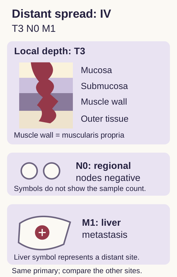
T describes local depth. Tis remains within the mucosa. T1 enters submucosa, T2 invades muscularis propria, and T3 extends through that muscle into pericolorectal tissue. T4 includes penetration of the visceral peritoneal surface or direct invasion of another organ. Crossing muscularis mucosae is not the same as invading muscularis propria. For an endoscopically excised T1 lesion, margins and histologic risk features help determine whether endoscopic treatment is sufficient. [3][19]
N describes regional spread; M describes distant spread. Regional node metastases and qualifying regional tumor deposits affect N. Separate liver, lung or peritoneal metastases affect M. Direct penetration of a primary through the visceral peritoneum is local T4a extension, not automatically a distant peritoneal deposit. The TNM categories are not points to add together. [3]
Before elective colon cancer treatment, cross-sectional imaging of the chest, abdomen and pelvis assesses distant extent. Pathology refines local depth and regional nodes after resection. Rectal cancer additionally needs a dedicated local assessment, usually rectal-protocol pelvic MRI, because surrounding pelvic planes influence treatment sequence and margin risk. Baseline CEA and tumor mismatch repair testing answer additional questions; neither replaces the anatomic workup. [3][4][6][18]
Transfer: after biopsy proves an ascending colon adenocarcinoma, a high CEA does not prove stage IV. After CT excludes distant disease in a low rectal tumor, the local MRI question can still be unanswered. State the missing information before ordering another test. [3][6][18]
Why can the same cancer produce different symptoms?
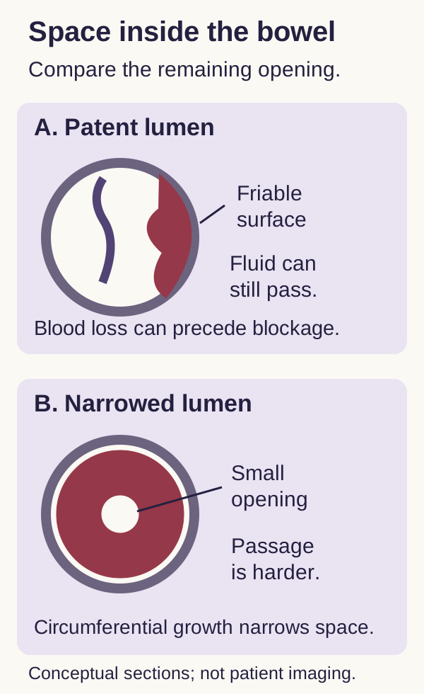
Imagine two equally friable lesions, one leaving a broad lumen and another almost closing it. Which patient is more likely to notice impaired passage first? Use the geometry below rather than memorizing a symptom as exclusive to one side. [3][9][13]
Compare residual space and lesion surface. The shapes illustrate mechanisms, not fixed right-versus-left diagnostic criteria.
Make a prediction: keep the bleeding surface but narrow the opening. Passage becomes more difficult even if the tumor sheds no additional blood. A right-sided lesion can bleed slowly while liquid contents pass through a relatively broad lumen, producing occult iron loss and fatigue without early obstruction. A left-sided annular lesion can obstruct passage of more formed contents, producing progressive constipation, cramping and distention. Stool caliber changes alone are nonspecific. [3][9][13][14]
Rectal involvement adds a local reservoir problem: irritation and reduced capacity can produce urgency, tenesmus and a sense of incomplete evacuation. Bleeding may be visible. None of these patterns proves cancer, its site, or its stage. A right-sided cancer can obstruct, a left-sided cancer can cause iron deficiency, and a proximal cancer can exist with normal hemoglobin. [9][14]
Recognize when the task changes. Inability to pass stool or flatus with progressive distention suggests obstruction. Rigidity, free air, fever or circulatory compromise raises concern for perforation and sepsis. Resuscitation, antibiotics when indicated, and urgent surgical assessment take priority over elective bowel preparation and a complete cancer workup. CT helps define a stable patient's complication, but imaging should not delay treatment of clear diffuse peritonitis. Stenting is not a universal answer, especially with perforation. [13]
Another presentation may be outside the bowel: Streptococcus gallolyticus subsp. gallolyticus bacteremia or endocarditis is associated with colorectal neoplasia. Arrange appropriate colon evaluation after stabilization, even without bowel symptoms. This association includes adenomas and cancer; it does not prove either diagnosis in a particular patient. [11]
Transfer: a normal distal examination does not inspect the proximal colon. A normal hemoglobin does not inspect any part of it. Identify both the clinical concern and the anatomic territory that remains unexamined. [9][11][14]
What changes the starting risk?
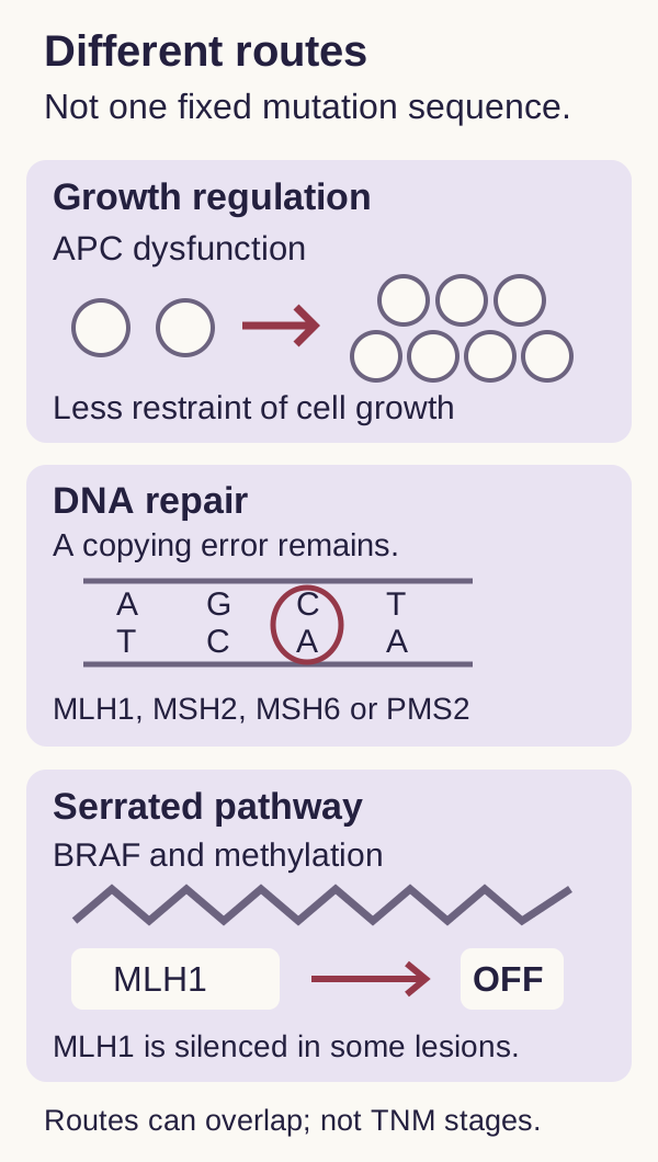
A single mutation sequence cannot explain every colorectal cancer. Compare the routes in the diagram, then ask which evidence would justify an inherited-cancer evaluation rather than assuming that every abnormal tumor test is inherited. [4]
These are overlapping biologic routes, not a universal ordered sequence and not TNM stages. A tumor alteration can be somatic or related to inherited susceptibility.
Sort the evidence: hundreds of early adenomas plus an affected parent suggest APC-associated familial adenomatous polyposis. Colorectal and related cancers across a family without extensive polyposis can suggest Lynch syndrome, involving mismatch repair genes such as MLH1, MSH2, MSH6 or PMS2, or certain EPCAM deletions. Both need a formal genetic assessment; a family story alone does not identify a pathogenic variant. [4]
New colorectal cancers should receive tumor mismatch repair or microsatellite instability assessment. Loss of MLH1 and PMS2 can arise from sporadic MLH1 promoter methylation; reflex methylation and, in appropriate pathways, BRAF testing help interpret the result. Tumor dMMR does not automatically establish Lynch syndrome, and a reassuring reflex result must still be considered alongside age and personal and family history. This same tumor biology can affect treatment selection. [4][7][8]
Risk also accumulates without a single inherited syndrome. Colorectal cancer is common and risk generally increases with age, but incidence has also risen in adults younger than 50. Prior cancer or advanced adenomas change future surveillance. Larger size, villous architecture and high-grade dysplasia make an adenoma more concerning. Longstanding ulcerative colitis or Crohn disease involving the colon adds inflammation-associated dysplasia risk; isolated small-bowel disease is not the same surveillance category. [3][4][10][15]
For many patients with colonic IBD, initial dysplasia surveillance begins around 8 to 10 years after diagnosis, with subsequent timing based on risk. Newly diagnosed primary sclerosing cholangitis warrants immediate assessment in this context. For a first-degree relative diagnosed with colorectal cancer before 60, colonoscopy generally starts at 40 or ten years before the relative's diagnosis, whichever is earlier, and repeats every five years after a normal examination. Syndrome-specific plans may start much earlier. [9][10]
Obesity, smoking and alcohol exposure increase risk; activity is protective. Processed-meat exposure is associated with risk, but no individual food history diagnoses cancer and a healthier lifestyle does not replace screening. For the mechanisms behind these decisions, compare molecular pathways, colorectal polyps and Lynch syndrome. [15][17]
Transfer: controlled colitis symptoms do not necessarily mean low dysplasia risk. Ask what has changed in disease duration, extent, prior pathology, or associated PSC before retaining an old surveillance schedule. [10]
How does extent change the treatment goal?
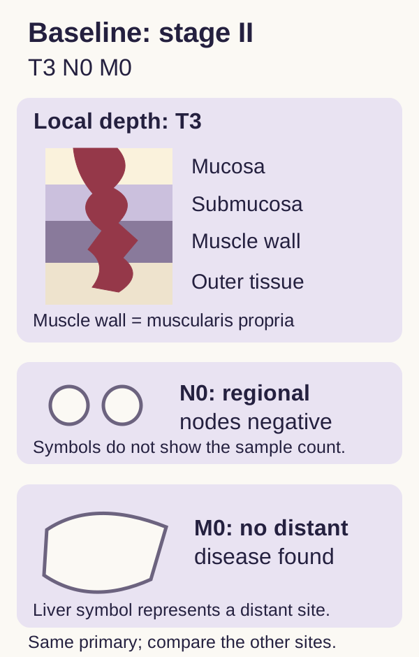
Two tumors can have identical wall depth but different treatment implications. Start with the T3N0M0 colon model below: the tumor extends beyond muscularis propria, regional nodes are negative, and no distant disease is identified. Before opening a comparison, predict which compartment changes and which decision must be reconsidered. [3][5]
Original staging model: T3N0M0, stage II. It illustrates compartment changes, not a complete staging calculator or a treatment prescription.
Keep wall depth fixed; change one finding
Choose either question to inspect a changed visual state. Both comparisons can stay open together. Close either answer to reset that comparison. All general staging and treatment principles remain visible below.
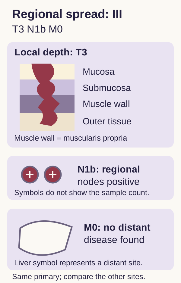
What changes if two regional nodes contain tumor?
Comparison answer: only regional spread changes. T3N1bM0 is stage III; a fit patient generally needs an adjuvant systemic-treatment discussion after complete resection.
The wall drawing is unchanged, but the N category is not. Regional nodal spread supports stage III rather than stage II. It does not by itself establish distant metastatic disease. [3]
What changes if a separate liver metastasis is confirmed?
Comparison answer: distant spread changes. T3N0M1 is stage IV; next assess resectability, biology and patient goals rather than assuming that local treatment is impossible.
The N category can remain negative while M becomes positive. The new decision is whether all disease can be treated locally and which systemic strategy is appropriate. [3][7]
Core colon cancer decisions, visible without opening either comparison
Extent
Usual treatment logic
ExtentStage I: T1 or T2, N0, M0
Usual treatment logicComplete definitive local treatment; routine adjuvant chemotherapy is not used. Selected T1 lesions require an endoscopic-curability assessment.
ExtentStage II: T3 or T4, N0, M0
Usual treatment logicResection; assess adverse features and mismatch repair before discussing adjuvant treatment.
ExtentStage III: regional spread, M0
Usual treatment logicResection plus an individualized, finite adjuvant systemic regimen for suitable patients.
ExtentStage IV: M1
Usual treatment logicAssess complete local treatability, tumor biomarkers, fitness and goals. Some limited metastases permit curative-intent treatment.
Stage II is not one risk category. T4 disease supports offering adjuvant chemotherapy; obstruction, perforation, adverse histology, vascular or perineural invasion, and fewer than twelve sampled nodes can also influence the discussion. A low-risk dMMR stage II tumor is not a routine indication for fluoropyrimidine-only adjuvant therapy. If high-risk dMMR disease leads to a chemotherapy decision, an oxaliplatin-containing approach is considered rather than assuming fluoropyrimidine monotherapy is adequate. Patient fitness and preferences remain part of the decision. [3][5][19]
For metastatic disease, limited liver or lung deposits may be amenable to complete treatment, whereas diffuse unresectable disease generally requires systemic care. MSI-H/dMMR can favor checkpoint inhibitor-based treatment; current options are not limited to a single agent. Conventional anti-EGFR selection requires attention to RAS status and sidedness. Molecularly defined combinations evolve, so the general pathway is not a substitute for current regimen review. Symptom relief and supportive care belong throughout treatment, not only after anticancer options end. [3][7][8]
Rectal exception: low or higher-risk locally advanced pMMR rectal cancer commonly receives total neoadjuvant therapy based on MRI assessment before definitive surgical reassessment. dMMR rectal disease has a distinct immunotherapy pathway requiring specialist planning. Do not copy a colon surgery-first sequence onto every rectal tumor. [6]
Transfer: a small primary with a confirmed liver metastasis remains stage IV. A deeply invasive primary with negative nodes may remain stage II. Explain which anatomic finding changed the group before describing the treatment intention. [3][7]
What does a follow-up result actually tell you?
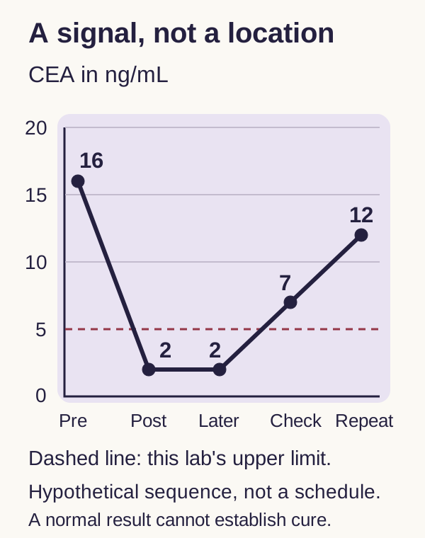
A falling marker can support a response assessment without proving cure. A rising marker can prompt investigation without proving recurrence. Read the CEA trend below and decide which conclusion is supported before deciding what to do. The points are a sequence, not a recommended testing timetable. [3]
Original graph with values. Normalization followed by a reproducible rise supports reassessment, not automatic restaging or chemotherapy. The laboratory limit is supplied for this example, not universal.
Predict the next question: is the increase reproducible, and does appropriate clinical and imaging evaluation show disease? CEA cannot localize a lesion, distinguish every malignant from benign cause of an increase, or replace cross-sectional imaging. Some colorectal cancers never produce an abnormal CEA; a normal baseline does not exempt a patient from appropriate follow-up. Surveillance intensity depends on stage, treatment history, health and whether detecting recurrence would change care. [3][18]
Colonoscopy answers a different follow-up question: are there metachronous lesions or relevant luminal findings? After curative resection and a complete clearing examination, the first surveillance colonoscopy is generally at one year. If normal, the next is three years later, then five years later if still appropriate. Findings, health, hereditary syndromes and rectal local-recurrence risks can change this plan. If obstruction prevented complete initial inspection, arrange clearing within three to six months after surgery rather than waiting for routine one-year surveillance. [12]
Treatment terminology also follows purpose. Induction is the initial phase used to obtain disease control or support later local treatment. Maintenance is a selected lower-intensity continuation after control in an appropriate metastatic strategy. Adjuvant treatment is a finite course after potentially curative definitive treatment to reduce recurrence risk. Persistent metastatic disease, response and accumulating toxicity can justify discussing maintenance, a break, or another individualized strategy; a response alone does not mean cure. [3][7][20]
Transfer: a new liver lesion deserves characterization even when CEA is normal. New symptoms deserve evaluation even when the next scheduled visit is months away. Return to the initial question: which problem does the available result actually answer, and which important problem remains? [3][7][12]
Apply the reasoning
These original educational cases are not patient records. Cover the options when useful, decide what the findings imply, and then compare the alternatives. Explanations address every option; none of the questions is required to continue reading.
Case 1
Show answer and explanations for case 1
A. Repeat FIT while starting oral iron (Why this does not fit)
FIT can be used repeatedly in an average-risk screening program. New unexplained iron deficiency requires evaluation despite a recent negative screen. A screening result does not settle a subsequent diagnostic problem.
Reasoning steps for option A
What new problem is established by hemoglobin 9.4, MCV 71 and ferritin 8?
These results establish iron-deficiency anemia in a man without an identified source of loss.
Would another negative FIT identify why this man has lost iron?
No. FIT is a screening assay; the newly unexplained anemia requires gastrointestinal source evaluation even after a negative sample.
B. Arrange upper endoscopy and colonoscopy (Best answer)
Bidirectional endoscopy evaluates upper and lower gastrointestinal sources of iron loss. Microcytic anemia with very low ferritin establishes iron deficiency in a man without another explanation. Replace iron while investigating its loss; neither step replaces the other.
Reasoning steps for option B
Which two gastrointestinal regions need initial assessment for this unexplained iron deficiency?
Both the upper tract and colon need assessment because no alternative source of iron loss is known.
How do upper endoscopy and colonoscopy differ from his recent FIT?
They investigate potential sources directly rather than using a screening result to dismiss the new anemia.
Can iron replacement replace either examination if his hemoglobin improves?
No. Replacing iron treats the deficit but does not identify its cause; treatment and source investigation proceed together.
C. Obtain capsule endoscopy before conventional endoscopy (Why this does not fit)
Capsule examination can investigate selected small-bowel sources after appropriate initial evaluation. Upper and lower gastrointestinal evaluation has not yet been performed. Do not bypass the initial endoscopic assessment.
Reasoning steps for option C
Which territory would a capsule chiefly investigate in this proposed sequence?
The small bowel, before the more appropriate initial upper and lower endoscopic evaluation has been completed.
What missing examinations make capsule endoscopy premature in this stable patient?
Upper endoscopy and colonoscopy have not been performed. Selected small-bowel investigation follows an appropriate initial evaluation rather than bypassing it.
D. Repeat the blood count after iron therapy (Why this does not fit)
Monitoring hemoglobin helps assess response to iron treatment. An improved count would not identify the source of the iron deficiency. Treatment response and etiologic diagnosis answer different questions.
Reasoning steps for option D
What would a higher hemoglobin after oral iron demonstrate?
It would show a response to replacement, not reveal why this man developed ferritin 8 with microcytic anemia.
Why is repeating the blood count alone insufficient despite his stability?
His unexplained iron loss still needs a source evaluation. Clinical stability permits organized investigation but does not make treatment response an etiologic diagnosis.
Takeaway: New unexplained iron deficiency requires a source evaluation even after negative screening.
A. Repeat FIT in twelve months (Why this does not fit)
Annual FIT is an accepted strategy when screening results are negative. The first positive result has not received the required follow-up examination. A later negative sample does not complete an abnormal screening episode.
Reasoning steps for option A
Which FIT result would justify simply continuing an annual screening schedule?
A negative screening episode without an unresolved positive result could support annual FIT; this patient already has a positive screen.
Why does the second negative FIT not justify waiting twelve months?
Bleeding can be intermittent. The later sample leaves the first positive result unevaluated and does not replace colonoscopy.
B. Obtain stool DNA-FIT this month (Why this does not fit)
Stool DNA-FIT is another screening option for appropriate asymptomatic adults. A second screening modality cannot determine the source of the positive FIT. Follow an abnormal screen with the indicated diagnostic examination.
Reasoning steps for option B
What does changing from FIT to stool DNA-FIT change about the testing method?
It substitutes another stool-based screening strategy rather than directly examining the source of the first positive FIT.
Would a negative stool DNA-FIT establish that her original positive FIT was benign?
No. A second screening assay cannot exclude the lesion responsible; the positive screening episode still requires colonoscopic follow-up.
C. Treat hemorrhoids and repeat FIT (Why this does not fit)
Hemorrhoids can account for some visible rectal bleeding. Their presence does not exclude a separate colorectal neoplasm or resolve the abnormal screening result. A plausible benign lesion does not cancel colorectal evaluation.
Reasoning steps for option C
Do small internal hemorrhoids exclude a separate colorectal neoplasm?
No. A benign anorectal lesion can coexist with neoplasia and cannot establish the source of this positive screening sample.
Why would treating the hemorrhoids and retesting still leave an unanswered question?
It would not examine the colon. With no prior colonoscopy, neither hemorrhoid treatment nor a later negative FIT completes the original positive screen.
D. Arrange a complete colonoscopy (Best answer)
Colonoscopy evaluates the lesion responsible for an abnormal stool screen and permits tissue sampling. Intermittent bleeding can produce discordant stool results, leaving the first positive test unresolved. Complete a positive FIT with colonoscopy rather than another stool test.
Reasoning steps for option D
How can FIT change from positive to negative over two weeks?
Intermittent bleeding means that a later stool sample may contain less blood even while a colorectal lesion remains.
Which test can inspect the colon and sample a lesion after this discordant screening sequence?
Complete colonoscopy can evaluate the unresolved positive result and obtain tissue; neither another stool test nor the hemorrhoid finding supplies that evaluation.
Takeaway: A positive stool screen remains unresolved until the appropriate colon examination.
A. FIT every year, with colonoscopy after a positive result (Best answer)
Annual FIT provides an accepted home-based screening strategy. His age, average-risk history and willingness to complete follow-up make this plan appropriate. The interval and the follow-up examination are both parts of screening.
Reasoning steps for option A
Why is this asymptomatic 46-year-old eligible for the named screening program?
He is over the USPSTF starting age of forty-five and has no supplied feature requiring a higher-risk pathway.
Which two commitments make annual home FIT a complete screening strategy?
He must repeat FIT every year and undergo colonoscopy after a positive result, both of which match his stated preferences and agreement.
B. FIT every five years, with colonoscopy after a positive result (Why this does not fit)
Longer intervals are appropriate for certain direct-visualization tests. Five years is not the recommended FIT interval. Do not transfer a colonography interval to a stool test.
Reasoning steps for option B
Which test uses the five-year interval that this option assigns to FIT?
CT colonography is repeated every five years in the named framework; FIT is a different test with an annual schedule.
Would willingness to repeat home testing make five-year FIT sufficient?
No. His preference supports a stool-based option, but it does not change the annual interval required for FIT.
C. Stool DNA-FIT every ten years, with colonoscopy after a positive result (Why this does not fit)
Stool DNA-FIT is an accepted home-based option. Ten years is a colonoscopy interval, not the USPSTF stool DNA-FIT interval of one to three years. Match each screening test to its own schedule.
Reasoning steps for option C
What is the USPSTF interval for the proposed stool DNA-FIT method?
One to three years, not the ten years offered in this option.
Why can a normal-colonoscopy interval not be transferred to his home stool test?
The methods have different recommended testing schedules. Ten years fits high-quality normal screening colonoscopy, not stool DNA-FIT.
D. FIT starting at fifty years, with annual testing thereafter (Why this does not fit)
Age fifty was a former routine starting threshold. This 46-year-old already meets the USPSTF age threshold of forty-five. Eligibility does not require symptoms or a family history.
Reasoning steps for option D
How far is age forty-six from the applicable screening starting threshold?
He is already one year beyond forty-five; waiting until fifty would postpone screening for an eligible average-risk adult.
Does the absence of symptoms or affected relatives justify delaying FIT until fifty?
No. Those findings describe his average-risk screening setting rather than a reason to defer the age-appropriate annual FIT program.
Takeaway: Choose a screening strategy the patient can complete, including its follow-up.
A. Offer routine screening to both on the same schedule (Why this does not fit)
Routine population screening applies through age seventy-five. At seventy-nine, health and previous screening materially change expected benefit. The older-adult decision is not determined by age alone.
Reasoning steps for option A
Does the routine screening recommendation through age seventy-five cover both 79-year-olds?
No. Both patients fall in the selective screening range of seventy-six through eighty-five.
What would an identical schedule ignore about A and B?
A is healthy and unscreened, whereas B has prior normal examinations and advanced heart failure. These differences change the expected benefit and burden.
B. End screening discussion for both because of their age (Why this does not fit)
Screening is discontinued after age eighty-five in this framework. Both patients are within the selective screening age range, and one has never been screened. Seventy-six through eighty-five is not an automatic stop interval.
Reasoning steps for option B
At what age does the named framework discontinue screening rather than consider it selectively?
After eighty-five. At seventy-nine, neither patient has reached that age-based stopping boundary.
Which facts about A argue against ending discussion solely because of age?
A has never been screened, is independent and has no major illness, leaving a potential screening benefit that deserves discussion.
C. Discuss screening for A; favor stopping for B (Best answer)
Selective screening weighs previous testing, overall health and preferences. A has more opportunity to benefit, whereas B has prior negative testing and substantial competing illness. Discuss the balance of benefit and harm rather than prescribing by birthday.
Reasoning steps for option C
Why might the healthy, never-screened A gain more from screening now?
A lacks the previous negative examinations that reduce the incremental benefit of further screening and has no supplied major competing illness.
Which two features favor stopping screening for B despite the same age?
B has already had normal high-quality colonoscopies and now has limited life expectancy from advanced heart failure. Those facts shift the balance away from further screening.
D. Favor screening for B and defer it for A (Why this does not fit)
Previous normal tests can guide future screening intervals. Prior adequate screening lowers the incremental benefit, while lack of screening can increase it. A reassuring screening history does not create greater need than never being screened.
Reasoning steps for option D
Does B having two previous normal colonoscopies create more unmet screening need than A?
No. Prior adequate screening generally lowers the additional expected benefit, while A has never been examined.
How does B having advanced heart failure affect the proposed preference for screening B?
Limited life expectancy further reduces the opportunity to benefit, so prioritizing B over healthy unscreened A reverses the supplied benefit comparison.
Takeaway: Prior screening and competing illness change the value of screening at the same age.
A. Begin colonoscopy at forty-five and repeat every ten years (Why this does not fit)
That age and interval fit a common average-risk colonoscopy plan. Her first-degree relative was diagnosed before age sixty. Family history can change both the starting age and interval.
Reasoning steps for option A
Which family finding prevents using a starting age of forty-five with ten-year repeats?
Her father is a first-degree relative with colorectal cancer diagnosed before sixty, so an average-risk starting age and interval do not fit.
What start and repeat interval follow from a paternal diagnosis at forty-nine?
Ten years earlier is thirty-nine, earlier than forty; colonoscopy begins now and generally repeats every five years after a normal examination.
B. Begin colonoscopy at forty and repeat every ten years (Why this does not fit)
Forty is one starting age used in family-history screening. Ten years before the affected relative is earlier here, and the usual interval for this history is five years. Use the earlier starting age and the appropriate interval together.
Reasoning steps for option B
Is forty earlier than ten years before her father developed cancer at forty-nine?
No. Forty-nine minus ten is thirty-nine, which is her current age and is the earlier starting threshold.
What second error remains even apart from waiting until forty?
A ten-year repeat interval is too long for this family-history pattern; the usual interval after a normal colonoscopy is five years.
C. Begin colonoscopy at forty-nine and repeat every five years (Why this does not fit)
A five-year interval is used for this family-history pattern. Waiting until the relative's diagnosis age misses the earlier starting recommendation. The relative's age sets an earlier threshold, not the same threshold.
Reasoning steps for option C
Which part of starting at forty-nine and repeating every five years fits this family history?
The five-year interval fits a first-degree relative diagnosed before sixty, but the starting age does not.
How does using the same diagnosis age as her father delay recommended screening?
It misses the ten-year lead time: screening is due at thirty-nine, not at the age when her father developed cancer.
D. Begin colonoscopy now and repeat every ten years (Why this does not fit)
Screening is due because she is ten years younger than her father at diagnosis. The ten-year repeat interval is too long for this first-degree-relative history. An appropriate starting date does not make an average-risk interval appropriate.
Reasoning steps for option D
Why is beginning colonoscopy now appropriate at age thirty-nine?
She is ten years younger than her father was at diagnosis, making thirty-nine earlier than the alternative starting age of forty.
Why does a correct starting age not justify ten-year repeat examinations?
Her first-degree-relative history still calls for a five-year interval after a normal examination; eligibility and repeat timing are separate decisions.
E. Begin colonoscopy now and repeat every five years (Best answer)
For a first-degree relative diagnosed before sixty, colonoscopy generally starts at forty or ten years earlier than diagnosis, whichever comes first. Forty-nine minus ten is thirty-nine, making screening due now. Confirm family details and revisit genetic assessment if additional features emerge.
Reasoning steps for option E
Which of forty and forty-nine minus ten is the earlier screening start?
Thirty-nine is earlier, so this patient is due for colonoscopy now.
What interval accompanies a first-degree relative diagnosed before sixty?
Generally five years after a normal high-quality colonoscopy, with subsequent findings modifying the plan.
Does a family-history screening plan itself identify a hereditary pathogenic variant?
No. Family details still need confirmation, and additional suggestive features can warrant genetic assessment rather than assuming a named syndrome.
Takeaway: For this family-history pattern, choose the earlier start and a five-year colonoscopy interval.
A. Start routine screening when he reaches forty-five (Why this does not fit)
Forty-five is the routine starting age for average-risk screening. Persistent bleeding, altered bowel habits and weight loss create a current diagnostic problem. Symptoms are not governed by the screening starting age.
Reasoning steps for option A
Which of his symptoms make waiting for an average-risk screening birthday inappropriate?
Six weeks of blood mixed with stool, persistent bowel-habit change and weight loss create a current diagnostic concern.
Why does being thirty-two not justify deferring that concern until forty-five?
Forty-five governs routine screening of asymptomatic average-risk adults, not investigation of ongoing alarm symptoms.
B. Arrange diagnostic colorectal evaluation now (Best answer)
Persistent colorectal symptoms warrant evaluation despite young age or a normal blood count. The hemorrhoid does not account sufficiently for the full symptom pattern. Evaluate the patient's findings rather than assigning all bleeding to the first benign lesion.
Reasoning steps for option B
Which findings remain inadequately explained by the small nonbleeding hemorrhoid?
Persistent bowel-habit change and unintentional weight loss accompanying blood mixed with stool remain unexplained.
Can hemoglobin 13.8 or the absence of affected relatives exclude important colorectal disease?
No. Neither resolves the symptom cluster, so diagnostic colorectal evaluation, including colonoscopy when appropriate, is needed now.
C. Use FIT to decide whether endoscopy is needed (Why this does not fit)
FIT can select follow-up in an asymptomatic screening program. An additional screening test would not adequately evaluate these ongoing concerning symptoms. A screening assay is not a substitute for investigating persistent alarm features.
Reasoning steps for option C
What would using FIT as a gatekeeper assume about this presentation?
It would treat persistent bleeding with weight loss and altered bowel habits as though this were an asymptomatic screening encounter.
Would a negative FIT resolve those six weeks of alarm symptoms?
No. In this supplied setting, another screening result would not replace direct diagnostic evaluation of the persistent unexplained symptoms.
D. Treat the hemorrhoid and defer further evaluation (Why this does not fit)
Hemorrhoid treatment is useful when the clinical presentation is adequately explained by it. Blood mixed with stool, persistent change and weight loss remain unexplained. Address benign disease without allowing it to conceal a separate diagnosis.
Reasoning steps for option D
What would hemorrhoid treatment address in this examination?
It would address the observed benign anorectal finding, but would not establish the cause of the full symptom pattern.
Which accompanying symptoms prevent attributing all the bleeding to that hemorrhoid?
Persistent bowel-habit change and weight loss, together with blood mixed with stool, require further colorectal investigation rather than deferral.
Takeaway: Diagnostic evaluation is driven by symptoms, not the routine screening birthday.
A bleeding surface can cause iron loss, while severe luminal narrowing impedes transit. A has a patent lumen and B has upstream dilation at a narrowed segment. Structural findings support a presentation pattern but do not establish a unique symptom profile.
Reasoning steps for option A
What can the friable cecal surface in A cause while a broad lumen remains open?
It can lose blood chronically without early obstruction, supporting occult iron loss despite continued passage.
Why does upstream dilation at the narrowed sigmoid segment favor obstruction in B?
The dilation shows impaired transit proximal to the circumferential narrowing, supporting progressive obstructive symptoms rather than a location-only rule.
Do these structural findings exclude bleeding in B or later obstruction in A?
No. They support the most consistent paired presentation without making either symptom exclusive to one side of the colon.
B. A: progressive obstructive symptoms; B: chronic occult blood loss (Why this does not fit)
Both obstruction and chronic bleeding can occur with colon cancer. The demonstrated impaired passage is in B, whereas A has a broad residual lumen without upstream dilation. Use the actual lumen and upstream findings rather than reversing the structural comparison.
Reasoning steps for option B
Which patient actually has narrowing accompanied by upstream dilation?
B has both findings, providing direct evidence of impaired passage; A has a broad residual lumen without proximal dilation.
Why is assigning obstruction chiefly to A a poorer match than assigning it to B?
It reverses the demonstrated structural evidence. Bleeding can occur in either patient, but the supplied obstruction pattern is in B.
C. A: urgency with tenesmus; B: large-volume secretory diarrhea (Why this does not fit)
Distal irritation can produce urgency, and some large secretory colorectal lesions cause watery diarrhea. The supplied lesions demonstrate a patent cecal lumen and a mechanically obstructing sigmoid segment, not rectal reservoir dysfunction or a secretory phenotype. Match the symptom pattern to the demonstrated lesion behavior.
Reasoning steps for option C
What site-related mechanism would better explain urgency and tenesmus than A's cecal lesion?
Rectal irritation and reservoir dysfunction can cause urgency and tenesmus; the described patent cecal lesion does not establish that mechanism.
What would large-volume secretory diarrhea require that B's scan does not show?
A secretory lesion phenotype, rather than the supplied mechanical narrowing with upstream dilation. The images support impaired passage in B.
D. A: chronic occult blood loss; B: sudden diffuse peritonitis (Why this does not fit)
A friable surface may bleed, and malignant obstruction can become complicated by perforation. Upstream dilation supports obstruction but no free air, acute systemic illness or peritoneal findings establish perforation here. Do not substitute a possible later complication for the currently supported consequence.
Reasoning steps for option D
Does the friability of A's lesion support the occult-blood-loss half of this pairing?
Yes. A friable surface can bleed despite a patent lumen, so that half is consistent with A's findings.
Which evidence needed for sudden diffuse peritonitis is absent from B's description?
No free air, peritoneal signs or acute systemic illness is supplied. Upstream dilation establishes obstruction, not an already perforated bowel.
Takeaway: Predict from the demonstrated structure; laterality alone cannot exclude bleeding or obstruction.
A. Complete bowel preparation for colonoscopy and tissue diagnosis (Why this does not fit)
Elective colonoscopy can establish tissue diagnosis and evaluate the remaining colon. Shock, peritoneal signs and free air indicate a perforation emergency rather than a routine endoscopic setting. Stabilization and source control take priority over elective diagnostic completeness.
Reasoning steps for option A
What do rigidity and free intraperitoneal air add to the sigmoid obstruction?
They establish concern for perforation with peritoneal contamination rather than uncomplicated luminal narrowing.
Why should bowel preparation and elective diagnostic colonoscopy not come first at 84/50 mm Hg?
She has circulatory compromise and perforation requiring resuscitation and urgent source control; elective diagnostic completeness would delay treatment of the immediate threat.
B. Place an endoscopic stent to relieve the sigmoid obstruction (Why this does not fit)
Stenting can be considered for selected malignant obstructions without perforation in an expert setting. Free air and diffuse peritonitis make this an inappropriate uncomplicated-obstruction strategy. A perforation changes the therapeutic problem.
Reasoning steps for option B
What would make stenting an option for selected malignant obstruction?
An expert setting with an appropriate obstruction and no perforation can support consideration of a stent.
Which CT and examination findings disqualify that uncomplicated-obstruction approach here?
Free intraperitoneal air and diffuse rigidity indicate perforation and peritonitis, so decompression by stent does not address the required urgent surgical source control.
C. Obtain staging MRI to guide subsequent operative planning (Why this does not fit)
MRI can clarify selected local or liver staging questions. Additional elective staging would delay treatment of shock and contamination. Do not postpone source control for a complete oncologic map.
Reasoning steps for option C
What kind of question could staging MRI answer after an emergency is controlled?
It could refine selected local or liver staging questions, not reverse this patient's shock or contaminated peritoneum.
Why does lactate 5.2 with hypotension make additional staging the wrong priority?
The patient needs immediate resuscitation and source control. Waiting for an elective oncologic map would delay treatment of severe circulatory compromise.
D. Provide resuscitation, antibiotics, and urgent surgical source control (Best answer)
Perforated bowel with septic physiology requires simultaneous resuscitation and urgent source-control assessment. Hypotension, high lactate, rigidity and free air establish the immediate threat. The definitive operation is individualized after emergency priorities are addressed.
Reasoning steps for option D
Which combination identifies the emergency beyond a blocked sigmoid lumen?
Free air and diffuse rigidity indicate perforation and peritonitis, while fever, hypotension and elevated lactate indicate severe septic physiology.
Why are resuscitation, antibiotics and surgical source control needed together?
They address circulatory compromise, infection and the perforated source of contamination. Tumor staging cannot substitute for any of those emergency priorities.
Does this emergency pattern specify one universal definitive operation?
No. It establishes the need for urgent surgical source control, with the operation individualized to the emergency findings and patient condition.
E. Begin systemic anticancer therapy to shrink the obstructing tumor (Why this does not fit)
Systemic treatment can shrink selected tumors over time. It cannot immediately control perforation with peritoneal contamination and shock. Separate time-sensitive complications from subsequent cancer therapy.
Reasoning steps for option E
On what time scale would systemic anticancer treatment address tumor-related narrowing?
Tumor shrinkage occurs over time; it is not immediate control of a perforated, contaminated bowel.
What untreated threats would persist while waiting for the tumor to shrink?
Peritoneal contamination and shock, already supported by free air, rigidity, blood pressure 84/50 and lactate 5.2, require emergency treatment first.
Takeaway: When perforation and shock accompany a tumor, emergency source control precedes elective staging.
A. Repeat flexible sigmoidoscopy after recovery (Why this does not fit)
Flexible sigmoidoscopy can assess distal colorectal lesions. It would leave the proximal colon unevaluated despite an organism associated with colorectal neoplasia. The required examination must cover the relevant anatomy.
Reasoning steps for option A
Which part of the colon was not examined by his prior flexible sigmoidoscopy?
The proximal colon was not covered by that distal examination.
Why would repeating the same limited examination be insufficient after this endocarditis?
S. gallolyticus subsp. gallolyticus is associated with colorectal neoplasia, including lesions outside the distal segment, so a complete colon evaluation is needed.
B. Continue annual FIT without endoscopic evaluation (Why this does not fit)
Annual FIT is a routine screening option for appropriate average-risk adults. This bloodstream isolate is a separate clinical indication for colorectal evaluation. Absence of bowel symptoms does not erase a disease-specific diagnostic signal.
Reasoning steps for option B
What new clinical finding takes this encounter beyond routine annual FIT screening?
Treated S. gallolyticus subsp. gallolyticus endocarditis creates a separate reason to investigate associated colorectal neoplasia.
Why do a negative FIT and no anemia not justify avoiding endoscopy here?
Neither excludes a colorectal lesion associated with this organism. After stabilization, full colon evaluation remains appropriate even without bowel symptoms.
C. Arrange complete colonoscopy after stabilization (Best answer)
Colonoscopy evaluates the full colon and permits sampling of neoplasia associated with this organism. Neither normal distal inspection nor a negative stool screen excludes an associated proximal lesion. The organism warrants evaluation; it does not itself prove cancer.
Reasoning steps for option C
Which disease association is relevant to the identified subspecies?
S. gallolyticus subsp. gallolyticus bacteremia or endocarditis is associated with colorectal adenomas and carcinoma.
How does colonoscopy close the gap left by a normal distal examination and negative FIT?
It inspects the full colon and permits sampling, including proximal lesions that the prior sigmoidoscopy did not assess.
Does the organism establish that this patient has colorectal cancer?
No. The association justifies investigation after stabilization, not a cancer diagnosis without lesion assessment.
D. Use serial CEA as the next colorectal assessment (Why this does not fit)
CEA can be informative during follow-up of selected treated colorectal cancers. It cannot screen reliably for a previously undiagnosed colorectal lesion. Use a tumor marker for its validated purpose, not as an endoscopic substitute.
Reasoning steps for option D
What established colorectal setting gives serial CEA a role?
CEA can contribute to follow-up of selected treated colorectal cancers, unlike this patient who has no diagnosed colorectal tumor.
Why would serial CEA fail to investigate the neoplasia concern raised by the endocarditis?
CEA cannot reliably screen for or localize a previously undiagnosed colorectal lesion and cannot replace the complete colon examination needed here.
Takeaway: S. gallolyticus subsp. gallolyticus infection warrants colorectal evaluation, not a presumption of cancer.
Chest CT evaluates pulmonary metastatic disease. The unresolved question is pelvic local anatomy, not thoracic spread. Choose the study that resolves the specific staging uncertainty.
Reasoning steps for option A
Which metastatic compartment did the existing chest CT already assess?
The chest was assessed for thoracic spread, and the supplied staging CT shows no distant metastases.
Would repeating chest CT map the relation of this low rectal tumor to the mesorectal fascia?
No. The unresolved question concerns local pelvic soft-tissue planes, outside the purpose of another chest examination.
B. Obtain a screening CT colonography (Why this does not fit)
CT colonography can assess the colonic lumen when conventional inspection is incomplete. The lesion is already diagnosed and the needed information concerns rectal soft-tissue planes. Luminal assessment and pelvic local staging are different tasks.
Reasoning steps for option B
What would CT colonography chiefly assess in contrast to a rectal-protocol MRI?
CT colonography assesses the colorectal lumen rather than supplying the dedicated soft-tissue map needed for rectal treatment planning.
What has already been established that makes another screening luminal study the wrong next task?
Colonoscopy and biopsy have identified rectal adenocarcinoma. The remaining question is the tumor's relationship to the mesorectal fascia.
C. Measure serum CEA twice (Why this does not fit)
CEA supplies a baseline for selected subsequent monitoring. A blood concentration does not show the mesorectal fascia or surgical margin relationship. A tumor marker cannot substitute for an anatomic map.
Reasoning steps for option C
What monitoring information might a baseline CEA provide after diagnosis?
It may provide a reference for subsequent follow-up, but not a picture of local tumor extent.
Why cannot two serum CEA measurements determine the threatened surgical plane?
A blood concentration cannot show the mesorectal fascia or measure the tumor's relationship to it; the question requires anatomic imaging.
D. Repeat superficial endoscopic biopsies (Why this does not fit)
Biopsy establishes histology and can supply tissue for molecular analysis. Histology is already established, and superficial samples cannot map the surrounding pelvic planes. Do not repeat a completed diagnostic task instead of staging.
Reasoning steps for option D
Which diagnostic conclusion has the existing endoscopic biopsy already supplied?
It has established adenocarcinoma in the lesion located 5 cm from the anal verge.
What limitation would persist after repeating superficial tissue samples?
Superficial biopsies would still not map the surrounding pelvic planes or the relationship to the mesorectal fascia needed for treatment selection.
E. Obtain a rectal-protocol pelvic MRI (Best answer)
Pelvic MRI assesses rectal tumor extent and its relationship to the mesorectal fascia. A low rectal tumor without distant disease still needs local risk assessment before treatment sequencing. Rectal treatment planning requires a dedicated local map.
Reasoning steps for option E
Why does a lesion 5 cm from the anal verge need a dedicated local pelvic assessment?
It is a low rectal cancer, where the surrounding pelvic planes and mesorectal fascia affect margin risk and treatment sequence.
What information does rectal-protocol MRI add despite negative distant staging?
It maps local tumor extent and its relation to the mesorectal fascia, answering the surgeon's unresolved planning question.
Takeaway: A negative distant staging scan does not supply the local MRI assessment required for rectal planning.
A. Stage II; discuss adjuvant treatment for high-risk disease (Why this does not fit)
Stage II requires local extension beyond the muscularis propria without regional or distant spread. This tumor does not extend beyond the muscularis propria. Use wall depth rather than tumor diameter to assign the T category.
Reasoning steps for option A
How far must an N0M0 colon primary extend to belong to stage II?
It must extend beyond muscularis propria, as T3 or T4 disease.
Why does invasion into, but not beyond, muscularis propria fail that stage II requirement?
It is T2 local depth. With nineteen negative regional nodes and no distant metastases, the supplied cancer belongs to stage I.
B. Stage I; proceed with postoperative surveillance (Best answer)
T2 disease with N0 and M0 belongs to stage I. Complete resection with adequate negative regional nodes supports surveillance without routine adjuvant chemotherapy. Adjuvant treatment is not determined by the word cancer alone.
Reasoning steps for option B
Which T category follows from tumor entering muscularis propria without passing through it?
T2. The deepest reported invasion is within the muscle, not beyond it.
What stage and postoperative plan follow from T2, nineteen negative nodes and M0?
T2N0M0 is stage I. After complete resection with clear margins, surveillance is appropriate without routine adjuvant chemotherapy.
C. Stage III; offer oxaliplatin-based adjuvant therapy (Why this does not fit)
Regional nodal involvement makes nonmetastatic colon cancer stage III. All examined regional nodes are negative. Do not infer nodal disease from invasion of the bowel muscle.
Reasoning steps for option C
What regional finding would support the proposed stage III adjuvant pathway?
Documented regional nodal disease would support stage III in the absence of distant metastases.
Do the nineteen examined nodes or the muscle invasion establish that regional spread?
No. All nineteen nodes are negative, and local invasion of bowel muscle is a T finding, not evidence of nodal metastasis.
D. Stage IV; begin biomarker-directed systemic therapy (Why this does not fit)
Distant metastatic disease establishes stage IV. No distant disease is present on the supplied evaluation. The M category is distinct from local wall invasion.
Reasoning steps for option D
Which category must be positive to assign this tumor to stage IV?
The M category must show distant metastatic disease.
Why do muscularis propria invasion and a known cancer diagnosis not support stage IV here?
Those establish the local primary, while the supplied imaging shows no distant disease. Local wall invasion cannot substitute for M-positive evidence.
Takeaway: Wall depth, regional nodes and distant disease must be interpreted separately before choosing treatment.
A. Give fluoropyrimidine monotherapy as routine adjuvant treatment (Why this does not fit)
Fluoropyrimidines are used in selected adjuvant colon cancer regimens. Low-risk stage II disease with deficient mismatch repair is not a setting for routine fluoropyrimidine-only adjuvant treatment. Tumor biology changes the adjuvant benefit assessment.
Reasoning steps for option A
How does dMMR affect routine fluoropyrimidine-only adjuvant treatment in this low-risk T3N0 setting?
It argues against routine fluoropyrimidine monotherapy rather than providing an indication for it.
Which pathology findings reinforce that treatment assessment?
Twenty-two negative nodes, clear margins and no supplied obstruction, perforation or vascular or perineural invasion support a low-risk stage II assessment.
B. Give oxaliplatin-based chemotherapy for regional node involvement (Why this does not fit)
Oxaliplatin-containing adjuvant regimens are standard options for fit patients with stage III colon cancer. No regional node contains tumor, and the supplied stage II risk features are favorable. Do not assign node-positive treatment solely because a tumor is invasive.
Reasoning steps for option B
Which finding would justify treating this as regional node-positive colon cancer?
A regional node containing tumor would supply the node-positive basis for a stage III adjuvant approach.
Why does T3 invasion not supply that indication when all twenty-two nodes are negative?
T3 describes wall depth, not regional spread. The supplied tumor is node-negative with favorable stage II risk features, not stage III.
C. Use surveillance without routine adjuvant chemotherapy (Best answer)
Low-risk stage II colon cancer is generally managed without routine adjuvant chemotherapy. Adequate negative node sampling, absence of adverse features and deficient mismatch repair support this approach. Individualize risk rather than treating all stage II tumors alike.
Reasoning steps for option C
What stage group follows from a T3 colon primary with negative nodes and no distant disease?
Stage II, because the tumor extends beyond muscularis propria but has no regional or distant spread.
What combined risk and repair findings support surveillance here?
Adequate negative node sampling, no supplied adverse features and deficient mismatch repair favor surveillance without routine adjuvant chemotherapy, particularly fluoropyrimidine monotherapy.
D. Give pelvic chemoradiation before further surveillance (Why this does not fit)
Pelvic chemoradiation is used in selected rectal treatment pathways. This is a completely resected colon primary without the rectal local-control problem. Keep colon and rectal treatment pathways distinct.
Reasoning steps for option D
Which anatomic treatment problem commonly gives pelvic chemoradiation a role?
Selected rectal cancers require pelvic local-control treatment because of their surrounding pelvic anatomy and recurrence risk.
Why does that pathway not fit this completely resected T3N0 primary?
The primary is in the colon, not the rectum, and the supplied findings do not present a rectal margin or local-control problem.
Takeaway: Low-risk stage II dMMR disease is not a routine fluoropyrimidine-only adjuvant indication.
A. High-risk stage II disease supports offering adjuvant chemotherapy (Best answer)
T4 disease is a high-risk feature within node-negative, nonmetastatic colon cancer. Penetration of the visceral peritoneal surface establishes T4a even with negative nodes. Negative nodes do not make every stage II tumor low risk.
Reasoning steps for option A
Which T category is established by direct penetration of the visceral peritoneal surface?
T4a describes this local extension. It does not require a separate peritoneal metastatic deposit.
What do eighteen negative regional nodes and negative distant staging make the overall group?
T4a with N0 and M0 is stage II, with T4 invasion providing a high-risk feature.
Why should this fit patient with retained mismatch repair proteins be offered an adjuvant discussion?
T4 disease supports offering adjuvant chemotherapy despite negative nodes; treatment selection still depends on individualized benefit and risk.
B. Stage I disease supports omitting an adjuvant discussion (Why this does not fit)
Stage I tumors are limited to submucosa or muscularis propria with N0 and M0. This tumor extends through the bowel wall to the peritoneal surface. The deepest invasion matters more than the absence of nodal disease alone.
Reasoning steps for option B
What depth limit would be compatible with stage I when nodes and distant sites are negative?
A primary limited to submucosa or muscularis propria, corresponding to T1 or T2.
Why does this specimen exceed that stage I boundary despite eighteen negative nodes?
The primary penetrates the visceral peritoneal surface, establishing T4a. Negative nodes do not reverse the greater local depth.
C. Stage III disease supports treatment for regional nodal spread (Why this does not fit)
Stage III requires regional nodal involvement or qualifying regional tumor deposits. No nodal disease or deposits are described. T4 invasion increases risk without itself creating N-positive disease.
Reasoning steps for option C
Which regional evidence would be needed to classify this nonmetastatic cancer as stage III?
Positive regional nodes or qualifying regional tumor deposits, rather than greater wall depth alone.
Does peritoneal surface penetration provide that regional evidence in this specimen?
No. All eighteen nodes are negative and no deposits are described. T4a raises stage II risk without creating an N-positive finding.
D. Stage IV disease supports treatment for peritoneal metastases (Why this does not fit)
Separate peritoneal metastases establish distant disease. Direct penetration of the tumor through visceral peritoneum is T4a, not a separate metastatic deposit. Distinguish direct extension from discontinuous distant spread.
Reasoning steps for option D
How does a separate peritoneal metastasis differ from the invasion reported here?
A separate metastatic deposit represents distant spread, whereas direct extension of the primary through the visceral peritoneal surface is local T4a invasion.
Why does this surface finding not establish stage IV?
No separate metastasis is reported and distant staging is negative; the observed penetration changes T, not M.
Takeaway: A T4 primary can be high-risk stage II without nodal or distant metastasis.
A. Surveillance without adjuvant therapy for stage I disease (Why this does not fit)
Muscle-confined disease can be stage I when nodes and distant staging are negative. Two positive regional nodes exclude stage I. A favorable T category does not erase the N category.
Reasoning steps for option A
Would muscle-confined colon cancer be stage I if regional nodes and distant sites were negative?
Yes. T2N0M0 can be stage I, but that requires the negative regional node condition.
Which finding prevents applying that surveillance-only stage I pathway here?
Two of twenty-one regional nodes contain adenocarcinoma, making the M0 tumor stage III despite its limited wall depth.
B. Pelvic chemoradiation for a localized colon primary (Why this does not fit)
Radiation is important in selected rectal local-control strategies. This is a colon primary with regional nodal spread, not a rectal margin problem. Choose site-appropriate systemic adjuvant therapy.
Reasoning steps for option B
Which colorectal location commonly requires a pelvic radiation strategy for local control?
Selected rectal cancers, where pelvic anatomy and margin risk guide local treatment.
Why is pelvic chemoradiation not the appropriate response to this colon tumor's two positive nodes?
The primary is in the colon and the demonstrated risk is regional spread, supporting a systemic adjuvant discussion rather than a rectal local-control pathway.
C. Palliative systemic therapy for distant metastatic disease (Why this does not fit)
Palliative-intent systemic treatment is used for many unresectable metastatic presentations. The involved nodes are regional and no distant disease is identified. Regional nodes and distant metastases imply different stage groups and intentions.
Reasoning steps for option C
Do two involved regional nodes establish the distant metastatic disease asserted by this option?
No. They establish regional spread; the supplied distant staging remains negative.
How do complete resection and M0 status affect the purpose of postoperative therapy?
Adjuvant therapy aims to reduce recurrence after potentially curative resection, rather than treating this as unresectable distant disease with palliative intent.
D. Further observation until CEA establishes residual disease (Why this does not fit)
CEA trends can contribute to follow-up after treatment. Regional nodal involvement already supplies the indication for an adjuvant discussion, independent of CEA. Do not wait for a surveillance marker to replace pathologic risk information.
Reasoning steps for option D
What already establishes postoperative recurrence risk without waiting for CEA?
Pathology has documented two positive regional nodes, supplying the stage III basis for an adjuvant discussion.
Why can observation until a CEA abnormality not replace that pathology-based decision?
CEA contributes to surveillance but cannot establish residual disease by itself or erase the risk already demonstrated by nodal metastases.
E. Finite oxaliplatin-based adjuvant therapy for stage III (Best answer)
Fit patients with resected stage III colon cancer are candidates for an oxaliplatin-containing adjuvant regimen. Regional nodal involvement makes this stage III despite limited wall penetration. Regimen and duration are individualized; adjuvant therapy is not indefinite maintenance.
Reasoning steps for option E
Which stage group results when a T2 colon primary has two positive regional nodes and no distant spread?
Stage III. The positive regional nodes change the group despite muscle-confined local invasion.
Which patient features support considering an oxaliplatin-containing adjuvant course?
Good performance status without major neuropathy or organ dysfunction supports treatment consideration after complete resection.
Why is the proposed course finite rather than indefinite maintenance?
Its purpose is to reduce recurrence after potentially curative surgery. Regimen and duration are individualized within an adjuvant strategy, not ongoing treatment of known unresectable disease.
Takeaway: Regional nodal disease can make a shallow colon tumor stage III and change postoperative therapy.
A. Seven negative nodes give the same staging confidence as twenty (Why this does not fit)
A negative adequately sampled node evaluation supports the N0 assignment. Only seven nodes were examined, below the commonly used twelve-node adequacy threshold. The number examined affects confidence in staging.
Reasoning steps for option A
How does examining seven nodes compare with the usual adequacy benchmark?
Seven is below the commonly used twelve-node threshold, so the regional assessment has less sampling support than a twenty-node examination.
Why cannot the same confidence be assigned merely because every sampled node is negative?
Negative findings apply to the nodes examined. A small sample leaves greater concern that regional disease could have been missed, without proving that it was.
B. Limited sampling adds risk; review pathology and discuss adjuvant therapy (Best answer)
Sampling fewer than twelve nodes is a high-risk feature used in stage II adjuvant assessment. Seven negative nodes leave greater concern for missed regional disease without proving that disease exists. Recognize staging uncertainty without inventing a positive node.
Reasoning steps for option B
What high-risk feature is present despite T3N0 findings and no obstruction or perforation?
Only seven nodes were examined, fewer than twelve, which is a risk feature used in stage II adjuvant assessment.
What should pathology review and the adjuvant discussion address about those seven nodes?
They should address sampling adequacy and the possibility of understaging while retaining that no positive regional node has been documented.
C. Limited sampling establishes stage III despite the negative node findings (Why this does not fit)
Positive regional nodes or qualifying tumor deposits can establish stage III. No positive node or tumor deposit is reported. Inadequate sampling is not equivalent to documented metastasis.
Reasoning steps for option C
What documented finding would support stage III instead of this node-negative classification?
A positive regional node or a qualifying regional tumor deposit would supply the required regional spread.
Why does a count of seven examined nodes not itself provide that finding?
A low count creates staging uncertainty, not a positive node. All reported nodes remain negative, so limited sampling alone cannot establish stage III.
D. Limited sampling makes CEA the preferred test for regional nodal staging (Why this does not fit)
CEA can provide a baseline for selected follow-up. A serum marker cannot determine whether a regional node contains cancer. Resolve pathology adequacy with pathology, not an unrelated marker.
Reasoning steps for option D
What would a CEA measurement reveal about whether a particular regional node contains tumor?
It cannot determine regional nodal involvement; CEA is a serum marker, not a pathologic examination of nodes.
Which assessment addresses the actual weakness of this seven-node specimen?
Review of the specimen and nodal sampling addresses staging adequacy. Substituting CEA would leave the pathology question unresolved.
Takeaway: A low node count increases uncertainty and risk; it does not prove nodal metastasis.
A. Observation until symptoms develop, because the deposits are limited (Why this does not fit)
Observation can be appropriate when treatment is not beneficial or is declined. Potentially treatable metastatic disease is already established in a fit patient. An asymptomatic presentation does not remove the need for oncologic planning.
Reasoning steps for option A
Which findings indicate an opportunity for treatment before symptoms develop?
The sigmoid primary is resectable, the two liver metastases are judged completely treatable, and the patient has good performance status.
Why is limited metastatic burden not a reason simply to observe this patient?
The limited burden supports considering complete treatment rather than waiting for symptoms. Neither lack of symptoms nor limited deposits removes the need for oncologic planning.
B. Colon resection alone, with no plan for the liver lesions (Why this does not fit)
Colon resection can control a localized primary or a primary-related complication. Leaving documented liver metastases untreated does not pursue clearance of all known disease. Treatment intent must account for every established disease site.
Reasoning steps for option B
What known disease would remain after colon resection alone?
Both documented liver metastases would remain outside the treatment plan.
Why must the hepatic sites be included when the goal is complete disease treatment?
The expert assessment supports clearing the liver disease with an adequate remnant, so treatment intent must address both the primary and the metastases.
C. Systemic palliation alone, because stage IV excludes curative local therapy (Why this does not fit)
Many patients with unresectable metastatic disease receive palliative-intent systemic treatment. This patient has limited disease judged fully resectable with preserved liver function. Metastatic stage does not by itself establish unresectability.
Reasoning steps for option C
What does stage IV establish about these two liver lesions?
It establishes distant spread from colorectal cancer, not whether that spread can be completely treated.
Which specialist finding contradicts the claim that only systemic palliation is possible?
The hepatobiliary surgeon judges all hepatic disease removable with an adequate functional liver remnant and no extrahepatic metastasis is identified.
D. Multidisciplinary plan for complete local treatment with curative intent (Best answer)
Selected liver-limited metastases can be addressed with surgery and an individualized systemic strategy. Both primary and metastatic disease are judged amenable to complete treatment in a fit patient. Potential cure is an intent, not a guarantee; resectability requires expert assessment.
Reasoning steps for option D
Which anatomic and functional facts support considering complete local treatment?
The primary is resectable, liver disease is limited to one lobe, no extrahepatic spread is found, and an adequate functional liver remnant can remain.
Why should the plan be multidisciplinary rather than limited to the colon operation?
Both primary and hepatic disease need a coordinated strategy, integrating local treatment and systemic therapy as appropriate.
Does curative intent guarantee cure for this stage IV patient?
No. It describes the goal made plausible by complete treatability and patient fitness; outcomes and the final strategy still require individualized expert assessment.
Takeaway: Stage IV describes distant spread; it does not settle whether complete treatment is feasible.
A. A first-line anti-EGFR antibody selected solely from the MSI result (Why this does not fit)
Anti-EGFR treatment is selected using factors including RAS status and tumor sidedness in an appropriate molecular setting. MSI-H/dMMR identifies a different treatment-sensitive group and does not establish anti-EGFR eligibility. Do not substitute one biomarker for another.
Reasoning steps for option A
Which missing molecular information matters for conventional anti-EGFR selection?
RAS status, together with tumor sidedness and the appropriate molecular setting, matters; the supplied MSI result does not provide those eligibility findings.
What treatment-sensitive group does her MSI-H/dMMR result actually identify?
It identifies a checkpoint-sensitive metastatic colorectal group, not proof that a conventional anti-EGFR antibody is appropriate.
B. Immediate colectomy as the sole treatment for all disease (Why this does not fit)
Surgery can be central for localized or completely resectable disease and for selected complications. The disease is diffusely metastatic and unresectable, without a stated primary-related emergency. Local treatment of the primary alone does not control diffuse distant disease.
Reasoning steps for option B
Which disease sites would immediate colectomy leave untreated?
The diffuse liver and lung metastases would remain, and their overall burden is judged unresectable.
Does the stem supply a primary-related emergency that makes colectomy the sole answer now?
No obstruction or other primary-related emergency is supplied. Removing the colon primary alone would not control the established distant disease.
C. A first-line immune checkpoint inhibitor-based strategy (Best answer)
MSI-H/dMMR metastatic colorectal cancer is an established checkpoint-sensitive treatment group. The patient has unresectable disease and no supplied major obstacle to considering this strategy. Choice among approved regimens includes individual benefit-risk assessment, not MSI status alone.
Reasoning steps for option C
Why is a systemic strategy needed rather than complete local treatment in this patient?
Her diffuse liver and lung disease is judged unresectable, so colectomy alone cannot treat all known cancer.
How does MSI-H/dMMR change the first-line treatment class under consideration?
It supports immune checkpoint inhibitor-based treatment. Her functional status and absence of a supplied transplant or severe active autoimmune disease support considering that strategy.
Does the biomarker specify a single mandatory checkpoint regimen?
No. Options include pembrolizumab and other approved approaches such as nivolumab with ipilimumab, with individual benefit and risk guiding selection.
D. Fluoropyrimidine monotherapy because dMMR predicts its superior effect (Why this does not fit)
Fluoropyrimidines have roles in several colorectal cancer regimens. Deficient mismatch repair is not a marker of superior benefit from fluoropyrimidine monotherapy. Use the actual predictive relationship rather than treating every abnormal biomarker as chemotherapy sensitivity.
Reasoning steps for option D
Does deficient mismatch repair predict superior benefit from fluoropyrimidine monotherapy?
No. dMMR is not a marker of superior effect from fluoropyrimidine alone, although fluoropyrimidines have roles in colorectal cancer regimens.
Which predictive relationship should instead drive comparison of the offered treatment classes?
The MSI-H/dMMR result supports checkpoint sensitivity in this unresectable metastatic setting, rather than the claimed enhanced sensitivity to fluoropyrimidine monotherapy.
Takeaway: MSI-H/dMMR status can change the first-line systemic treatment class in unresectable metastatic disease.
A. B sustains downstream RAS signaling despite EGFR blockade (Best answer)
Activating RAS signaling can bypass dependence on upstream EGFR activation. B has an activating KRAS mutation, whereas A meets the RAS wild-type requirement relevant to conventional anti-EGFR selection. Wild-type status permits consideration but does not guarantee response.
Reasoning steps for option A
Where is KRAS relative to the receptor blocked by cetuximab?
KRAS acts downstream of EGFR, so an activating alteration can maintain signaling without dependence on the receptor being activated.
Why does the activating KRAS mutation in B argue against conventional cetuximab-based selection?
The activated downstream signal can persist despite upstream EGFR blockade, unlike the RAS wild-type eligibility setting in A.
Does wild-type KRAS and NRAS guarantee a response for A?
No. It permits consideration of conventional anti-EGFR treatment in the supplied left-sided setting but is not a guarantee of benefit.
B. B has increased dependence on ligand binding to EGFR (Why this does not fit)
An upstream ligand-dependent pathway can be interrupted by blocking its receptor. Activating downstream KRAS reduces that dependence rather than increasing it. Locate the altered component relative to the drug target.
Reasoning steps for option B
What would increased dependence on EGFR ligand binding imply about blocking the receptor?
A ligand-dependent signal could be interrupted upstream by EGFR blockade, which is the opposite of bypassing that dependence.
Why does B's activating KRAS alteration not support that increased-dependence explanation?
The alteration is downstream of EGFR and reduces reliance on upstream receptor activation rather than increasing it.
C. A has deficient DNA mismatch repair from the RAS result (Why this does not fit)
DNA mismatch repair status can predict sensitivity to checkpoint inhibitors. Both tumors are repair-proficient, and RAS wild-type status does not diagnose repair deficiency. RAS and mismatch repair answer different predictive questions.
Reasoning steps for option C
What repair status is explicitly supplied for A, independent of its RAS result?
A is mismatch repair proficient, as is B; neither is described as repair deficient.
Why cannot wild-type RAS be used to infer deficient DNA mismatch repair?
RAS growth signaling and DNA mismatch repair are different molecular systems. Absence of an activating RAS mutation does not establish a repair defect.
D. A has no malignant signaling because RAS is wild-type (Why this does not fit)
A tumor can lack a particular oncogenic RAS mutation. Wild-type RAS does not mean that the tumor lacks other growth-promoting alterations. Absence of one mutation is not absence of cancer biology.
Reasoning steps for option D
What does the wild-type RAS result exclude in A?
It excludes the tested activating KRAS and NRAS alterations, not every possible growth-promoting mechanism in the tumor.
Why is that negative mutation result compatible with A having metastatic cancer?
Other malignant signaling alterations can exist despite wild-type RAS, so absence of that mutation is not absence of cancer biology.
Takeaway: An activating downstream RAS mutation can defeat conventional upstream EGFR blockade.
A. Proceed directly to local transanal excision (Why this does not fit)
Local excision is an option for carefully selected early rectal lesions. T3 extension, suspicious nodes and a threatened fascia exceed that low-risk setting. Local excision selection requires local staging, not location alone.
Reasoning steps for option A
What kind of rectal lesion can be considered for local transanal excision?
A carefully selected early lesion without the advanced local-risk pattern supplied here.
Which MRI findings make immediate limited excision inadequate for this low tumor?
T3 extension, suspicious mesorectal nodes and a threatened mesorectal fascia exceed the low-risk local-excision setting.
B. Total neoadjuvant therapy, then surgical reassessment (Best answer)
Chemotherapy plus pelvic chemoradiation before surgery is supported for higher-risk pMMR locally advanced rectal cancer. The low location and MRI risk features make immediate limited local treatment inadequate. Treatment sequence reflects pelvic recurrence risk as well as distant stage.
Reasoning steps for option B
What important risk remains despite the absence of distant metastases?
The low rectal tumor has high pelvic local-control risk, with T3 extension, suspicious nodes and a threatened mesorectal fascia.
How do pMMR status and that MRI pattern support the proposed treatment sequence?
They fit a higher-risk locally advanced rectal pathway using total neoadjuvant therapy, including chemotherapy and pelvic chemoradiation, before definitive surgical reassessment.
Why should a colon surgery-first sequence not be copied onto this case?
Rectal pelvic anatomy and margin risk alter the initial strategy; negative distant staging does not remove those local treatment concerns.
C. Use postoperative surveillance after biopsy alone (Why this does not fit)
Surveillance follows appropriate definitive treatment or a carefully established nonoperative response protocol. A diagnostic biopsy has not eradicated this locally advanced tumor. Observation requires a defined treatment or response context.
Reasoning steps for option C
What has the diagnostic biopsy accomplished for this T3 rectal tumor?
It has established histology, not eradicated the primary or treated the suspicious mesorectal nodes.
What would need to precede a surveillance-based strategy rather than observation after biopsy alone?
Appropriate definitive treatment or an established nonoperative response protocol would be needed. No such treatment or response is supplied here.
D. Choose checkpoint monotherapy on the basis of low rectal location (Why this does not fit)
Checkpoint therapy has a particular role in MSI-H/dMMR rectal cancer. The tumor is repair-proficient, and location does not supply the missing biomarker. Site and predictive biology must both be considered.
Reasoning steps for option D
Which repair finding supports the distinct checkpoint-directed rectal pathway?
MSI-H or deficient mismatch repair, not low rectal location by itself.
Why does this tumor's stated pMMR status defeat selection of checkpoint monotherapy solely from its location?
The required predictive repair-deficiency finding is absent. A tumor 4 cm from the anal verge still needs treatment chosen using both its MRI risk and its molecular status.
Takeaway: High-risk pMMR rectal cancer requires a pelvic treatment strategy, not an automatic colon surgery-first plan.
A. Adjuvant therapy after complete cancer resection (Why this does not fit)
Adjuvant therapy follows potentially curative definitive treatment to reduce recurrence risk. She still has unresectable metastatic disease and has not had complete resection. Persistent measurable metastatic disease is not an adjuvant context.
Reasoning steps for option A
What disease state does adjuvant therapy follow in the proposed definition?
Potentially curative definitive treatment, such as complete cancer resection, with treatment aimed at reducing recurrence risk.
Which restaging finding prevents describing this patient's continuation regimen as adjuvant?
Diffuse unresectable metastases persist after a partial response, and no complete resection has occurred.
B. Neoadjuvant therapy before a planned curative resection (Why this does not fit)
Neoadjuvant treatment precedes definitive local therapy in a planned strategy. No curative resection is currently planned because disease remains diffuse and unresectable. Do not assume a surgical goal from a tumor response alone.
Reasoning steps for option B
What planned event gives neoadjuvant therapy its purpose?
Definitive local treatment, such as a planned curative resection, follows the preoperative strategy.
Why does a partial response not establish that neoadjuvant purpose here?
Restaging still shows diffuse unresectable disease and no curative resection is planned. Tumor shrinkage alone does not create a surgical treatment goal.
C. Induction therapy to obtain an initial disease response (Why this does not fit)
Induction describes the initial treatment phase used to gain disease control. The initial response is already established and the plan is to reduce treatment intensity. Distinguish gaining initial control from continuing it with lower burden.
Reasoning steps for option C
Which phase of this patient's treatment was intended to obtain the initial response?
The preceding combination systemic treatment served the initial disease-control purpose described as induction.
How does the new lower-intensity plan differ from obtaining that first response?
An initial partial response is already present. The new plan continues control with less treatment burden in response to cumulative neuropathy.
D. Maintenance therapy after response to initial treatment (Best answer)
Maintenance is a planned lower-intensity continuation after disease control in an appropriate metastatic strategy. Persistent unresectable disease, partial response and cumulative toxicity explain the change in intensity. Maintenance is neither a declaration of cure nor an automatic requirement for every regimen.
Reasoning steps for option D
What do persistent metastases and partial response indicate about the treatment goal?
Disease control has been achieved without eradication; unresectable disease still needs an individualized ongoing strategy.
Why does reducing intensity after response because of sensory neuropathy fit maintenance?
Maintenance describes a lower-intensity continuation intended to preserve disease control while reducing treatment burden after the initial phase.
Does calling this maintenance mean that every responding patient must receive it?
No. It describes the agreed plan in this case, not proof of cure or a universal requirement instead of a treatment break or another individualized strategy.
Takeaway: Name the treatment phase from its timing and purpose, not simply from the drug being given.
A. Reassess clinically and obtain cross-sectional imaging (Best answer)
A reproducible CEA rise after normalization can signal recurrence and deserves evaluation. The serial pattern is concerning even without symptoms, but it does not show the location or prove the cause. Localize and assess suspected disease before choosing new cancer treatment.
Reasoning steps for option A
How do values of 7 and then 12 compare with the post-treatment CEA of 2 and laboratory limit of 5?
They show a reproducible rise above the limit after prior normalization, not a single isolated borderline measurement.
What can that rising marker not establish about possible recurrence?
It cannot prove the cause of the rise or show the location and distribution of disease.
Why do clinical reassessment and cross-sectional imaging fit this asymptomatic, treatment-fit patient?
They investigate and localize suspected recurrence before selecting treatment; feeling well does not resolve the repeated marker abnormality.
B. Restart the previous chemotherapy regimen from the CEA trend (Why this does not fit)
Systemic therapy may be appropriate for confirmed recurrent disease. CEA alone has not established recurrence, its distribution, or the best treatment strategy. A surveillance signal is not a complete treatment indication.
Reasoning steps for option B
What evidence is missing before selecting the previous chemotherapy regimen again?
Recurrence has not been confirmed or localized, so its distribution and the appropriate treatment strategy are unknown.
Why is the sequence 2, 7 and 12 insufficient to choose chemotherapy by itself?
It raises suspicion but does not establish that cancer caused the increase or determine whether recurrent disease would need systemic treatment, local treatment or another approach.
C. Return to annual stool screening because he feels well (Why this does not fit)
Stool testing is used in routine screening of selected asymptomatic adults. He has a post-treatment marker abnormality requiring diagnostic follow-up, not a new average-risk screening episode. Surveillance and screening are not interchangeable.
Reasoning steps for option C
Does feeling well turn this previously treated stage III patient into an average-risk screening patient?
No. He remains in post-treatment surveillance with a new reproducible CEA abnormality requiring evaluation.
What part of the recurrence question would annual stool screening fail to answer?
It would not localize or assess the suspected recurrent disease signaled by the marker rise, including disease outside the colorectal lumen.
D. Classify the rise as benign and continue the same follow-up (Why this does not fit)
Nonmalignant conditions can produce CEA increases. Two increasing measurements after prior normalization require evaluation rather than an unsupported benign attribution. The possibility of a false positive does not justify ignoring a reproducible signal.
Reasoning steps for option D
Can a CEA increase have a nonmalignant cause?
Yes, so the rise alone is not a confirmed recurrence diagnosis.
Why does that possibility not justify labeling the rise benign without reassessment?
Two increasing measurements after normalization are unexplained. The pattern warrants clinical and imaging evaluation rather than assuming either a benign cause or recurrent cancer.
Takeaway: CEA can prompt a recurrence evaluation; it cannot localize disease or choose treatment by itself.
A. Exclude recurrence because the tumor marker remains normal (Why this does not fit)
CEA can be followed as one component of surveillance. Her original cancer did not produce an abnormal CEA, and a normal value cannot exclude the new lesion being malignant. A marker with limited sensitivity cannot veto an anatomic finding.
Reasoning steps for option A
Was CEA elevated when this patient's original colon cancer was present?
No. It was normal even before treatment, so her known cancer history already shows the marker's limited sensitivity in this patient.
Why can the current CEA of 2 not exclude malignancy in the new liver lesion?
A normal marker does not characterize the structural finding or rule out recurrence, especially after a marker-negative primary.
B. Treat the lesion as confirmed metastasis without further assessment (Why this does not fit)
A new liver lesion after colorectal cancer can represent metastatic disease. The CT description is indeterminate and does not establish its identity or optimal treatment. A plausible recurrence still needs appropriate characterization.
Reasoning steps for option B
Does a new liver lesion two years after colon cancer treatment raise concern for metastasis?
Yes. The history makes recurrence a plausible explanation for a lesion absent six months earlier.
Which description prevents treating that possibility as a confirmed diagnosis now?
The CT calls the lesion indeterminate. Its identity and treatment implications need characterization rather than assuming metastatic disease without further assessment.
C. Replace imaging follow-up with more frequent CEA measurements (Why this does not fit)
Serial marker values can contribute information when a marker is informative. They cannot characterize a new lesion, especially after a marker-negative primary. More measurements do not fix a test's missing capability.
Reasoning steps for option C
Would more frequent CEA measurements show what the 2.4-cm lesion contains?
No. Serial serum values cannot characterize a liver lesion, and the original tumor did not elevate this marker.
Why would replacing imaging with CEA leave the central uncertainty unresolved?
The lesion's identity is an anatomic diagnostic question. Repeating an insensitive marker does not supply the missing lesion assessment.
D. Obtain targeted liver imaging; assess the need for confirmation (Best answer)
Dedicated liver imaging can clarify an indeterminate lesion and guide whether tissue confirmation is needed. The new structural abnormality matters despite a normal CEA. Interpret imaging, pathology and markers together rather than letting one negate the others.
Reasoning steps for option D
Which new finding needs investigation independently of the normal CEA?
A 2.4-cm indeterminate liver lesion that was absent on CT six months earlier needs characterization.
What can targeted liver imaging add before deciding whether confirmation is needed?
It can clarify the lesion's nature and guide whether tissue or other confirmation is necessary; a normal CEA does not remove that need for assessment.
Takeaway: A normal CEA cannot dismiss a new suspicious structural finding.
A. Lynch syndrome favored; paired protein loss establishes inheritance (Why this does not fit)
Loss of mismatch repair proteins can initiate an inherited-cancer evaluation. Tumor protein loss alone does not establish a germline pathogenic variant, and methylation favors a sporadic explanation here. A tumor screening result and an inherited diagnosis are different levels of evidence.
Reasoning steps for option A
Does loss of MLH1 and PMS2 in the tumor identify a germline pathogenic variant?
No. It demonstrates abnormal tumor repair protein expression and can trigger inherited-risk assessment, but it does not itself establish Lynch syndrome.
What does demonstrated MLH1 promoter methylation add to this older patient's interpretation?
It supplies a sporadic mechanism for MLH1 silencing, making an inherited diagnosis from the two absent proteins alone unjustified.
B. Familial adenomatous polyposis favored; MLH1 methylation establishes the syndrome (Why this does not fit)
Familial adenomatous polyposis is associated with inherited APC dysfunction and a polyposis phenotype. The supplied finding involves mismatch repair regulation rather than an APC-associated polyposis diagnosis. Separate the molecular pathway from an unrelated inherited syndrome.
Reasoning steps for option B
Which gene and phenotype characterize the familial adenomatous polyposis diagnosis claimed here?
APC-associated dysfunction and extensive adenomatous polyposis, rather than MLH1 promoter methylation.
Why does the supplied methylation result not establish that polyposis syndrome?
It concerns mismatch repair gene regulation in this tumor. No APC defect or polyposis phenotype is established by the MLH1 finding.
Promoter methylation can silence MLH1 and secondarily lead to PMS2 loss in a sporadic tumor. Her older presentation and supplied history support that explanation without making genetics categorically unnecessary. Interpret reflex tumor tests alongside age and personal and family history.
Reasoning steps for option C
How can MLH1 silencing explain the paired MLH1 and PMS2 protein loss?
Promoter methylation can reduce MLH1 expression, with associated loss of PMS2, producing the deficient repair pattern seen in this tumor.
Which history makes a sporadic mechanism a better fit in this case?
Presentation at seventy-six without a reported suggestive personal or family history supports the sporadic interpretation of the methylation result.
Why should inherited risk still be assessed clinically rather than dismissed categorically?
Reflex tumor testing and germline risk assessment answer different questions. Age and personal and family history still guide whether further genetic evaluation is indicated.
D. Restored mismatch repair favored; MLH1 methylation restores expression (Why this does not fit)
An intact repair system requires functional expression of its relevant proteins. Promoter methylation explains loss of expression rather than restoring it. An explanatory test does not reverse the abnormal biology.
Reasoning steps for option D
What does promoter methylation do to MLH1 expression in the proposed mechanism?
It silences expression rather than restoring functional repair protein production.
Why does explaining the paired protein loss not make the tumor repair proficient?
The explanation accounts for the absent MLH1 and PMS2 proteins; it does not reverse the deficient mismatch repair demonstrated by the tumor testing.
Takeaway: Tumor dMMR is not synonymous with Lynch syndrome.
A. MLH1 dysfunction with impaired correction of replication errors (Why this does not fit)
Inherited MLH1 dysfunction is a cause of Lynch syndrome. Hundreds of adenomas and familial early colectomy are more characteristic of a polyposis syndrome. Adenoma burden helps distinguish inherited colorectal pathways.
Reasoning steps for option A
Which inherited colorectal syndrome can result from MLH1 dysfunction?
Lynch syndrome, involving impaired correction of DNA replication errors.
Why is that repair defect a poorer fit than APC dysfunction for this family?
More than 150 colorectal adenomas, duodenal adenomas and an affected parent with early extensive polyposis point toward classic familial adenomatous polyposis rather than the usual Lynch phenotype.
B. An activating KRAS alteration with persistent downstream growth signaling (Why this does not fit)
KRAS activation can contribute to progression of a conventional colorectal neoplasm. A typical somatic progression event does not best explain this autosomal dominant polyposis pattern. Do not equate a common tumor alteration with the inherited syndrome.
Reasoning steps for option B
What role can activating KRAS have within a conventional colorectal neoplasm?
It can sustain downstream growth signaling during tumor progression.
Why does that common tumor event not best explain adenoma burdens across two generations?
A somatic progression alteration does not establish the inherited polyposis defect suggested by this young patient and his father's early colectomy.
C. BRAF activation with a serrated methylation-associated pathway (Why this does not fit)
BRAF alterations are associated with a major serrated route to colorectal neoplasia. The supplied numerous conventional adenomas and family history fit a different inherited route. Combine polyp phenotype with inheritance rather than using one tumor pathway for every family.
Reasoning steps for option C
Which colorectal route is associated with BRAF activation and methylation?
A major serrated pathway to colorectal neoplasia, rather than the classic APC-associated polyposis phenotype.
What supplied polyp and family findings argue against using that route to explain this syndrome?
The numerous conventional colorectal and duodenal adenomas plus extensive early polyposis in his father favor an inherited adenomatous polyposis syndrome.
D. TP53 dysfunction with impaired response to genomic damage (Why this does not fit)
TP53 dysfunction can contribute to malignant progression. It does not best explain the classic inherited colorectal and duodenal adenoma burden described here. A later progression alteration is not necessarily the initiating familial defect.
Reasoning steps for option D
What cellular function is impaired by the TP53 defect described in this option?
The response to genomic damage, which can contribute to malignant progression.
Why does that progression-related function not best account for this inherited adenoma pattern?
The key phenotype is extensive early colorectal and duodenal polyposis across generations, which points to APC-associated growth control rather than a later progression alteration.
E. APC dysfunction with reduced restraint of beta-catenin signaling (Best answer)
Inherited APC dysfunction underlies classic familial adenomatous polyposis. The extensive early adenomas, duodenal involvement and affected parent support that syndrome and pathway. Confirm the inherited variant through appropriate genetic evaluation rather than tumor appearance alone.
Reasoning steps for option E
Which syndrome is suggested by more than 150 colorectal adenomas and an affected parent?
Classic familial adenomatous polyposis, further supported by duodenal adenomas and early extensive polyposis in the father.
What growth-control relationship does APC dysfunction disrupt?
APC-dependent restraint of beta-catenin signaling is reduced, linking the inherited defect to adenoma formation.
Does this characteristic phenotype by itself identify the exact inherited variant?
No. Appropriate genetic evaluation is still needed to confirm the inherited defect rather than relying on polyp appearance and family history alone.
Takeaway: A familial polyposis phenotype points toward APC-associated growth control, not simply any colorectal mutation.
A. Wait until eight to ten years after colitis diagnosis (Why this does not fit)
That interval is used for the initial dysplasia examination in many patients with colonic IBD. The new diagnosis of primary sclerosing cholangitis is an exception that warrants evaluation now. A new risk factor can supersede a previously appropriate timetable.
Reasoning steps for option A
What would waiting eight to ten years represent for many patients with colonic IBD?
It reflects the usual timing of an initial dysplasia surveillance examination based on colitis duration.
Which newly established diagnosis overrides that duration-based wait in this patient?
Primary sclerosing cholangitis warrants dysplasia assessment now in the setting of colonic IBD, even though her pancolitis was diagnosed only three years ago.
B. Colonoscopy now, followed by high-risk surveillance (Best answer)
Colonic IBD with newly diagnosed primary sclerosing cholangitis warrants immediate dysplasia assessment. The absence of bowel symptoms and short colitis duration do not eliminate the additional risk. Subsequent intervals should follow the high-risk IBD and PSC pathway.
Reasoning steps for option B
How does new PSC change risk assessment despite controlled pancolitis symptoms?
It adds colorectal dysplasia risk that is not removed by quiet bowel symptoms or the short duration of colitis.
What should change in the surveillance plan at recognition of these combined diagnoses?
Arrange colonoscopy now and use the high-risk IBD and PSC follow-up pathway rather than waiting for the usual duration threshold.
C. Switch to routine FIT because bowel symptoms are controlled (Why this does not fit)
FIT is useful in an average-risk screening program. Controlled symptoms do not make colonic IBD with PSC an average-risk state. Inflammatory risk surveillance is distinct from symptom monitoring.
Reasoning steps for option C
Does symptomatic control make ulcerative pancolitis with PSC an average-risk screening setting?
No. Both diagnoses remain relevant to dysplasia surveillance even without current bleeding.
Why would switching to routine FIT fail to address the actual surveillance task?
FIT is an average-risk stool-screening method, not a substitute for the endoscopic dysplasia assessment required by her colonic IBD and PSC risk.
D. Delay colorectal evaluation until a liver transplant is scheduled (Why this does not fit)
Transplant planning can trigger additional comprehensive assessment. PSC-associated colorectal risk warrants attention before a transplant indication exists. Evaluate the risk when it is recognized, not only during a later unrelated milestone.
Reasoning steps for option D
Must PSC-associated colorectal risk wait until liver disease warrants transplantation?
No. The risk becomes relevant with the recognized PSC and colonic IBD, not only during a later transplant assessment.
What would delaying until a transplant is scheduled miss in this asymptomatic patient?
It would postpone the immediate dysplasia evaluation indicated now despite controlled bowel symptoms and no current bleeding.
Takeaway: PSC changes colorectal surveillance timing in colonic IBD even when bowel symptoms are quiet.
A. One year after this normal surveillance colonoscopy (Why this does not fit)
Short intervals may be appropriate for selected findings or special high-risk circumstances. No finding requiring another annual examination is supplied. Do not extend an initial one-year schedule indefinitely without a reason.
Reasoning steps for option A
What findings might justify a shorter colonoscopy interval after cancer resection?
Selected abnormal findings or special high-risk circumstances can justify closer follow-up.
Are any such reasons supplied for repeating this normal one-year examination after another year?
No. He has complete preoperative clearing, a normal first surveillance examination and no new polyps, symptoms or hereditary syndrome requiring the proposed annual repeat.
B. Ten years after this normal surveillance colonoscopy (Why this does not fit)
Ten years is an average-risk screening colonoscopy interval after a normal examination. He has a personal history of colorectal cancer and is following postoperative surveillance. A normal follow-up examination does not erase the cancer history.
Reasoning steps for option B
Which setting generally uses ten years after a normal high-quality colonoscopy?
Average-risk screening, not post-resection surveillance in someone with a personal history of colorectal cancer.
Why do his two normal examinations not transfer him to that ten-year screening schedule?
His previous cancer still determines the surveillance pathway. Normal clearing and one-year follow-up support the next postoperative interval, not erasure of that history.
C. Three years after this normal surveillance colonoscopy (Best answer)
After a normal one-year postoperative examination, the usual next interval is three years. Complete clearing and a normal first surveillance examination support that schedule here. The later interval is generally five years if findings and health continue to support it.
Reasoning steps for option C
Which completed examinations allow use of the routine next postoperative surveillance interval?
A complete high-quality preoperative clearing examination and a normal one-year postoperative colonoscopy have both been performed.
When does a three-year interval from the current examination place the next colonoscopy?
Three years from now, approximately four years after surgery, not three years from the operation.
What interval generally follows if that later examination is also normal and health permits?
Five years, with future findings, health and risk modifiers still able to change the plan.
D. Only after FIT or CEA becomes abnormal (Why this does not fit)
Abnormal markers or symptoms may prompt additional investigation. Neither FIT nor CEA replaces scheduled colonoscopic surveillance for metachronous neoplasia. Scheduled colonoscopy and recurrence monitoring have different purposes.
Reasoning steps for option D
What does scheduled colonoscopy look for that FIT or CEA cannot replace?
It directly assesses metachronous neoplasia and relevant luminal findings after colorectal cancer treatment.
Why should the next examination not depend solely on FIT or CEA becoming abnormal?
An abnormal marker can prompt additional investigation, but a normal result does not cancel the scheduled colonoscopic surveillance indicated by his cancer history.
Takeaway: After a normal one-year postoperative colonoscopy, the usual next interval is three years.
A. Clearing colonoscopy three to six months after surgery (Best answer)
Early postoperative clearing is used when obstruction prevented complete preoperative colonoscopy. The unseen proximal colon may contain synchronous neoplasia despite successful resection of the obstructing lesion. Complete the missed baseline assessment before relying on routine surveillance.
Reasoning steps for option A
Which part of the baseline evaluation was left unfinished by the obstructing sigmoid lesion?
The proximal colon was not fully inspected, leaving possible synchronous neoplasia unevaluated.
Why does successful resection not turn this into routine surveillance of an already cleared colon?
Removing the obstructing lesion does not examine the previously inaccessible bowel, and no alternative complete evaluation has been performed.
What timing addresses that missed baseline once she has recovered?
Clearing colonoscopy within three to six months after surgery, with timing individualized to recovery and treatment needs.
B. Wait until the routine one-year surveillance examination (Why this does not fit)
A one-year examination is part of surveillance after an adequate clearing evaluation. The required complete baseline evaluation has not yet occurred. Clearing an unexamined colon and surveilling a cleared colon are separate tasks.
Reasoning steps for option B
What prior evaluation does the routine one-year surveillance schedule assume?
Appropriate complete perioperative clearing of the colon.
Why is that assumption unmet after this patient's urgent sigmoid resection?
The scope could not pass the obstruction and the proximal colon has not been assessed by another method. Waiting a year would delay completion of the missing baseline examination.
C. Inspect only the surgical anastomosis at one year (Why this does not fit)
Anastomotic inspection may assess a local postoperative site. It would leave the previously unseen proximal colon unevaluated. The purpose of clearing is a complete examination, not only local inspection.
Reasoning steps for option C
What would an inspection limited to the surgical anastomosis evaluate?
It would assess the local postoperative site rather than the entire colon.
Which unresolved territory would that limited inspection still leave unseen?
The proximal colon that could not be examined before surgery, where synchronous neoplasia still needs to be excluded by complete clearing.
D. Use annual FIT while the postoperative CEA remains normal (Why this does not fit)
Stool screening and CEA each have limited, different roles. Neither provides complete direct evaluation of the previously inaccessible proximal colon. A negative indirect test cannot complete a missing structural assessment.
Reasoning steps for option D
Can a normal postoperative CEA demonstrate that the unseen proximal colon contains no lesion?
No. A serum marker cannot substitute for direct assessment of that unexamined territory.
Would annual FIT complete the structural evaluation that the obstruction prevented?
No. Stool screening does not provide the complete colon examination needed to investigate synchronous lesions after an incomplete baseline assessment.
Takeaway: An incomplete preoperative examination requires early clearing, not simply the routine one-year visit.
A. Repeat endoscopic biopsy to estimate the M category (Why this does not fit)
Biopsy can establish histology and supply tissue for biomarker testing. The diagnosis is established, and a biopsy of the primary cannot evaluate distant organs. Histologic diagnosis and metastatic staging are different tasks.
Reasoning steps for option A
What has the existing biopsy of the ascending colon lesion already established?
It has established adenocarcinoma and can provide tissue for relevant biomarker testing.
Why would repeating a biopsy of the same primary not assign the M category?
Primary-site tissue sampling does not survey distant organs. The unresolved question is the distribution of disease beyond the known colon lesion.
B. Obtain staging CT of the chest, abdomen and pelvis (Best answer)
Cross-sectional staging evaluates common sites of spread before elective treatment. Neither the primary lesion's diameter nor a CEA of eighteen establishes distant disease. Determine extent with appropriate imaging rather than inferring it from a marker.
Reasoning steps for option B
Do the 4-cm diameter and CEA of 18 establish distant metastatic disease?
No. Neither establishes the M category, and no baseline staging imaging has been performed.
What information does CT of the chest, abdomen and pelvis add before elective treatment?
It assesses common sites of distant spread and can identify disease that changes the treatment plan; his renal function permits appropriate intravenous contrast.
C. Repeat CEA after one month before choosing imaging (Why this does not fit)
Serial CEA may help during selected follow-up situations. Waiting for another marker measurement would not provide the baseline anatomic staging required now. A trend cannot substitute for the first staging examination.
Reasoning steps for option C
Would a second CEA measurement in one month show where this cancer has spread?
No. A marker trend cannot provide an anatomic map or establish the metastatic distribution.
Why should baseline staging not be deferred while awaiting that trend?
Biopsy-proven cancer already requires assessment of extent before elective planning, and the patient can undergo contrast imaging now.
D. Obtain pelvic MRI as the sole staging examination (Why this does not fit)
Pelvic MRI has a central role in local staging of rectal cancer. This is an ascending colon primary, and the question concerns distant disease throughout the chest and abdomen. Match the field and purpose of imaging to the cancer site.
Reasoning steps for option D
What local staging task is a rectal-protocol pelvic MRI designed to answer?
It evaluates rectal tumor extent and surrounding pelvic soft-tissue relationships.
Why would pelvic MRI alone leave this ascending colon cancer's staging question unanswered?
The needed assessment includes distant disease in the chest and abdomen, not just pelvic local anatomy. The field and purpose of that sole examination would be inadequate.
Takeaway: Colonoscopy proves and maps luminal disease; staging imaging asks where else it is present.
A. Stage I, because invasion remains within the submucosa (Why this does not fit)
T1 disease can be stage I when both nodes and distant sites are negative. This patient has regional nodal and confirmed distant disease. A favorable local depth cannot negate documented spread.
Reasoning steps for option A
Under what N and M conditions could a submucosal T1 primary belong to stage I?
Both regional nodes and distant sites would need to be negative.
Which two findings prevent applying that stage I label to this small primary?
One regional node contains cancer and the liver biopsy confirms distant colorectal metastasis; shallow local invasion cannot negate either form of spread.
B. Stage II, because the primary is smaller than two centimeters (Why this does not fit)
Stage II describes deeper local invasion without nodal or distant disease. Diameter does not define the colorectal T category, and spread is already documented. Size alone does not determine colorectal stage.
Reasoning steps for option B
Does a primary diameter below two centimeters define stage II colorectal cancer?
No. Colorectal T categories use depth of invasion, and stage II requires T3 or T4 disease without nodal or distant spread.
Why is the 1.6-cm measurement insufficient to support the proposed stage II interpretation?
The primary is submucosal and both regional and distant spread are documented. Tumor diameter does not override those TNM findings.
C. Stage III, because one regional node contains cancer (Why this does not fit)
Regional nodal spread makes M0 colorectal cancer stage III. The confirmed liver metastasis changes the overall group beyond node-positive M0 disease. Interpret M status before finalizing the overall group.
Reasoning steps for option C
What overall group would a positive regional node support if distant staging were M0?
Stage III, because regional spread in the absence of distant metastasis fits that group.
Which result makes that M0 condition false in this patient?
The liver biopsy contains metastatic colorectal adenocarcinoma, establishing distant spread and changing the overall group to stage IV.
D. Indeterminate stage, because more primary tumor size data are needed (Why this does not fit)
Some tumors require size thresholds for staging decisions. The primary size is already known and a distant colorectal metastasis establishes the overall metastatic group. Do not import another cancer's size-based rule into colorectal staging.
Reasoning steps for option D
Is primary tumor size still unknown in this case?
No. It is already reported as 1.6 cm.
Why is additional primary size information unnecessary to establish the overall metastatic group?
The confirmed colorectal liver metastasis establishes stage IV regardless of the primary size; a size threshold from another tumor type is not the relevant rule.
E. Stage IV, because a distant colorectal metastasis is established (Best answer)
Any confirmed distant metastasis places colorectal cancer in stage IV regardless of local T or regional N category. The liver biopsy identifies colorectal spread rather than an indeterminate unrelated lesion. Resectability and treatment intent still require a separate assessment.
Reasoning steps for option E
What does the liver biopsy resolve that an indeterminate scan alone did not?
It confirms that the separate liver lesion is metastatic colorectal adenocarcinoma rather than an unidentified or unrelated lesion.
How does that confirmed distant spread affect the overall group despite T1 depth and one regional node?
It establishes stage IV regardless of the favorable local depth or small 1.6-cm diameter.
Does assigning stage IV alone determine whether all disease can be treated locally?
No. Resectability and treatment intent require a separate assessment from the overall stage group.
Takeaway: Confirmed distant spread determines stage IV even when the primary tumor is small.
A. T1 invasion; review resection quality and histologic nodal risk (Best answer)
Invasion into submucosa establishes T1 colorectal cancer. The missing pathologic risk details are needed before judging whether endoscopic treatment is sufficient. T1 does not automatically mean either curative endoscopy or mandatory surgery for every patient.
Reasoning steps for option A
Which deepest invaded compartment follows from crossing muscularis mucosae but sparing muscularis propria?
The submucosa, which establishes T1 invasion rather than Tis, T2 or T3.
Which missing report details prevent judging this endoscopic resection sufficient yet?
Margin clearance, lymphovascular invasion, differentiation and tumor budding are missing and are relevant to resection quality and histologic nodal risk.
Why does a T1 label alone mandate neither surveillance after endoscopy nor surgery for every patient?
Endoscopic curability requires the complete pathology risk assessment, with potential surgical morbidity considered in the individualized treatment decision.
B. Tis disease; no invasive-cancer risk assessment is needed (Why this does not fit)
Carcinoma confined to the mucosa falls within the in situ category. The glands have crossed the muscularis mucosae into submucosa. Submucosal invasion changes the risk assessment.
Reasoning steps for option B
What boundary would a lesion need to respect to remain in the Tis category?
It would need to remain confined to the mucosa rather than invade the submucosa.
Which microscopic finding disproves the proposed Tis interpretation?
Malignant glands have crossed muscularis mucosae and entered the submucosa, requiring an invasive-cancer risk assessment.
C. T2 invasion; bowel muscle invasion is already established (Why this does not fit)
T2 requires invasion of the muscularis propria. Crossing the muscularis mucosae is not the same as invading muscularis propria. Name the muscle layer before assigning the T category.
Reasoning steps for option C
Which muscle layer must be invaded for colorectal T2 disease?
The muscularis propria, not merely the muscularis mucosae.
Why does crossing the muscle layer named first in this report not establish T2?
The crossed layer is muscularis mucosae. The report explicitly says muscularis propria is not invaded, so the described depth is submucosal T1.
D. T3 invasion; pericolic tissue extension is already established (Why this does not fit)
T3 requires extension through muscularis propria into pericolorectal tissue. The described invasion is confined to submucosa. Do not skip anatomic layers when interpreting pathology.
Reasoning steps for option D
How far must invasion extend to qualify as T3 colorectal disease?
Through muscularis propria into the surrounding pericolorectal tissue.
Which intact boundary rules out the proposed pericolic extension in this specimen?
The muscularis propria is not invaded, and the deepest reported malignant glands are in submucosa. Pericolic extension beyond that muscle is therefore not established.
Takeaway: A T1 diagnosis requires a complete pathology risk assessment before deciding that endoscopic treatment is sufficient.