Hepatitis serologies: what the result can actually tell you
Interpret hepatitis antibody patterns, distinguish exposure from replication, and choose the next test or prevention step in pregnancy and immunosuppression.
A positive hepatitis antibody does not answer a single universal question. It may reflect vaccination, previous infection, recent infection, or a false-positive result. The central question is: does this result describe the virus now, the immune response, or the patient's history? By the end, you should be able to interpret an HBV panel, select a test that resolves uncertainty, and recognize when pregnancy or immunosuppression changes the next action.
Give each marker one job
Start with an adult whose HBsAg is negative and anti-HBs is positive. Is that enough to distinguish vaccination from recovery? No. Surface antibody can follow either. Add total anti-HBc: a negative result supports vaccine-derived immunity; a positive result supports previous natural infection. This distinction matters before immunosuppression, even when both patients appear well. [1][2]
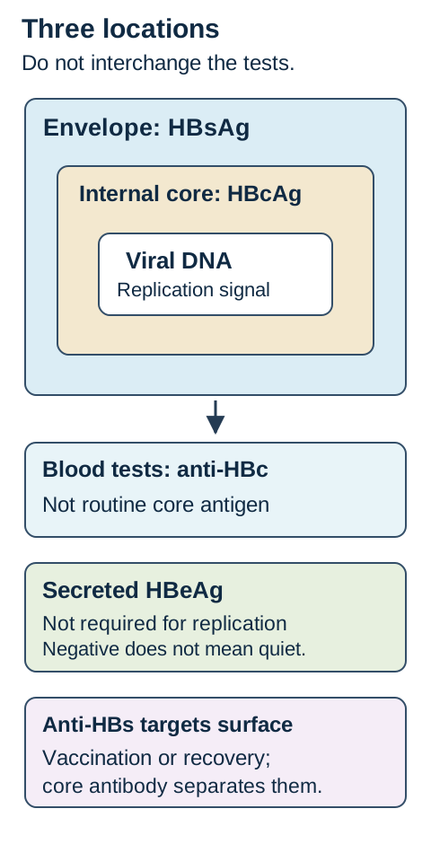
The companion diagram, Three locations, three questions, separates the viral envelope, the core, and a secreted protein. HBsAg is surface antigen in the envelope and in abundant noninfectious surface particles. Its presence usually signals current HBV infection, but it does not measure the amount of intact infectious virus. Anti-HBs binds surface antigen and is the principal neutralizing antibody measured after vaccination. HBcAg lies inside the virus; routine blood testing measures antibody to core, not circulating core antigen. HBeAg is a secreted protein related to the core gene region, not an essential structural component of the virion. [3]
Map each blood test to its target before inferring infection or immunity. [1][3]
The first three tests answer different questions
Test
Main question
Important limit
TestHBsAg
Main questionIs surface antigen detectable now?
Important limitPersistence establishes chronicity; a single result does not.
TestAnti-HBs
Main questionIs surface antibody detectable?
Important limitIt does not distinguish vaccination from recovery.
TestTotal anti-HBc
Main questionHas natural HBV exposure occurred?
Important limitIt combines IgM and IgG and does not date exposure.
Trace each of those three tests to its target in the diagram. Then compare two people with anti-HBs of 42 mIU/mL: only the person with core antibody has serologic evidence of natural exposure. Total anti-HBc usually persists for life after infection and does not result from vaccination. IgM anti-HBc is a separate timing test when acute infection is suspected. It usually supports recent infection, but can also appear during severe chronic flares or reactivation. The history must decide between those possibilities. [1][2]
Transfer this distinction to a healthy vaccinated student: a positive surface antibody is expected, whereas a newly positive core antibody deserves an explanation beyond vaccination. Conversely, lack of measurable surface antibody years after a documented vaccine response does not automatically erase immune memory.
Read a pattern, then place it in time
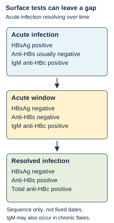
Can two negative surface tests mean that infection never occurred? During the acute HBV window, HBsAg has disappeared but anti-HBs is not yet detectable. Core antibody, particularly IgM anti-HBc, supplies the missing evidence. The companion diagram, Surface tests can leave a gap, shows this as a schematic sequence, not a calendar with fixed transition dates. [1][3]
Trace why two negative surface tests do not exclude the acute window. [1][3]
Typical HBV patterns, interpreted with the history
State
HBsAg
Anti-HBs
Total anti-HBc
IgM anti-HBc
StateSusceptible
HBsAgNegative
Anti-HBsNegative
Total anti-HBcNegative
IgM anti-HBcNegative
StateAcute infection
HBsAgPositive
Anti-HBsUsually negative
Total anti-HBcPositive
IgM anti-HBcPositive
StateAcute window
HBsAgNegative
Anti-HBsNegative
Total anti-HBcPositive
IgM anti-HBcPositive
StateChronic infection
HBsAgPositive
Anti-HBsUsually negative
Total anti-HBcPositive
IgM anti-HBcUsually negative
StateResolved infection
HBsAgNegative
Anti-HBsPositive
Total anti-HBcPositive
IgM anti-HBcNegative
StateVaccination
HBsAgNegative
Anti-HBsPositive
Total anti-HBcNegative
IgM anti-HBcNegative
Do not let this table replace chronology. HBsAg present for at least six months supports chronic HBV. IgM positivity alone cannot overturn documented years of HBsAg positivity. Very early infection can precede a complete antibody response; recent vaccination can transiently produce HBsAg positivity. A surprising result needs its exposure date, vaccination date, symptoms, and confirmatory testing. [1][2]
Marker comparison: change one piece of evidence
Begin with a patient whose HBsAg is negative and anti-HBs is positive. The core result is initially unavailable. Open either comparison below to add one result to that same starting panel. Both comparisons can stay open; close them to reset. Nothing else in the lesson depends on the choice.
Add a negative total anti-HBc result
Panel: HBsAg negative; anti-HBs positive; total anti-HBc negative. The new result supports vaccination rather than recovery from natural infection.
Add a positive total anti-HBc result
Panel: HBsAg negative; anti-HBs positive; total anti-HBc positive. The new result supports resolved natural infection and preserves a reason to assess reactivation risk before immunosuppression.
Worked comparison: the surface results did not change. The added core result changed the inferred exposure history. It did not by itself establish current replication. This is why replacing a triple panel with surface antibody alone can miss clinically consequential history. [1][2]
Now transfer the method to a patient with jaundice, negative HBsAg, negative anti-HBs, and positive total anti-HBc. Ask for IgM anti-HBc rather than calling the patient susceptible. Positive IgM with a compatible recent exposure supports the acute window; the same isolated core pattern in a well patient can have a different explanation.
Use thresholds only for the decision they belong to
Six months, 10 mIU/mL, 200,000 IU/mL, and 28 weeks are not interchangeable measures of severity. Assign each number to its own question: duration of HBsAg positivity, vaccine response, maternal viral load, or gestational timing. Adding these values together has no clinical meaning.
Anti-HBs of at least 10 mIU/mL, measured one to two months after completing a vaccine series when postvaccination testing is indicated, documents an adequate response. An immunocompetent person with a documented response may later have anti-HBs below that threshold while retaining protection. Do not generalize this to a person with no reliable series or response documentation, or to special protocols such as hemodialysis. HBIG can provide passively acquired anti-HBs, so testing soon afterward cannot establish the person's own vaccine response. [2]
Pregnancy requires a different comparison. Screen for HBsAg during every pregnancy, preferably in the first trimester, regardless of vaccination or previous testing. If positive, obtain HBV DNA. In the current AASLD/IDSA recommendation, HBV DNA above 200,000 IU/mL at any point in pregnancy supports TDF or TAF beginning at gestational week 28 for prevention of transmission, regardless of HBeAg status. TDF has the longer pregnancy safety record. Assess separate maternal treatment indications rather than assuming all therapy is prevention-only. [4][5]
Compare two pregnant patients at 26 weeks: one has negative HBeAg and DNA 850,000 IU/mL; the other has positive HBeAg and DNA 80,000 IU/mL. For this prevention threshold, the first patient's DNA crosses the threshold despite negative HBeAg. The second patient's positive HBeAg does not substitute for the DNA threshold; maternal liver disease may still independently require treatment. Arrange specialist care and postpartum monitoring, particularly when prevention-only therapy is stopped. [4]
Maternal antivirals do not replace newborn prophylaxis. An infant born to an HBsAg-positive parent needs hepatitis B vaccine and HBIG within 12 hours of birth, at different injection sites, followed by completion of the vaccine series. Postvaccination testing uses HBsAg and anti-HBs at age 9 to 12 months, or one to two months after the last dose if the series is delayed. Do not test too early, and do not use anti-HBc for this purpose: maternal core antibody may remain detectable for up to 24 months. [5]
For transfer, imagine the mother's DNA becomes undetectable before delivery. The newborn plan still follows maternal infection status, not a reassuring single viral load. Later, a positive infant core antibody alone would not diagnose infant infection.
Separate replication from antigen labels
Does anti-HBe mean that the virus has stopped replicating? HBeAg often accompanies higher replication and infectivity, and anti-HBe often appears as replication declines. Neither is a direct substitute for HBV DNA. Precore or basal core promoter variants can reduce HBeAg production while replication continues. [3]
Compare two HBeAg-negative patients with anti-HBe: one has repeatedly low HBV DNA and normal ALT; the other has HBV DNA 2.5 million IU/mL and increased ALT. Their e-marker labels match, but their replication and liver injury do not. HBV DNA estimates circulating viral burden; ALT reflects hepatocyte injury. A normal ALT at one visit does not prove that high DNA is harmless, and an increased ALT does not identify its cause without clinical assessment. Stage chronic disease using serial results and fibrosis assessment rather than one serologic label. [3][4]
Next, consider isolated total anti-HBc: HBsAg negative, anti-HBs negative, core antibody positive. The possibilities include resolved infection with waning surface antibody, an acute window, a false-positive core result, occult infection, and occasionally an HBsAg variant missed by an assay. The next test follows the situation. Recent symptoms and exposure favor IgM testing. A low-risk, well person needs confirmation of an unexpected result. Planned immunosuppression warrants HBV DNA assessment and a prevention plan, not dismissal of the panel. [1][2][6]
Occult HBV refers to persistence of replication-competent HBV DNA in the liver despite negative HBsAg, with or without detectable circulating DNA. In routine blood work, HBsAg-negative but HBV-DNA-positive results raise this possibility after assay confirmation and contextual evaluation. A single negative blood DNA result cannot prove absence of a hepatic reservoir. [6][10]
Try the counterfactual: replace a negative DNA result with a clearly detectable result while leaving the antibody panel unchanged. The new result establishes evidence of viral replication that the antibodies could not provide. Now replace high DNA with undetectable DNA during antiviral therapy. That establishes suppression within the assay's sensitivity, not eradication of every infected hepatocyte.
Why resolved infection still matters before immunosuppression
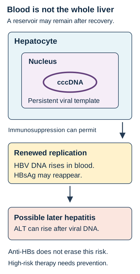
If surface antigen disappears, why can infection return? The companion diagram, Blood tests and the nuclear reservoir, places HBV covalently closed circular DNA, or cccDNA, inside a hepatocyte nucleus. It can persist after apparent clinical resolution. Antibody patterns describe the host response; they are not a direct inventory of every viral template in the liver. HBV is a DNA virus whose replication includes an RNA intermediate and reverse transcription. Nucleos(t)ide analogues suppress viral DNA production without reliably eliminating cccDNA. [3][10]
Relate retained nuclear templates to reactivation before biochemical hepatitis. [6][10]
Trace the diagram from retained template to renewed circulating DNA and then to possible liver injury. Viral replication can increase before ALT rises. Reactivation includes a rise or reappearance of HBV DNA; in a previously HBsAg-negative, core-positive person, HBsAg may also reappear. A biochemical hepatitis flare is a possible consequence, not a prerequisite for recognizing virologic reactivation. [6]
Before immunosuppression, establish HBsAg and total anti-HBc status; the triple panel also identifies immunity and susceptibility. An HBsAg-negative, core-positive patient scheduled for rituximab remains at substantial risk even with anti-HBs and undetectable baseline DNA. Anti-CD20 therapy and stem-cell transplantation are settings in which prophylactic antiviral therapy is recommended rather than waiting for hepatitis. Entecavir, TDF, or TAF are high-resistance-barrier options selected with the patient's comorbidities in mind. Start before, or at the latest with, immunosuppression; anti-CD20 prophylaxis continues for at least 12 months after treatment, with follow-up tailored because delayed reactivation can occur. [6]
Compare that patient with someone receiving a lower-risk regimen for whom reliable monitoring is feasible. Some such patients can be followed with ALT, HBV DNA, and HBsAg and treated promptly if reactivation occurs; this is a risk-specific plan, not an interchangeable alternative for rituximab. Explain aloud why the same baseline antibody panel can lead to different prevention choices when the immunosuppressive drug changes. [6]
For transfer, a patient develops detectable DNA during immunosuppression while ALT remains normal. Do not wait for jaundice to acknowledge reactivation. Conversely, a rising ALT with stable HBV markers requires evaluation for other liver injury rather than automatic attribution to HBV.
Choose the question before choosing the virus test
For an adult's initial HBV screen, use HBsAg, anti-HBs, and total anti-HBc together. Add IgM anti-HBc for suspected acute disease and DNA when replication will determine staging, pregnancy prevention, occult-infection evaluation, or management around immunosuppression. Vaccination should not be unnecessarily delayed while awaiting screening results; collect the blood first when practical. [1][2]
HCV requires a different two-test sequence. A reactive HCV antibody means exposure, not necessarily current infection or protection. Obtain reflex HCV RNA. Detectable RNA identifies current infection; antibody positive with RNA undetectable usually means no current infection, reflecting resolved or successfully treated infection or a false-positive antibody result. Recent exposure, ongoing clinical concern, or a specimen problem can justify repeat RNA testing. Confirm RNA positivity on a subsequent specimen before treatment. [7]
Compare an exposure two weeks ago with one two years ago. HCV RNA can become detectable about one to two weeks after exposure, whereas antibody usually takes eight to eleven weeks. A negative antibody does not exclude recent infection; use RNA after a recent exposure or when immune suppression could impair antibody formation. After cure, antibody may remain positive and does not prevent reinfection, so new exposure is evaluated with RNA. More than half of acute HCV infections become chronic without clearance or treatment; there is no HCV vaccine. [7][11]
HAV is usually an acute, fecal-oral infection and does not become chronic. In a compatible symptomatic illness, IgM anti-HAV supports recent infection. Total anti-HAV positive with IgM negative indicates previous infection or vaccination, not the cause of new jaundice. Indiscriminate IgM testing can produce misleading positive results through cross-reactivity or recent vaccination. An early negative result does not end evaluation when clinical suspicion remains high. Use the illness timeline and consider repeat testing as appropriate. [8][12]
HDV uses HBV surface proteins for particle assembly and transmission. Anti-HDV identifies exposure; HDV RNA establishes active infection. Simultaneous HBV and HDV acquisition is coinfection. HDV acquired by someone with established HBV is superinfection and has a greater tendency toward persistent, severe disease. Positive HDV testing does not erase the HBV diagnosis. HBV vaccination prevents HDV acquisition in people not yet infected with HBV, but it does not protect an already HBV-infected person from HDV superinfection. [9]
Make the final comparison: HBV core antibody can preserve exposure history, HCV antibody needs RNA to establish current infection, and HAV IgM needs a compatible acute syndrome. Transfer the method to any unfamiliar panel: identify the analyte, locate it in time, ask what remains uncertain, and choose the next test or prevention action that answers that uncertainty.
Practice and explain your decision
For each case, commit to an interpretation before reading the choices if helpful. After answering, explain which finding separates your choice from its nearest competitor. Review is optional and never restricts access to the lesson.
Case 1
Show answer and explanations for case 1
A. Prophylaxis for B, not A (Best answer)
B's positive total anti-HBc and remote hepatitis B records establish natural exposure; A's negative core antibody after vaccination supports vaccine-only immunity. Rituximab can permit reactivation of retained HBV in B, so B needs antiviral prophylaxis; A has no evidence of prior infection to reactivate.
Reasoning steps for option A
Which patient's core-antibody result identifies prior infection rather than vaccine-only immunity?
B's positive total anti-HBc and remote hepatitis B records establish natural exposure; A's negative core antibody after vaccination supports vaccine-only immunity.
How does that exposure difference change prophylaxis before their shared rituximab regimen?
Rituximab can permit reactivation of retained HBV in B, so B needs antiviral prophylaxis; A has no evidence of prior infection to reactivate.
B. Prophylaxis for both A and B (Why this does not fit)
A's results support vaccine-derived immunity, not a previous HBV infection with a retained reservoir. Rituximab raises reactivation risk when HBV has previously infected the patient; B has that history, whereas A does not, so treating both ignores the exposure distinction.
Reasoning steps for option B
Does A's documented vaccination and negative total anti-HBc supply a reason for HBV reactivation prophylaxis?
A's results support vaccine-derived immunity, not a previous HBV infection with a retained reservoir.
Why does receiving the same B-cell-depleting drug not justify prophylaxis for both patients?
Rituximab raises reactivation risk when HBV has previously infected the patient; B has that history, whereas A does not, so treating both ignores the exposure distinction.
C. DNA surveillance alone for A and B (Why this does not fit)
B has no measurable circulating HBV DNA at baseline, but the core-positive natural-infection history still permits a hepatic reservoir. Monitoring without prophylaxis is used in selected lower-risk settings, but rituximab warrants prophylaxis in B despite undetectable baseline DNA.
Reasoning steps for option C
What does undetectable baseline DNA tell us about B before rituximab, and what does it leave unresolved?
B has no measurable circulating HBV DNA at baseline, but the core-positive natural-infection history still permits a hepatic reservoir.
Is DNA surveillance alone adequate for the core-positive patient receiving rituximab?
Monitoring without prophylaxis is used in selected lower-risk settings, but rituximab warrants prophylaxis in B despite undetectable baseline DNA.
D. Prophylaxis for neither A nor B (Why this does not fit)
B's surface antibody does not remove the reservoir-related risk associated with prior natural infection. Withholding prophylaxis would leave B unprotected during high-risk B-cell depletion; withholding it from vaccine-only patient A does not create the same omission.
Reasoning steps for option D
Can B's positive anti-HBs erase the reactivation significance of positive total anti-HBc?
B's surface antibody does not remove the reservoir-related risk associated with prior natural infection.
Which patient would be left without indicated prevention by withholding prophylaxis from both?
Withholding prophylaxis would leave B unprotected during high-risk B-cell depletion; withholding it from vaccine-only patient A does not create the same omission.
Takeaway: Interpret the source of immunity before applying drug-specific reactivation prevention.
A. Classify primary nonresponse and repeat the series (Why this does not fit)
The documented concentration of 86 mIU/mL exceeded the 10 mIU/mL response threshold at the appropriate postseries testing time. Later antibody waning does not undo the demonstrated original response in this immunocompetent nurse, so primary nonresponse and a repeat series are not supported.
Reasoning steps for option A
Was the nurse's anti-HBs of 86 mIU/mL one month after series completion a primary vaccine response?
The documented concentration of 86 mIU/mL exceeded the 10 mIU/mL response threshold at the appropriate postseries testing time.
Can a concentration of 4 mIU/mL twelve years later reclassify that original response as primary nonresponse?
Later antibody waning does not undo the demonstrated original response in this immunocompetent nurse, so primary nonresponse and a repeat series are not supported.
B. Replace responder status with susceptibility pending a challenge dose (Why this does not fit)
Her completed vaccine series and anti-HBs of 86 mIU/mL one month later already document an adequate response. She is immunocompetent and has no recent exposure; a late low titer alone does not establish susceptibility or require a challenge to recover her documented responder status.
Reasoning steps for option B
What information is already documented that a challenge dose might otherwise help establish?
Her completed vaccine series and anti-HBs of 86 mIU/mL one month later already document an adequate response.
Does her current immune status justify replacing responder status with susceptibility while awaiting a challenge?
She is immunocompetent and has no recent exposure; a late low titer alone does not establish susceptibility or require a challenge to recover her documented responder status.
C. Retain documented responder status (Best answer)
The value of 86 mIU/mL one month after the completed series was appropriately timed and exceeded 10 mIU/mL; the value of 4 was measured twelve years later. An immunocompetent documented responder may retain immune memory after measurable antibody wanes, so the later low value does not erase her prior response.
Reasoning steps for option C
Which of the nurse's two anti-HBs measurements was obtained at the time used to document vaccine response?
The value of 86 mIU/mL one month after the completed series was appropriately timed and exceeded 10 mIU/mL; the value of 4 was measured twelve years later.
Why can the occupational record retain responder status despite that later fall?
An immunocompetent documented responder may retain immune memory after measurable antibody wanes, so the later low value does not erase her prior response.
D. Use the annual dialysis-patient titer protocol (Why this does not fit)
The stem describes an immunocompetent nurse working in the unit, not a patient receiving hemodialysis. That patient-specific protocol does not apply merely because of her workplace; her documented adequate vaccine response and immunocompetent status govern interpretation of the late low titer.
Reasoning steps for option D
Does employment in a dialysis unit make this nurse a hemodialysis patient?
The stem describes an immunocompetent nurse working in the unit, not a patient receiving hemodialysis.
Why is the annual dialysis-patient titer protocol the wrong basis for her occupational classification?
That patient-specific protocol does not apply merely because of her workplace; her documented adequate vaccine response and immunocompetent status govern interpretation of the late low titer.
Takeaway: Use correctly timed historical vaccine response and present immune status together.
A. A: acute window; B: assume susceptibility without confirming core reactivity (Why this does not fit)
A's recent documented HBsAg-positive illness followed by negative surface tests and positive IgM anti-HBc supports the acute window. B's total anti-HBc is reactive rather than negative, so the panel is not an ordinary susceptible pattern; confirmation and history review are needed to assess false positivity versus prior exposure.
Reasoning steps for option A
Does A's antigen clearance six weeks after hepatitis, with core IgM retained, fit the acute window?
A's recent documented HBsAg-positive illness followed by negative surface tests and positive IgM anti-HBc supports the acute window.
Why can B not be called susceptible before confirming the first reactive core result?
B's total anti-HBc is reactive rather than negative, so the panel is not an ordinary susceptible pattern; confirmation and history review are needed to assess false positivity versus prior exposure.
B. A: acute window; B: confirm the core result and reassess history (Best answer)
A has cleared detectable HBsAg before anti-HBs has appeared, leaving IgM anti-HBc as evidence of the recent infection during the acute window. B has no supplied recent illness or recognized exposure and only a first isolated-core result, so repeating the core test and reassessing history address the unresolved result without assuming an acute window.
Reasoning steps for option B
How do A's six-week history and positive core IgM explain the two negative surface tests?
A has cleared detectable HBsAg before anti-HBs has appeared, leaving IgM anti-HBc as evidence of the recent infection during the acute window.
Why does B's IgM-negative, asymptomatic presentation call for confirmation rather than the same acute-stage label?
B has no supplied recent illness or recognized exposure and only a first isolated-core result, so repeating the core test and reassessing history address the unresolved result without assuming an acute window.
C. A: susceptible; B: confirm the core result and reassess history (Why this does not fit)
A first isolated total anti-HBc result in an asymptomatic, low-risk, IgM-negative patient can have more than one explanation and warrants confirmation. A's documented hepatitis B six weeks earlier and current core IgM demonstrate recent natural infection despite both surface tests being negative; the pattern supports a window, not susceptibility.
Reasoning steps for option C
What supports confirming B's core result and revisiting exposure history?
A first isolated total anti-HBc result in an asymptomatic, low-risk, IgM-negative patient can have more than one explanation and warrants confirmation.
Which evidence prevents applying a susceptible label to A?
A's documented hepatitis B six weeks earlier and current core IgM demonstrate recent natural infection despite both surface tests being negative; the pattern supports a window, not susceptibility.
D. A: susceptible; B: assume susceptibility without confirming core reactivity (Why this does not fit)
A's recent HBsAg-positive hepatitis and retained IgM anti-HBc establish an acute-infection trajectory that a susceptible label would ignore. B's unexpected total anti-HBc reactivity still needs confirmation; neither absent surface antigen nor absent surface antibody explains whether that core result is spurious or reflects prior exposure.
Reasoning steps for option D
What infection history would be lost by treating A's negative surface tests as an all-negative panel?
A's recent HBsAg-positive hepatitis and retained IgM anti-HBc establish an acute-infection trajectory that a susceptible label would ignore.
What unresolved laboratory finding also blocks an immediate susceptible label for B?
B's unexpected total anti-HBc reactivity still needs confirmation; neither absent surface antigen nor absent surface antibody explains whether that core result is spurious or reflects prior exposure.
Takeaway: Use chronology and IgM to distinguish an acute window from an unresolved isolated-core screen.
A. IgM production during newly acquired primary HBV infection (Why this does not fit)
HBsAg positivity documented eighteen months apart exceeds the six-month chronicity criterion and establishes preexisting chronic HBV. IgM anti-HBc can accompany a chronic flare, so the new IgM result does not turn this established infection into a newly acquired primary infection.
Reasoning steps for option A
How does the eighteen-month record of HBsAg positivity constrain a newly acquired primary HBV diagnosis?
HBsAg positivity documented eighteen months apart exceeds the six-month chronicity criterion and establishes preexisting chronic HBV.
Does weakly positive core IgM override that documented chronology?
IgM anti-HBc can accompany a chronic flare, so the new IgM result does not turn this established infection into a newly acquired primary infection.
B. Incidental core IgM during liver injury unrelated to HBV activity (Why this does not fit)
HBV DNA has risen from prior measurements at the same time that ALT has increased, providing evidence of renewed or increased viral activity. Other causes of liver injury can coexist, but chronic HBsAg positivity together with rising DNA and ALT makes a flare a better integrated explanation than dismissing the IgM as incidental.
Reasoning steps for option B
Which changing viral measurement argues against treating the core IgM as unrelated to HBV activity?
HBV DNA has risen from prior measurements at the same time that ALT has increased, providing evidence of renewed or increased viral activity.
Why is an incidental-IgM explanation weaker than a chronic-flare explanation in this patient?
Other causes of liver injury can coexist, but chronic HBsAg positivity together with rising DNA and ALT makes a flare a better integrated explanation than dismissing the IgM as incidental.
C. Core IgM persisting through resolved HBV without renewed replication (Why this does not fit)
HBsAg remains positive after eighteen months, which conflicts with labeling this episode resolved HBV. The measured HBV DNA has increased, so the case contains evidence of increased replication rather than only a residual antibody after recovery.
Reasoning steps for option C
Does the current HBsAg result support the proposed resolved-infection state?
HBsAg remains positive after eighteen months, which conflicts with labeling this episode resolved HBV.
What contradicts the claim that core IgM persists without renewed replication?
The measured HBV DNA has increased, so the case contains evidence of increased replication rather than only a residual antibody after recovery.
D. IgM production during a flare of established chronic HBV (Best answer)
The HBsAg-positive records eighteen months apart establish chronic HBV rather than a first acute acquisition. Rising viral DNA with biochemical liver injury supports an active chronic flare, during which IgM anti-HBc can become positive.
Reasoning steps for option D
Which longitudinal finding establishes the chronic infection underlying the new IgM result?
The HBsAg-positive records eighteen months apart establish chronic HBV rather than a first acute acquisition.
How do rising HBV DNA and ALT explain core IgM within that chronic history?
Rising viral DNA with biochemical liver injury supports an active chronic flare, during which IgM anti-HBc can become positive.
Takeaway: Give documented chronicity priority over an isolated acute-associated antibody result.
A. HBsAg persists; DNA and IgM anti-HBc become positive (Why this does not fit)
A surface-antigen vaccine does not supply replicating HBV, so new viral DNA would argue for natural infection rather than transient vaccine antigenemia. Core IgM would add evidence of a recent natural immune response to HBV; together with persistent HBsAg and detectable DNA, it would oppose a vaccine-only explanation.
Reasoning steps for option A
Would newly detectable HBV DNA after the donor's vaccine be explained by vaccine surface antigen alone?
A surface-antigen vaccine does not supply replicating HBV, so new viral DNA would argue for natural infection rather than transient vaccine antigenemia.
How would new IgM anti-HBc change the meaning of persistent HBsAg in this follow-up pattern?
Core IgM would add evidence of a recent natural immune response to HBV; together with persistent HBsAg and detectable DNA, it would oppose a vaccine-only explanation.
B. HBsAg disappears; total anti-HBc and anti-HBs become positive (Why this does not fit)
Surface antigen can disappear after either transient vaccine antigenemia or a resolving natural infection, so its loss alone does not distinguish the two. Vaccination can produce anti-HBs but not anti-HBc; new core antibody would favor natural exposure with recovery rather than a surface-antigen-only vaccine effect.
Reasoning steps for option B
Is disappearance of the donor's HBsAg sufficient to identify the earlier reactivity as vaccine-derived?
Surface antigen can disappear after either transient vaccine antigenemia or a resolving natural infection, so its loss alone does not distinguish the two.
What would new total anti-HBc alongside anti-HBs imply after that antigen loss?
Vaccination can produce anti-HBs but not anti-HBc; new core antibody would favor natural exposure with recovery rather than a surface-antigen-only vaccine effect.
C. HBsAg disappears; DNA and anti-HBc remain negative (Best answer)
The immediately preceding vaccine supplies surface antigen, while the negative prevaccination panel and current absence of DNA and core antibody support a vaccine-associated explanation. Disappearance of HBsAg while HBV DNA and anti-HBc remain negative supports transient vaccine antigenemia rather than an evolving natural infection.
Reasoning steps for option C
Why is low-level HBsAg one day after vaccination compatible with a noninfectious antigen signal?
The immediately preceding vaccine supplies surface antigen, while the negative prevaccination panel and current absence of DNA and core antibody support a vaccine-associated explanation.
Which follow-up findings would strengthen that explanation without showing viral replication or core seroconversion?
Disappearance of HBsAg while HBV DNA and anti-HBc remain negative supports transient vaccine antigenemia rather than an evolving natural infection.
D. HBsAg disappears; IgM anti-HBc remains positive without anti-HBs (Why this does not fit)
HBV vaccination does not generate anti-HBc, so core IgM would require an explanation beyond the vaccine's surface antigen. That combination can occur during the acute window, when surface antigen has cleared before surface antibody appears; it therefore does not support transient vaccine antigenemia.
Reasoning steps for option D
Would core IgM positivity after HBsAg disappearance be an expected vaccine-only antibody response?
HBV vaccination does not generate anti-HBc, so core IgM would require an explanation beyond the vaccine's surface antigen.
What natural-infection stage could combine absent HBsAg, absent anti-HBs, and positive core IgM?
That combination can occur during the acute window, when surface antigen has cleared before surface antibody appears; it therefore does not support transient vaccine antigenemia.
Takeaway: Use a longitudinal panel to test a postvaccination-antigenemia hypothesis.
A. Reduced e-antigen expression with preserved replication (Best answer)
The high DNA concentration demonstrates substantial ongoing replication, so the negative e-antigen result cannot be read as viral inactivity. Precore or basal core promoter variation can reduce e-antigen expression while preserving HBV replication, matching the discordance in this chronic infection.
Reasoning steps for option A
What does HBV DNA of 7,800,000 IU/mL show despite this patient's negative HBeAg?
The high DNA concentration demonstrates substantial ongoing replication, so the negative e-antigen result cannot be read as viral inactivity.
Which expression change can reconcile that viral load with absent HBeAg and positive anti-HBe?
Precore or basal core promoter variation can reduce e-antigen expression while preserving HBV replication, matching the discordance in this chronic infection.
B. Loss of surface-antigen synthesis with preserved replication (Why this does not fit)
HBsAg remains present after three years of chronic infection, rather than being absent as a loss of surface-antigen production would suggest. The missing marker is HBeAg, not HBsAg; a surface-antigen synthesis defect does not explain selective e-antigen negativity with high DNA.
Reasoning steps for option B
Which surface-antigen finding conflicts with loss of surface-antigen synthesis in this patient?
HBsAg remains present after three years of chronic infection, rather than being absent as a loss of surface-antigen production would suggest.
Which antigen's expression actually needs explanation alongside the 7,800,000 IU/mL DNA result?
The missing marker is HBeAg, not HBsAg; a surface-antigen synthesis defect does not explain selective e-antigen negativity with high DNA.
C. Loss of reverse transcription with preserved e-antigen synthesis (Why this does not fit)
HBV uses reverse transcription during DNA production, so loss of that process would reduce new viral DNA rather than explain abundant circulating DNA. The patient has absent HBeAg and HBV DNA of 7,800,000 IU/mL, the opposite of the proposed preserved e-antigen production with impaired DNA synthesis.
Reasoning steps for option C
How would losing reverse transcription affect production of new HBV DNA?
HBV uses reverse transcription during DNA production, so loss of that process would reduce new viral DNA rather than explain abundant circulating DNA.
Do preserved e-antigen synthesis and lost reverse transcription match this patient's paired measurements?
The patient has absent HBeAg and HBV DNA of 7,800,000 IU/mL, the opposite of the proposed preserved e-antigen production with impaired DNA synthesis.
D. Reduced DNA synthesis with increased e-protein secretion (Why this does not fit)
Reduced DNA synthesis would favor less DNA production, whereas the measured 7,800,000 IU/mL shows substantial replication. Increased e-protein secretion would favor detectable e antigen, not the absent HBeAg reported here; both parts of this proposed change point away from the measured pattern.
Reasoning steps for option D
What DNA pattern would reduced synthesis predict, and how does the measured viral load compare?
Reduced DNA synthesis would favor less DNA production, whereas the measured 7,800,000 IU/mL shows substantial replication.
Does increased e-protein secretion explain the negative HBeAg assay?
Increased e-protein secretion would favor detectable e antigen, not the absent HBeAg reported here; both parts of this proposed change point away from the measured pattern.
Takeaway: Explain why e-antigen expression and HBV DNA replication can diverge.
A. ALT rises first because hepatocyte injury restarts viral replication (Why this does not fit)
ALT is a marker of hepatocyte injury, not a mechanism that initiates viral replication when treatment is interrupted. Persistent viral templates can support renewed DNA production before biochemical hepatitis, so an injury-driven, ALT-first sequence is not the required mechanism of recurrence.
Reasoning steps for option A
Is ALT the mechanism that restarts HBV replication after interruption of antiviral suppression?
ALT is a marker of hepatocyte injury, not a mechanism that initiates viral replication when treatment is interrupted.
Could renewed viral DNA become detectable before an ALT rise in this still-HBsAg-positive patient?
Persistent viral templates can support renewed DNA production before biochemical hepatitis, so an injury-driven, ALT-first sequence is not the required mechanism of recurrence.
B. Core antibody falls because renewed replication requires loss of anti-HBc (Why this does not fit)
Total anti-HBc commonly persists during reactivation and its disappearance is not required for HBV DNA production to resume. Anti-HBc records natural exposure rather than serving as the principal measured neutralizing surface antibody; retained viral templates can support recurrence without a fall in core antibody.
Reasoning steps for option B
Must total anti-HBc disappear before this treated chronic infection can resume replication?
Total anti-HBc commonly persists during reactivation and its disappearance is not required for HBV DNA production to resume.
Why does treating core antibody loss as the cause of recurrence misidentify its role?
Anti-HBc records natural exposure rather than serving as the principal measured neutralizing surface antibody; retained viral templates can support recurrence without a fall in core antibody.
C. HBsAg clears because interruption eliminates residual antigen production (Why this does not fit)
Treatment interruption does not itself eliminate infected hepatocytes or the residual viral templates supporting antigen production. Persistent HBsAg already shows that suppression has not achieved surface-antigen loss; withdrawal of suppression does not supply an eradication mechanism and can instead permit renewed DNA production.
Reasoning steps for option C
Does stopping suppression remove the infected hepatocytes that can still produce HBsAg?
Treatment interruption does not itself eliminate infected hepatocytes or the residual viral templates supporting antigen production.
What makes HBsAg clearance from interruption an unsupported prediction when HBsAg remains positive on therapy?
Persistent HBsAg already shows that suppression has not achieved surface-antigen loss; withdrawal of suppression does not supply an eradication mechanism and can instead permit renewed DNA production.
D. HBV DNA returns before ALT rises because a reservoir persists (Best answer)
Undetectable circulating DNA demonstrates suppression within assay sensitivity, not elimination of persistent nuclear viral templates. Retained templates can support renewed replication before an ALT rise becomes evident, making DNA-first recurrence biologically possible without implying that interruption is safe or recurrence inevitable.
Reasoning steps for option D
What does undetectable serum DNA on treatment fail to establish about this chronic HBV reservoir?
Undetectable circulating DNA demonstrates suppression within assay sensitivity, not elimination of persistent nuclear viral templates.
How could loss of that suppression produce DNA recurrence before biochemical hepatitis?
Retained templates can support renewed replication before an ALT rise becomes evident, making DNA-first recurrence biologically possible without implying that interruption is safe or recurrence inevitable.
Takeaway: Predict possible virologic recurrence without equating serum suppression with eradication.
A. Use serial IgM anti-HBc alone to establish an acute window (Why this does not fit)
There is no supplied recent exposure or acute illness, and IgM anti-HBc is negative, so an acute window is not established by the two negative surface markers. IgM testing addresses recent infection but does not by itself distinguish a false-positive total core result from remote natural exposure, leaving the immediate isolated-core uncertainty unresolved.
Reasoning steps for option A
What acute-window evidence is missing in this asymptomatic patient with negative core IgM?
There is no supplied recent exposure or acute illness, and IgM anti-HBc is negative, so an acute window is not established by the two negative surface markers.
Would serial core IgM alone resolve why the first total anti-HBc screen was reactive?
IgM testing addresses recent infection but does not by itself distinguish a false-positive total core result from remote natural exposure, leaving the immediate isolated-core uncertainty unresolved.
B. Repeat total anti-HBc with the same assay and reassess exposure history (Best answer)
Isolated core reactivity can reflect previous natural infection or false positivity, and the absence of an acute syndrome does not select a single explanation. Repeat testing checks whether the unexpected core reactivity is reproducible, while history review adjusts its clinical meaning before a definitive infection label is assigned.
Reasoning steps for option B
Why is the first isolated total anti-HBc result not enough to assign a definite infection category in this low-risk adult?
Isolated core reactivity can reflect previous natural infection or false positivity, and the absence of an acute syndrome does not select a single explanation.
What does repeating total anti-HBc with the same assay add to reassessing the exposure history?
Repeat testing checks whether the unexpected core reactivity is reproducible, while history review adjusts its clinical meaning before a definitive infection label is assigned.
C. Follow surface markers alone without confirming the unexpected core result (Why this does not fit)
The reactive total anti-HBc result would remain unconfirmed even if both surface markers were followed over time. Surface-marker trends may provide context, but they do not test the reproducibility of the core reactivity that could represent either false positivity or prior exposure.
Reasoning steps for option C
Which unexpected result would remain unconfirmed if follow-up measured only HBsAg and anti-HBs?
The reactive total anti-HBc result would remain unconfirmed even if both surface markers were followed over time.
Why do additional surface-marker results not directly settle the low-risk isolated-core finding?
Surface-marker trends may provide context, but they do not test the reproducibility of the core reactivity that could represent either false positivity or prior exposure.
D. Use one negative blood DNA assay to exclude all retained HBV (Why this does not fit)
It would show no detectable circulating DNA in that specimen, not directly inventory persistent HBV templates in the liver. A hepatic reservoir can remain without measurable serum DNA, and a negative DNA result does not independently determine whether the isolated anti-HBc reactivity was spurious.
Reasoning steps for option D
Which compartment would a single negative blood HBV DNA assay actually assess?
It would show no detectable circulating DNA in that specimen, not directly inventory persistent HBV templates in the liver.
Can that negative blood result establish both absence of retained HBV and false positivity of the core screen?
A hepatic reservoir can remain without measurable serum DNA, and a negative DNA result does not independently determine whether the isolated anti-HBc reactivity was spurious.
Takeaway: Confirm an unexpected isolated-core result before assigning its cause in a low-risk person.
A. Occult HBV remains relevant despite the later negative serum assay (Best answer)
Repeated HBV DNA detection despite negative HBsAg supports occult HBV rather than dismissing the core-positive history as antibody alone. Low or intermittent circulating DNA can fall below detection while hepatic viral templates persist, so the later negative specimen does not demonstrate eradication of the reservoir.
Reasoning steps for option A
What do two confirmed low-level DNA detections mean when the transplant candidate's HBsAg remains negative?
Repeated HBV DNA detection despite negative HBsAg supports occult HBV rather than dismissing the core-positive history as antibody alone.
Why does an undetectable serum DNA result one month later not remove that concern?
Low or intermittent circulating DNA can fall below detection while hepatic viral templates persist, so the later negative specimen does not demonstrate eradication of the reservoir.
B. Reclassify the earlier DNA results as contamination based on the third result (Why this does not fit)
One later negative result does not by itself invalidate DNA detection confirmed on two separate specimens. Occult HBV can involve low or intermittently detectable viremia, so changing serum detectability can coexist with persistent hepatic infection.
Reasoning steps for option B
Does the third specimen's negative DNA result prove contamination of both earlier positive specimens?
One later negative result does not by itself invalidate DNA detection confirmed on two separate specimens.
What biological explanation preserves the earlier evidence without assuming laboratory contamination?
Occult HBV can involve low or intermittently detectable viremia, so changing serum detectability can coexist with persistent hepatic infection.
C. Classify HBV as eradicated after the negative follow-up serum assay (Why this does not fit)
It establishes that blood DNA was below the assay's detection limit in that specimen, not that every infected hepatocyte had lost HBV. Occult HBV concerns persistent replication-competent DNA in the liver, which may remain even when circulating DNA is no longer measurable.
Reasoning steps for option C
What would the follow-up negative serum assay establish about circulating HBV at that sampling time?
It establishes that blood DNA was below the assay's detection limit in that specimen, not that every infected hepatocyte had lost HBV.
Which reservoir-related limitation prevents declaring eradication after the two earlier positive tests?
Occult HBV concerns persistent replication-competent DNA in the liver, which may remain even when circulating DNA is no longer measurable.
D. The earlier results establish an acute window that has now resolved (Why this does not fit)
IgM anti-HBc is negative and no recent acute-infection trajectory is supplied, so an acute window is not established by HBsAg negativity and core positivity alone. It cannot date the earlier infection or erase two confirmed DNA-positive specimens; intermittent serum DNA in occult HBV remains a relevant explanation.
Reasoning steps for option D
Which evidence needed to identify a recent acute window is absent from this candidate's history?
IgM anti-HBc is negative and no recent acute-infection trajectory is supplied, so an acute window is not established by HBsAg negativity and core positivity alone.
Why does a later negative DNA assay not prove that a proposed acute window has resolved?
It cannot date the earlier infection or erase two confirmed DNA-positive specimens; intermittent serum DNA in occult HBV remains a relevant explanation.
Takeaway: Recognize occult HBV and the limits of a later negative blood DNA assay.
A. Monitor DNA, HBsAg and ALT without prophylaxis, treating if reactivation occurs (Why this does not fit)
The planned drug is rituximab, a B-cell-depleting anti-CD20 therapy for which prior natural HBV exposure warrants antiviral prophylaxis. Neither surface antibody nor an undetectable baseline blood viral load eliminates the reservoir-related risk signaled by positive total anti-HBc before rituximab.
Reasoning steps for option A
Which feature makes monitoring alone less appropriate here than in a lower-risk immunosuppression regimen?
The planned drug is rituximab, a B-cell-depleting anti-CD20 therapy for which prior natural HBV exposure warrants antiviral prophylaxis.
Do anti-HBs of 72 mIU/mL and undetectable baseline DNA remove that prophylaxis indication?
Neither surface antibody nor an undetectable baseline blood viral load eliminates the reservoir-related risk signaled by positive total anti-HBc before rituximab.
B. Monitor ALT alone and start an antiviral after biochemical hepatitis (Why this does not fit)
HBV DNA can reappear or increase before biochemical hepatitis produces an ALT rise. Waiting would omit indicated prophylaxis in a high-risk anti-CD20 setting and delay recognition until liver injury rather than addressing reactivation risk before treatment.
Reasoning steps for option B
What early virologic change could ALT-only monitoring miss in this core-positive patient?
HBV DNA can reappear or increase before biochemical hepatitis produces an ALT rise.
Why is waiting for biochemical hepatitis an inadequate prevention plan before rituximab?
Waiting would omit indicated prophylaxis in a high-risk anti-CD20 setting and delay recognition until liver injury rather than addressing reactivation risk before treatment.
C. Give a vaccine booster and follow the anti-HBs response instead (Why this does not fit)
A vaccine booster does not eliminate the persistent hepatic reservoir that can remain after a core-positive natural infection. The surface-antibody response does not substitute for suppressing HBV replication during rituximab, so the booster-and-titer plan leaves the reactivation risk unaddressed.
Reasoning steps for option C
Would boosting an existing anti-HBs level of 72 mIU/mL remove viral templates left by natural infection?
A vaccine booster does not eliminate the persistent hepatic reservoir that can remain after a core-positive natural infection.
Can tracking a booster-induced anti-HBs response replace antiviral prophylaxis during B-cell depletion?
The surface-antibody response does not substitute for suppressing HBV replication during rituximab, so the booster-and-titer plan leaves the reactivation risk unaddressed.
D. Start a high-barrier antiviral before or with rituximab (Best answer)
Positive total anti-HBc identifies prior natural HBV exposure and a potential reservoir that the negative circulating markers do not exclude. High-risk B-cell depletion warrants a high-resistance-barrier antiviral started before or at the start of rituximab rather than waiting for DNA recurrence or hepatitis.
Reasoning steps for option D
Which marker establishes prior natural exposure even though HBsAg and baseline DNA are negative?
Positive total anti-HBc identifies prior natural HBV exposure and a potential reservoir that the negative circulating markers do not exclude.
How should the planned rituximab regimen determine the timing and type of prevention?
High-risk B-cell depletion warrants a high-resistance-barrier antiviral started before or at the start of rituximab rather than waiting for DNA recurrence or hepatitis.
Takeaway: Prefer prophylaxis over a monitoring-only plan for prior HBV exposure before anti-CD20 therapy.
A. Virologic reactivation absent; biochemical hepatitis present (Why this does not fit)
Reappearance of HBsAg and detectable, rising DNA after previously resolved HBV demonstrates virologic reactivation. ALT remains within the laboratory range, so a biochemical hepatitis flare is not demonstrated; this option reverses the virologic and biochemical findings.
Reasoning steps for option A
Do reappearing HBsAg and newly rising DNA fit the proposed absence of virologic reactivation?
Reappearance of HBsAg and detectable, rising DNA after previously resolved HBV demonstrates virologic reactivation.
Which biochemical evidence is missing from the proposed hepatitis-only interpretation?
ALT remains within the laboratory range, so a biochemical hepatitis flare is not demonstrated; this option reverses the virologic and biochemical findings.
B. Virologic reactivation present; biochemical hepatitis absent at this time (Best answer)
Newly detectable, rising HBV DNA with HBsAg seroreversion establishes virologic reactivation of the previously resolved infection. Normal ALT means current biochemical hepatitis has not been demonstrated, even though virologic recurrence is already evident and can precede a later flare.
Reasoning steps for option B
What do the changes from negative baseline DNA and HBsAg to positivity establish during immunosuppression?
Newly detectable, rising HBV DNA with HBsAg seroreversion establishes virologic reactivation of the previously resolved infection.
How does the normal ALT qualify the interpretation of that reactivation at this visit?
Normal ALT means current biochemical hepatitis has not been demonstrated, even though virologic recurrence is already evident and can precede a later flare.
C. Virologic reactivation present; biochemical hepatitis present (Why this does not fit)
The return of HBsAg and rising HBV DNA supports the virologic-reactivation component. A normal ALT does not demonstrate a concurrent biochemical flare, so the laboratory evidence supports reactivation without established biochemical hepatitis rather than both.
Reasoning steps for option C
Which half of a combined reactivation-and-hepatitis diagnosis is supported by the new HBV tests?
The return of HBsAg and rising HBV DNA supports the virologic-reactivation component.
Does an ALT still within the reference range establish the biochemical-hepatitis component too?
A normal ALT does not demonstrate a concurrent biochemical flare, so the laboratory evidence supports reactivation without established biochemical hepatitis rather than both.
D. Virologic reactivation absent; biochemical hepatitis absent (Why this does not fit)
The normal ALT supports the absence of a demonstrated biochemical hepatitis flare at this visit. HBsAg has reappeared and HBV DNA is newly detectable and rising; those changes establish reactivation independently of the ALT result.
Reasoning steps for option D
What does normal ALT support in the proposed absence of both reactivation and hepatitis?
The normal ALT supports the absence of a demonstrated biochemical hepatitis flare at this visit.
Which two new positive measurements contradict declaring that virologic reactivation is also absent?
HBsAg has reappeared and HBV DNA is newly detectable and rising; those changes establish reactivation independently of the ALT result.
Takeaway: Separate virologic reactivation from its potentially later biochemical consequence.
A. No maternal prevention; newborn vaccine plus HBIG (Why this does not fit)
The measured DNA exceeds 200,000 IU/mL, so negative HBeAg and normal ALT do not remove the high-viral-load indication for antenatal transmission prevention. The newborn measures are indicated, but the maternal viral load also supports tenofovir beginning at week 28; omitting maternal prevention leaves that separate indication untreated.
Reasoning steps for option A
Does DNA of 960,000 IU/mL cross the maternal prevention threshold despite negative HBeAg and normal ALT?
The measured DNA exceeds 200,000 IU/mL, so negative HBeAg and normal ALT do not remove the high-viral-load indication for antenatal transmission prevention.
Why is newborn vaccine plus HBIG alone an incomplete plan at 27 weeks in this patient?
The newborn measures are indicated, but the maternal viral load also supports tenofovir beginning at week 28; omitting maternal prevention leaves that separate indication untreated.
B. Tenofovir from week 28; newborn vaccine without HBIG (Why this does not fit)
At 27 weeks with DNA of 960,000 IU/mL, planning week-28 tenofovir matches the high-viral-load maternal prevention recommendation. Maternal antiviral prevention complements rather than replaces neonatal prophylaxis; an infant born to an HBsAg-positive parent still needs both vaccine and HBIG.
Reasoning steps for option B
Is starting tenofovir at week 28 appropriate for the mother's current gestational age and DNA level?
At 27 weeks with DNA of 960,000 IU/mL, planning week-28 tenofovir matches the high-viral-load maternal prevention recommendation.
Does maternal tenofovir justify replacing newborn vaccine plus HBIG with vaccine alone?
Maternal antiviral prevention complements rather than replaces neonatal prophylaxis; an infant born to an HBsAg-positive parent still needs both vaccine and HBIG.
C. Tenofovir from week 28; newborn vaccine plus HBIG (Best answer)
DNA of 960,000 IU/mL is above the 200,000 IU/mL threshold and supports tenofovir from gestational week 28 regardless of the negative HBeAg result. Maternal HBsAg positivity requires newborn hepatitis B vaccine and HBIG within 12 hours of birth, even when maternal antiviral prevention is given.
Reasoning steps for option C
Which measured value selects maternal transmission prevention, and when should it begin from this 27-week visit?
DNA of 960,000 IU/mL is above the 200,000 IU/mL threshold and supports tenofovir from gestational week 28 regardless of the negative HBeAg result.
Which maternal result independently determines the newborn's need for combined prophylaxis?
Maternal HBsAg positivity requires newborn hepatitis B vaccine and HBIG within 12 hours of birth, even when maternal antiviral prevention is given.
D. Tenofovir only after delivery; newborn vaccine plus HBIG (Why this does not fit)
The mother's high HBV DNA supports antenatal prevention beginning at week 28, which addresses transmission risk before delivery rather than only afterward. Newborn vaccine and HBIG remain necessary, but they do not change the separate indication for appropriately timed maternal prevention in this high-viral-load pregnancy.
Reasoning steps for option D
What prevention opportunity is missed by waiting until after delivery to start tenofovir?
The mother's high HBV DNA supports antenatal prevention beginning at week 28, which addresses transmission risk before delivery rather than only afterward.
Does retaining the correct newborn vaccine-and-HBIG plan compensate for that missed maternal timing?
Newborn vaccine and HBIG remain necessary, but they do not change the separate indication for appropriately timed maternal prevention in this high-viral-load pregnancy.
Takeaway: Integrate maternal DNA, gestational timing and independent newborn prophylaxis.
A. Infant HBV infection despite an adequate surface-antibody response (Why this does not fit)
Maternal core antibody can remain detectable in an infant for up to 24 months, so the extra positive core test does not by itself establish infant infection. HBsAg is negative and anti-HBs is 64 mIU/mL, above the 10 mIU/mL response threshold at a valid testing age; these results support vaccine response rather than the proposed infant infection.
Reasoning steps for option A
Can positive total anti-HBc at ten months prove that the infant, rather than the parent, generated core antibody?
Maternal core antibody can remain detectable in an infant for up to 24 months, so the extra positive core test does not by itself establish infant infection.
What do the recommended surface tests show two months after this infant completed vaccination?
HBsAg is negative and anti-HBs is 64 mIU/mL, above the 10 mIU/mL response threshold at a valid testing age; these results support vaccine response rather than the proposed infant infection.
B. Failed vaccination because core antibody remains detectable (Why this does not fit)
Anti-HBs measures the response, and 64 mIU/mL exceeds the required 10 mIU/mL threshold; core antibody is not a marker of vaccine failure. Transferred maternal anti-HBc can still be present at this age, while the appropriately timed HBsAg and anti-HBs panel documents the desired postvaccination result.
Reasoning steps for option B
Which result measures vaccine response here: the incidental core antibody or anti-HBs of 64 mIU/mL?
Anti-HBs measures the response, and 64 mIU/mL exceeds the required 10 mIU/mL threshold; core antibody is not a marker of vaccine failure.
Why does persistent core antibody not overturn the negative HBsAg and adequate anti-HBs result at ten months?
Transferred maternal anti-HBc can still be present at this age, while the appropriately timed HBsAg and anti-HBs panel documents the desired postvaccination result.
C. Uninterpretable response because birth HBIG necessarily persists (Why this does not fit)
The infant was tested at an appropriate postvaccination time within the recommended 9-to-12-month age range. Birth HBIG can confound early testing, but it does not justify dismissing this appropriately timed HBsAg-negative, anti-HBs-positive result as necessarily passive antibody.
Reasoning steps for option C
Is testing at ten months, two months after the final dose, within the recommended infant response-testing window?
The infant was tested at an appropriate postvaccination time within the recommended 9-to-12-month age range.
Does birth HBIG make that ten-month surface-antibody result necessarily uninterpretable?
Birth HBIG can confound early testing, but it does not justify dismissing this appropriately timed HBsAg-negative, anti-HBs-positive result as necessarily passive antibody.
D. Documented vaccine response; core antibody may still reflect maternal transfer (Best answer)
Negative HBsAg with anti-HBs above 10 mIU/mL after the completed series documents an adequate vaccine response without detected surface antigen. Total anti-HBc may still reflect maternal antibody at ten months, so it should not be used to relabel this valid vaccine response as infant infection or vaccine failure.
Reasoning steps for option D
How do anti-HBs of 64 mIU/mL and negative HBsAg classify the valid ten-month postvaccination panel?
Negative HBsAg with anti-HBs above 10 mIU/mL after the completed series documents an adequate vaccine response without detected surface antigen.
What age-specific explanation should be retained for the extra positive core-antibody result?
Total anti-HBc may still reflect maternal antibody at ten months, so it should not be used to relabel this valid vaccine response as infant infection or vaccine failure.
A. End follow-up because negative HBsAg establishes protection (Why this does not fit)
Negative HBsAg means surface antigen is not detected; it does not measure the infant's protective vaccine response. The measured surface antibody is below 10 mIU/mL after a completed series at a valid testing age, so protection has not been documented and follow-up remains necessary.
Reasoning steps for option A
What does negative HBsAg establish at this infant's ten-month visit, and what does it not measure?
Negative HBsAg means surface antigen is not detected; it does not measure the infant's protective vaccine response.
Why does anti-HBs of 3 mIU/mL prevent ending vaccine-response follow-up?
The measured surface antibody is below 10 mIU/mL after a completed series at a valid testing age, so protection has not been documented and follow-up remains necessary.
B. Arrange revaccination and repeat response testing (Best answer)
The panel does not demonstrate infection by HBsAg, but the anti-HBs value is below the 10 mIU/mL vaccine-response threshold at ten months of age. Guideline-based revaccination followed by repeat response testing addresses the low postseries anti-HBs result instead of declaring protection or starting HBV treatment.
Reasoning steps for option B
How should the negative HBsAg and anti-HBs of 3 mIU/mL be interpreted separately after series completion?
The panel does not demonstrate infection by HBsAg, but the anti-HBs value is below the 10 mIU/mL vaccine-response threshold at ten months of age.
Which follow-up addresses an inadequate response rather than treating an infection that has not been demonstrated?
Guideline-based revaccination followed by repeat response testing addresses the low postseries anti-HBs result instead of declaring protection or starting HBV treatment.
C. Start HBV treatment because the anti-HBs concentration is low (Why this does not fit)
Anti-HBs of 3 mIU/mL indicates an inadequate documented vaccine response, not the presence of replicating HBV; HBsAg is negative. The supplied tests do not establish infant HBV infection or an antiviral indication, whereas they do identify a need for revaccination and subsequent response assessment.
Reasoning steps for option C
Does a low anti-HBs concentration establish HBV infection in this HBsAg-negative infant?
Anti-HBs of 3 mIU/mL indicates an inadequate documented vaccine response, not the presence of replicating HBV; HBsAg is negative.
Why is antiviral treatment not the appropriate response to these ten-month results?
The supplied tests do not establish infant HBV infection or an antiviral indication, whereas they do identify a need for revaccination and subsequent response assessment.
D. Replace response testing with total anti-HBc confirmation (Why this does not fit)
Transferred maternal core antibody may still be detectable, which limits its use in infant postvaccination assessment. Core testing does not document a vaccine response or replace the recommended HBsAg and anti-HBs assessment, so it cannot resolve this infant's inadequate surface-antibody result.
Reasoning steps for option D
What can a total anti-HBc test detect at ten months that would not represent the infant's own infection?
Transferred maternal core antibody may still be detectable, which limits its use in infant postvaccination assessment.
Would replacing anti-HBs follow-up with core-antibody confirmation resolve the measured response of 3 mIU/mL?
Core testing does not document a vaccine response or replace the recommended HBsAg and anti-HBs assessment, so it cannot resolve this infant's inadequate surface-antibody result.
Takeaway: Distinguish absent detected infant infection from an inadequate documented vaccine response.
A. Start maternal antiviral prevention and omit newborn HBIG (Why this does not fit)
The measured DNA is below the 200,000 IU/mL threshold, and the specialist found no independent maternal treatment indication, so e-antigen positivity alone does not establish the stated regimen's indication. Maternal antivirals do not replace newborn HBIG; established maternal HBV still requires vaccine plus HBIG for the infant, making that omission a separate error.
Reasoning steps for option A
Does positive HBeAg alone justify maternal high-viral-load prevention when the available DNA result is 80,000 IU/mL?
The measured DNA is below the 200,000 IU/mL threshold, and the specialist found no independent maternal treatment indication, so e-antigen positivity alone does not establish the stated regimen's indication.
Would starting a maternal antiviral permit omitting HBIG for the infant of this chronically infected parent?
Maternal antivirals do not replace newborn HBIG; established maternal HBV still requires vaccine plus HBIG for the infant, making that omission a separate error.
B. Use the DNA result to omit both maternal and newborn prevention (Why this does not fit)
That comparison addresses the maternal high-viral-load transmission-prevention regimen; the stated DNA level does not meet its threshold. Newborn vaccine and HBIG are indicated by the parent's established HBV infection, not by crossing the maternal antiviral threshold.
Reasoning steps for option B
Which prevention decision actually uses the comparison between 80,000 and 200,000 IU/mL?
That comparison addresses the maternal high-viral-load transmission-prevention regimen; the stated DNA level does not meet its threshold.
Why can the same below-threshold result not be used to cancel newborn prophylaxis?
Newborn vaccine and HBIG are indicated by the parent's established HBV infection, not by crossing the maternal antiviral threshold.
C. No maternal therapy for HBeAg alone; newborn vaccine plus HBIG remains required (Best answer)
HBV DNA of 80,000 IU/mL is below the high-viral-load threshold, so positive HBeAg alone does not justify maternal treatment under the stated prevention framework. The infant still needs hepatitis B vaccine and HBIG because the parent has established HBV infection; the maternal DNA threshold and newborn prophylaxis indication are separate decisions.
Reasoning steps for option C
With measured DNA available and no separate maternal indication, should HBeAg positivity itself trigger this prevention regimen?
HBV DNA of 80,000 IU/mL is below the high-viral-load threshold, so positive HBeAg alone does not justify maternal treatment under the stated prevention framework.
What neonatal prevention remains necessary despite that maternal decision?
The infant still needs hepatitis B vaccine and HBIG because the parent has established HBV infection; the maternal DNA threshold and newborn prophylaxis indication are separate decisions.
D. Start maternal antiviral prevention solely because HBeAg is positive (Why this does not fit)
HBeAg can accompany greater replication, but this patient already has a measured HBV DNA concentration of 80,000 IU/mL. The stated maternal prevention recommendation uses DNA above 200,000 IU/mL regardless of HBeAg, and no separate maternal treatment indication is supplied.
Reasoning steps for option D
What does HBeAg suggest about replication, and which more direct result is already available in this pregnancy?
HBeAg can accompany greater replication, but this patient already has a measured HBV DNA concentration of 80,000 IU/mL.
Why should that e-antigen result not substitute for the available DNA value as the sole treatment trigger?
The stated maternal prevention recommendation uses DNA above 200,000 IU/mL regardless of HBeAg, and no separate maternal treatment indication is supplied.
Takeaway: Apply maternal DNA and newborn exposure criteria as separate prevention decisions.
A. Give the month-6 dose; test anti-HBs at month 12 (Best answer)
The newly detected antibody may be passive HBIG rather than a completed-series response, so the final dose of the stated three-dose series is still needed. Month 12 is seven months after the month-5 HBIG, beyond the four-to-six-month passive-antibody interval; it avoids that interference while retaining series completion, making it the appropriate offered plan.
Reasoning steps for option A
Can anti-HBs detected one week after month-5 HBIG replace the unfinished month-6 vaccine dose?
The newly detected antibody may be passive HBIG rather than a completed-series response, so the final dose of the stated three-dose series is still needed.
How far after HBIG is the proposed month-12 response test, and why does that timing help?
Month 12 is seven months after the month-5 HBIG, beyond the four-to-six-month passive-antibody interval; it avoids that interference while retaining series completion, making it the appropriate offered plan.
B. Give the month-6 dose; test anti-HBs at month 7 (Why this does not fit)
Giving the month-6 dose completes the stated three-dose series rather than treating the antibody detected after HBIG as proof of completion. Month 7 is only two months after HBIG, so the usual one-month postseries test can still detect passive antibody and cannot reliably identify the patient's active response alone.
Reasoning steps for option B
Which part of giving the month-6 dose and testing at month 7 correctly addresses vaccine completion?
Giving the month-6 dose completes the stated three-dose series rather than treating the antibody detected after HBIG as proof of completion.
Why is month 7 still too early for an uncontaminated response assessment after this patient's month-5 HBIG?
Month 7 is only two months after HBIG, so the usual one-month postseries test can still detect passive antibody and cannot reliably identify the patient's active response alone.
C. Omit the month-6 dose; test anti-HBs at month 12 (Why this does not fit)
Testing seven months after the month-5 HBIG avoids the four-to-six-month period in which passively transferred surface antibody can confuse response assessment. The positive antibody measured just after HBIG does not document an adequate response to a completed series, and delaying the test does not complete the missing month-6 dose.
Reasoning steps for option C
Would a month-12 anti-HBs test avoid the recent-HBIG interference present at month 7?
Testing seven months after the month-5 HBIG avoids the four-to-six-month period in which passively transferred surface antibody can confuse response assessment.
Why does that later testing date not justify omitting the scheduled third vaccine dose?
The positive antibody measured just after HBIG does not document an adequate response to a completed series, and delaying the test does not complete the missing month-6 dose.
D. Omit the month-6 dose; test anti-HBs at month 7 (Why this does not fit)
It assumes passive anti-HBs proves a completed vaccine response even though only two of the planned three doses have been administered. Month 7 is only two months after HBIG and can still reflect transferred antibody, so this plan both leaves the vaccine series incomplete and tests before passive interference has cleared.
Reasoning steps for option D
What unsupported assumption leads to omitting the final dose after the week-after-HBIG positive antibody test?
It assumes passive anti-HBs proves a completed vaccine response even though only two of the planned three doses have been administered.
What separate timing error remains in using month 7 to assess the patient's own antibody response?
Month 7 is only two months after HBIG and can still reflect transferred antibody, so this plan both leaves the vaccine series incomplete and tests before passive interference has cleared.
Takeaway: Independently account for vaccine-series completion and recent HBIG interference.
A. HBsAg positive; anti-HBs negative; total anti-HBc positive (Why this does not fit)
Persistent HBsAg would indicate ongoing detectable surface antigen rather than the expected surface-antigen loss after resolution. Core antibody can remain during either ongoing infection or recovery, so its presence does not overcome persistent HBsAg and absent anti-HBs in this proposed panel.
Reasoning steps for option A
Would persistent HBsAg in the proposed follow-up panel demonstrate the resolution specified in this case?
Persistent HBsAg would indicate ongoing detectable surface antigen rather than the expected surface-antigen loss after resolution.
Does retained total anti-HBc make an HBsAg-positive follow-up pattern equivalent to recovery?
Core antibody can remain during either ongoing infection or recovery, so its presence does not overcome persistent HBsAg and absent anti-HBs in this proposed panel.
B. HBsAg negative; anti-HBs positive; total anti-HBc positive (Best answer)
Detectable HBsAg should disappear and anti-HBs generally becomes detectable as the patient recovers from the natural infection. Total anti-HBc usually persists after natural infection, so the expected resolved pattern is HBsAg negative with both anti-HBs and total anti-HBc positive.
Reasoning steps for option B
What change in surface markers is expected if the newly acquired HBV infection resolves?
Detectable HBsAg should disappear and anti-HBs generally becomes detectable as the patient recovers from the natural infection.
Which antibody should retain evidence of the documented natural exposure after that recovery?
Total anti-HBc usually persists after natural infection, so the expected resolved pattern is HBsAg negative with both anti-HBs and total anti-HBc positive.
C. HBsAg negative; anti-HBs positive; total anti-HBc negative (Why this does not fit)
HBsAg-negative, anti-HBs-positive, anti-HBc-negative serology usually supports vaccine-derived immunity rather than recovery from natural infection. The patient has developed total anti-HBc and IgM anti-HBc during natural HBV infection, and total core antibody would ordinarily remain after recovery.
Reasoning steps for option C
What kind of immunity does anti-HBs positivity without core antibody usually describe?
HBsAg-negative, anti-HBs-positive, anti-HBc-negative serology usually supports vaccine-derived immunity rather than recovery from natural infection.
Which already documented immune response makes that core-negative endpoint unlikely for this patient?
The patient has developed total anti-HBc and IgM anti-HBc during natural HBV infection, and total core antibody would ordinarily remain after recovery.
D. HBsAg negative; anti-HBs negative; total anti-HBc negative (Why this does not fit)
The initial all-negative panel described susceptibility before infection, not the expected immune record left by recovery from natural HBV. Natural infection usually leaves total anti-HBc and recovery generally produces anti-HBs, so loss of both antibodies is not the expected endpoint.
Reasoning steps for option D
Would resolution normally return this patient to the completely negative panel documented two months earlier?
The initial all-negative panel described susceptibility before infection, not the expected immune record left by recovery from natural HBV.
Which antibodies make an all-negative postrecovery panel a poor prediction?
Natural infection usually leaves total anti-HBc and recovery generally produces anti-HBs, so loss of both antibodies is not the expected endpoint.
Takeaway: Use recent baseline testing to establish acute acquisition and predict recovery markers.
A. A: link to current-infection care; B: arrange later repeat RNA (Best answer)
Confirmed detectable HCV RNA establishes A's current infection despite negative antibody, which can lag behind RNA detection early after exposure. B's persistent antibody cannot distinguish reinfection, and RNA testing at two days is too early to exclude acquisition, so appropriately timed repeat RNA is needed.
Reasoning steps for option A
Do A's two positive RNA specimens at three weeks establish infection before antibody seroconversion?
Confirmed detectable HCV RNA establishes A's current infection despite negative antibody, which can lag behind RNA detection early after exposure.
Why does B still need later RNA testing after cure and a negative result two days into a new exposure timeline?
B's persistent antibody cannot distinguish reinfection, and RNA testing at two days is too early to exclude acquisition, so appropriately timed repeat RNA is needed.
B. A: link to current-infection care; B: end virologic follow-up (Why this does not fit)
A has HCV RNA confirmed on two specimens after a negative baseline, providing evidence of current infection without waiting for antibody formation. B's RNA was measured only two days after the new exposure, before the usual early detection interval, so that negative result cannot close the reinfection evaluation.
Reasoning steps for option B
Is linking A to current-infection care justified while A's antibody remains negative?
A has HCV RNA confirmed on two specimens after a negative baseline, providing evidence of current infection without waiting for antibody formation.
Which timing limitation makes ending B's virologic follow-up premature?
B's RNA was measured only two days after the new exposure, before the usual early detection interval, so that negative result cannot close the reinfection evaluation.
C. A: wait for antibody before linkage; B: arrange later repeat RNA (Why this does not fit)
B already retains reactive antibody after cure, and the negative RNA result was obtained too soon after reexposure to exclude a new infection. Two positive RNA specimens at three weeks already confirm current HCV infection in A; delayed antibody seroconversion does not invalidate that virologic evidence.
Reasoning steps for option C
Why is later repeat RNA, rather than another antibody result, appropriate for B?
B already retains reactive antibody after cure, and the negative RNA result was obtained too soon after reexposure to exclude a new infection.
What diagnostic evidence makes waiting for A's antibody before linkage unnecessary?
Two positive RNA specimens at three weeks already confirm current HCV infection in A; delayed antibody seroconversion does not invalidate that virologic evidence.
D. A: wait for antibody before linkage; B: end virologic follow-up (Why this does not fit)
HCV RNA can become detectable before antibody, and A's repeated RNA positivity already establishes current infection. The antibody reflects a prior infection and cannot date reinfection, while the RNA test was too early to exclude acquisition from the new exposure.
Reasoning steps for option D
Why should A's negative antibody not postpone care after RNA was positive on two specimens?
HCV RNA can become detectable before antibody, and A's repeated RNA positivity already establishes current infection.
Why should B's old positive antibody and new day-two negative RNA not end follow-up?
The antibody reflects a prior infection and cannot date reinfection, while the RNA test was too early to exclude acquisition from the new exposure.
Takeaway: Distinguish confirmed early current HCV from an unresolved very recent reexposure after cure.
A. Repeat HCV antibody to look for a second seroconversion (Why this does not fit)
The antibody is already positive, so another reactive result would not represent a new seroconversion or distinguish the recent exposure from the old infection. HCV RNA can identify current viremia, but it must be reassessed at an appropriate interval because the supplied negative test was obtained only two days after exposure.
Reasoning steps for option A
Can a patient whose HCV antibody stayed reactive after cure undergo a newly observable negative-to-positive antibody conversion?
The antibody is already positive, so another reactive result would not represent a new seroconversion or distinguish the recent exposure from the old infection.
Which analyte can instead address whether this new injection exposure produced current infection?
HCV RNA can identify current viremia, but it must be reassessed at an appropriate interval because the supplied negative test was obtained only two days after exposure.
B. End testing because negative RNA at two days excludes infection (Why this does not fit)
RNA is the appropriate viral analyte, but detectable viremia is not guaranteed two days after acquisition; RNA commonly becomes detectable about one to two weeks after exposure. It was obtained too early to exclude reinfection, and persistent antibody after cure neither excludes nor establishes a new episode, leaving a need for later RNA follow-up.
Reasoning steps for option B
Does using a direct viral RNA assay eliminate the early detection interval after this new exposure?
RNA is the appropriate viral analyte, but detectable viremia is not guaranteed two days after acquisition; RNA commonly becomes detectable about one to two weeks after exposure.
Why does that day-two negative result not justify ending this previously cured patient's testing?
It was obtained too early to exclude reinfection, and persistent antibody after cure neither excludes nor establishes a new episode, leaving a need for later RNA follow-up.
C. Repeat HCV RNA after sufficient time for detectability (Best answer)
The antibody may remain reactive from the old infection and cannot distinguish a new infection caused by the recent injection exposure. RNA can become detectable about one to two weeks after exposure, so the day-two negative result is too early to exclude acquisition and warrants appropriately timed repeat RNA.
Reasoning steps for option C
Why is persistent HCV antibody after cure insufficient to resolve the current reinfection question?
The antibody may remain reactive from the old infection and cannot distinguish a new infection caused by the recent injection exposure.
How does the two-day sampling time determine the need for another RNA test?
RNA can become detectable about one to two weeks after exposure, so the day-two negative result is too early to exclude acquisition and warrants appropriately timed repeat RNA.
D. Wait for symptoms and use ALT alone to diagnose reinfection (Why this does not fit)
HCV infection can be asymptomatic, so waiting for symptoms can miss a newly acquired infection. ALT indicates liver injury rather than the infecting virus, so it cannot diagnose reinfection or replace properly timed HCV RNA testing after this early negative result.
Reasoning steps for option D
Could this patient acquire HCV again without first developing symptoms that trigger testing?
HCV infection can be asymptomatic, so waiting for symptoms can miss a newly acquired infection.
Would an ALT rise alone identify HCV as the cause of a later illness?
ALT indicates liver injury rather than the infecting virus, so it cannot diagnose reinfection or replace properly timed HCV RNA testing after this early negative result.
Takeaway: Account for both persistent antibody after cure and very early RNA testing after reexposure.
A. Current chronic infection; repeat RNA solely to quantify its severity (Why this does not fit)
Reactive HCV antibody does not establish current infection, and two undetectable RNA tests with no interval exposure support no current viremia. No current infection has been demonstrated to quantify; the unresolved question concerns remote exposure versus false-positive antibody, not the severity of established viremia.
Reasoning steps for option A
Do reactive antibodies establish current chronic HCV when RNA was undetectable twice months apart?
Reactive HCV antibody does not establish current infection, and two undetectable RNA tests with no interval exposure support no current viremia.
Why is quantifying an assumed chronic infection the wrong purpose for another RNA test here?
No current infection has been demonstrated to quantify; the unresolved question concerns remote exposure versus false-positive antibody, not the severity of established viremia.
B. No past exposure; repeat the same antibody assay proves clearance (Why this does not fit)
RNA may be undetectable after a resolved or treated infection as well as after no infection, so negative RNA cannot erase the possibility of past exposure. The same assay can reproduce the same reactivity without resolving its cause; a different antibody assay, rather than another result from the same assay, can help address that optional distinction.
Reasoning steps for option B
Can two negative RNA tests prove that this patient was never exposed to HCV?
RNA may be undetectable after a resolved or treated infection as well as after no infection, so negative RNA cannot erase the possibility of past exposure.
Would repeating the same reactive antibody assay prove clearance or distinguish its false positivity from remote infection?
The same assay can reproduce the same reactivity without resolving its cause; a different antibody assay, rather than another result from the same assay, can help address that optional distinction.
C. No current viremia; another RNA assay will distinguish cleared infection from false-positive antibody (Why this does not fit)
Both explanations can produce reactive antibody with no current viremia, so the existing RNA results do not distinguish them. Another negative RNA would still fit both explanations and would not identify the source of the antibody reactivity; a different antibody assay addresses that remaining question more directly.
Reasoning steps for option C
Would both remote cleared HCV and a biologic false-positive antibody be compatible with the two negative RNA results?
Both explanations can produce reactive antibody with no current viremia, so the existing RNA results do not distinguish them.
What new distinction would a third negative RNA assay add in the absence of another exposure or specimen concern?
Another negative RNA would still fit both explanations and would not identify the source of the antibody reactivity; a different antibody assay addresses that remaining question more directly.
D. No current viremia; a different antibody assay may clarify past exposure (Best answer)
The repeated RNA results support no current viremia, while leaving remote cleared infection and a false-positive antibody as possible explanations for the reactive screen. Testing with a different HCV antibody assay can help clarify whether the reactivity supports past exposure rather than biologic false positivity; repeat RNA alone cannot resolve that distinction.
Reasoning steps for option D
What current-infection conclusion follows from two undetectable RNA tests months apart without interval exposure?
The repeated RNA results support no current viremia, while leaving remote cleared infection and a false-positive antibody as possible explanations for the reactive screen.
Which optional change in antibody testing can help distinguish those remaining explanations?
Testing with a different HCV antibody assay can help clarify whether the reactivity supports past exposure rather than biologic false positivity; repeat RNA alone cannot resolve that distinction.
Takeaway: Separate current-infection assessment from the optional distinction between cleared infection and false-positive HCV antibody.
A. Recognize prior immunity and evaluate other causes of hepatitis (Best answer)
The positive total antibody with negative IgM is compatible with prior vaccine-derived immunity rather than evidence that HAV is causing the current illness. The panel does not establish acute HAV as the cause of the new hepatitis, so other causes still require evaluation rather than attributing the injury to total antibody positivity.
Reasoning steps for option A
How does documented HAV vaccination years earlier explain total anti-HAV positivity with negative IgM?
The positive total antibody with negative IgM is compatible with prior vaccine-derived immunity rather than evidence that HAV is causing the current illness.
Does that immune-history result explain the new jaundice and ALT of 720 U/L?
The panel does not establish acute HAV as the cause of the new hepatitis, so other causes still require evaluation rather than attributing the injury to total antibody positivity.
B. Diagnose acute HAV and stop testing for other causes (Why this does not fit)
These findings demonstrate a hepatitis syndrome but do not identify which cause is responsible. Total anti-HAV can reflect the documented prior vaccination, and the IgM result is negative; that pattern does not establish acute HAV or justify ending evaluation for other causes.
Reasoning steps for option B
Do jaundice and ALT of 720 U/L identify HAV specifically as the cause of the hepatitis?
These findings demonstrate a hepatitis syndrome but do not identify which cause is responsible.
Why do the supplied HAV tests not justify diagnosing acute HAV and stopping the broader workup?
Total anti-HAV can reflect the documented prior vaccination, and the IgM result is negative; that pattern does not establish acute HAV or justify ending evaluation for other causes.
C. Use serial total anti-HAV titers to determine whether immunity came from infection (Why this does not fit)
Total anti-HAV can remain positive after either vaccination or natural infection, and its amount does not reliably identify which produced the immunity. Those titers do not establish the cause of new hepatitis; the negative IgM and documented vaccine history leave a need to evaluate the current liver injury independently of the immune-history marker.
Reasoning steps for option C
Can a serial total anti-HAV titer reliably distinguish previous vaccination from prior natural infection?
Total anti-HAV can remain positive after either vaccination or natural infection, and its amount does not reliably identify which produced the immunity.
Would determining total-antibody titers explain this patient's new ALT elevation?
Those titers do not establish the cause of new hepatitis; the negative IgM and documented vaccine history leave a need to evaluate the current liver injury independently of the immune-history marker.
D. Restrict evaluation to HAV RNA before investigating other causes (Why this does not fit)
The supplied panel provides total antibody positivity after known vaccination with negative IgM, not evidence establishing acute HAV as the cause. HAV RNA can help in selected suspected cases, but that possibility does not justify withholding a broader evaluation of the current hepatitis on the basis of this immunity-compatible panel.
Reasoning steps for option D
What acute-HAV evidence would justify making HAV RNA the sole focus before any other evaluation?
The supplied panel provides total antibody positivity after known vaccination with negative IgM, not evidence establishing acute HAV as the cause.
Why should selected use of HAV RNA not delay assessment of other causes of this new jaundice?
HAV RNA can help in selected suspected cases, but that possibility does not justify withholding a broader evaluation of the current hepatitis on the basis of this immunity-compatible panel.
Takeaway: Separate evidence of HAV immunity from the cause of a new hepatitis syndrome.
A. Diagnose acute HAV because IgM defines disease independently of symptoms (Why this does not fit)
The patient is asymptomatic, has ALT of 24 U/L within a 40 U/L upper limit, and reports no recognized exposure, so a compatible acute-hepatitis presentation is absent. Recent vaccination can be associated with IgM reactivity, and false-positive or cross-reactive results are also possible; the isolated result must be interpreted with the clinical history rather than defining acute HAV independently of it.
Reasoning steps for option A
What clinical support for acute hepatitis accompanies this isolated IgM result?
The patient is asymptomatic, has ALT of 24 U/L within a 40 U/L upper limit, and reports no recognized exposure, so a compatible acute-hepatitis presentation is absent.
Why does vaccination five days earlier prevent treating IgM positivity as disease-defining by itself?
Recent vaccination can be associated with IgM reactivity, and false-positive or cross-reactive results are also possible; the isolated result must be interpreted with the clinical history rather than defining acute HAV independently of it.
B. Use the normal ALT to dismiss the IgM result without clinical reassessment (Why this does not fit)
The normal ALT reduces evidence for current biochemical hepatitis but does not identify why the IgM assay is reactive. The recent vaccine, any evolving illness, and the exposure history still need review to distinguish vaccine-associated or false-positive reactivity from a clinically relevant infection.
Reasoning steps for option B
What does ALT of 24 U/L contribute to assessing the unexpected HAV IgM result?
The normal ALT reduces evidence for current biochemical hepatitis but does not identify why the IgM assay is reactive.
Which unresolved timing and exposure questions remain if the result is dismissed without reassessment?
The recent vaccine, any evolving illness, and the exposure history still need review to distinguish vaccine-associated or false-positive reactivity from a clinically relevant infection.
C. Reassess the result in context before labeling acute HAV (Best answer)
Those findings make an isolated positive IgM result less convincing as evidence of acute HAV; they do not independently establish an acute illness. Vaccine-associated IgM, false positivity, and cross-reactivity should be considered alongside the illness and exposure timeline, rather than assigning acute HAV from the antibody result alone.
Reasoning steps for option C
How do absent symptoms, normal ALT, and recent vaccination change the diagnostic meaning of this positive IgM screen?
Those findings make an isolated positive IgM result less convincing as evidence of acute HAV; they do not independently establish an acute illness.
Which explanations should be considered before applying an acute-HAV label after the vaccine five days earlier?
Vaccine-associated IgM, false positivity, and cross-reactivity should be considered alongside the illness and exposure timeline, rather than assigning acute HAV from the antibody result alone.
D. Use total anti-HAV alone to confirm that the IgM reflects acute disease (Why this does not fit)
Total anti-HAV can become positive after vaccination and can also reflect prior natural infection, so positivity is not specific to a current acute episode. The total-antibody test does not date the infection or supply the missing compatible illness, so it cannot settle whether the IgM reflects acute HAV rather than vaccine-associated or misleading reactivity.
Reasoning steps for option D
Would a positive total anti-HAV result be unexpected after HAV vaccination?
Total anti-HAV can become positive after vaccination and can also reflect prior natural infection, so positivity is not specific to a current acute episode.
Why would adding total anti-HAV not independently confirm that this patient's isolated IgM means acute disease?
The total-antibody test does not date the infection or supply the missing compatible illness, so it cannot settle whether the IgM reflects acute HAV rather than vaccine-associated or misleading reactivity.
Takeaway: Interpret a positive HAV IgM using pretest context and recent vaccination.
A. Repeat anti-HDV alone to establish active infection (Why this does not fit)
Anti-HDV identifies exposure but does not by itself establish current delta-virus replication. A second reactive antibody result would still not demonstrate current replication; HDV RNA is needed to investigate active infection.
Reasoning steps for option A
What has the newly reactive anti-HDV test established in this patient with treated HBV?
Anti-HDV identifies exposure but does not by itself establish current delta-virus replication.
Would repeating that antibody test alone answer whether active HDV is causing the new hepatitis?
A second reactive antibody result would still not demonstrate current replication; HDV RNA is needed to investigate active infection.
B. Repeat HBV DNA alone and stop if it remains low (Why this does not fit)
HBV DNA measures hepatitis B viral burden, not HDV replication, so low HBV DNA does not exclude a delta-virus explanation for the liver injury. The reactive anti-HDV screen would still lack RNA assessment, leaving active HDV untested despite the new hepatitis and persistent HBsAg.
Reasoning steps for option B
Which virus's replication is assessed by the repeatedly low HBV DNA measurements?
HBV DNA measures hepatitis B viral burden, not HDV replication, so low HBV DNA does not exclude a delta-virus explanation for the liver injury.
What remains unresolved if testing stops after another low HBV DNA result?
The reactive anti-HDV screen would still lack RNA assessment, leaving active HDV untested despite the new hepatitis and persistent HBsAg.
C. Proceed directly to biopsy before testing for viral RNA (Why this does not fit)
The reactive anti-HDV result has not been followed by an HDV RNA test to determine whether current delta infection is present. A noninvasive serum RNA test directly addresses the active-HDV possibility; biopsy can help with unresolved injury but need not precede this targeted virologic investigation.
Reasoning steps for option C
Which specific virologic question remains unanswered before proceeding to an invasive liver biopsy?
The reactive anti-HDV result has not been followed by an HDV RNA test to determine whether current delta infection is present.
Why is going directly to biopsy less appropriate than resolving that RNA question first?
A noninvasive serum RNA test directly addresses the active-HDV possibility; biopsy can help with unresolved injury but need not precede this targeted virologic investigation.
D. Measure serum HDV RNA to assess active delta-virus replication (Best answer)
HBV DNA does not measure HDV activity, and anti-HDV records exposure rather than proving current delta infection. HDV RNA tests for active delta infection and can identify a viral explanation for the hepatitis despite suppressed HBV replication.
Reasoning steps for option D
Why does suppressed HBV DNA not settle the cause of hepatitis when anti-HDV is newly reactive?
HBV DNA does not measure HDV activity, and anti-HDV records exposure rather than proving current delta infection.
What would serum HDV RNA add to the existing antibody and HBV DNA results?
HDV RNA tests for active delta infection and can identify a viral explanation for the hepatitis despite suppressed HBV replication.
Takeaway: Investigate active HDV when hepatitis is not explained by suppressed HBV DNA.
A. A lower tendency toward persistent HDV because HBV predates it (Why this does not fit)
Established HBV does not protect against HDV superinfection; it supplies surface proteins that HDV uses for particle assembly and transmission. HDV superinfection has a greater, not lower, tendency toward persistence than simultaneous HBV-HDV coinfection.
Reasoning steps for option A
Does having HBV for four years protect this patient from acquiring HDV after the new exposure?
Established HBV does not protect against HDV superinfection; it supplies surface proteins that HDV uses for particle assembly and transmission.
How does acquiring HDV on established HBV affect persistence risk relative to simultaneous first acquisition?
HDV superinfection has a greater, not lower, tendency toward persistence than simultaneous HBV-HDV coinfection.
B. A greater tendency toward persistent HDV and severe liver disease (Best answer)
The chronology identifies HDV superinfection on established HBV rather than simultaneous first acquisition of both viruses. Superinfection has a greater tendency toward persistent HDV and severe liver disease than simultaneous HBV-HDV coinfection.
Reasoning steps for option B
Which acquisition pattern follows from four years of HBV followed by newly positive HDV antibody and RNA?
The chronology identifies HDV superinfection on established HBV rather than simultaneous first acquisition of both viruses.
What persistence and liver-disease risks are more concerning with that acquisition pattern?
Superinfection has a greater tendency toward persistent HDV and severe liver disease than simultaneous HBV-HDV coinfection.
C. Greater persistence but milder liver disease because prior core antibody is protective (Why this does not fit)
Greater HDV persistence is a concern because this is superinfection of a patient who already has HBV. Core antibody does not protect an HBsAg-positive patient against HDV superinfection, which can cause severe rather than predictably milder liver disease.
Reasoning steps for option C
Which part of the prediction of greater persistence but milder disease fits HDV acquired after chronic HBV?
Greater HDV persistence is a concern because this is superinfection of a patient who already has HBV.
Does prior HBV core antibody supply protection that would make the resulting liver disease predictably milder?
Core antibody does not protect an HBsAg-positive patient against HDV superinfection, which can cause severe rather than predictably milder liver disease.
D. Similar persistence risk because only the current HDV RNA result determines prognosis (Why this does not fit)
HDV RNA establishes current delta infection but does not by itself distinguish simultaneous coinfection from superinfection on longstanding HBV. They establish that HDV arrived after HBV was already present, and that superinfection chronology carries greater persistence risk than simultaneous acquisition rather than an equivalent risk based on RNA positivity alone.
Reasoning steps for option D
What does the positive HDV RNA establish without describing the order in which the two viruses were acquired?
HDV RNA establishes current delta infection but does not by itself distinguish simultaneous coinfection from superinfection on longstanding HBV.
Why do the four-year HBV history and prior negative HDV test still matter for the persistence comparison?
They establish that HDV arrived after HBV was already present, and that superinfection chronology carries greater persistence risk than simultaneous acquisition rather than an equivalent risk based on RNA positivity alone.
Takeaway: Use HBV and HDV chronology to distinguish superinfection and predict its greater persistence risk.
A. It protects both A and B from HDV by eliminating circulating HBsAg (Why this does not fit)
HBV vaccination is preventive rather than treatment for an existing infection, so it does not eliminate B's established source of surface antigen. Vaccination can prevent HBV acquisition in susceptible contact A, but it does not remove the existing HBV support for HDV superinfection in contact B.
Reasoning steps for option A
Would HBV vaccination eliminate the established HBsAg positivity in contact B?
HBV vaccination is preventive rather than treatment for an existing infection, so it does not eliminate B's established source of surface antigen.
Why does that limitation defeat the claim that vaccination protects both contacts from HDV by clearing HBsAg?
Vaccination can prevent HBV acquisition in susceptible contact A, but it does not remove the existing HBV support for HDV superinfection in contact B.
B. It benefits B alone because A has no detectable viral antigens (Why this does not fit)
A lacks evidence of infection or immunity and is susceptible, making A a candidate for HBV vaccination rather than someone with no preventive benefit. Vaccination does not treat B's established HBV or prevent HDV superinfection on that basis, so the proposed restriction reverses the relevant benefit for these two contacts.
Reasoning steps for option B
What does contact A's entirely negative HBV triple panel imply about the need for vaccination?
A lacks evidence of infection or immunity and is susceptible, making A a candidate for HBV vaccination rather than someone with no preventive benefit.
Why is chronic HBsAg positivity in B not a reason to direct the vaccine benefit to B alone?
Vaccination does not treat B's established HBV or prevent HDV superinfection on that basis, so the proposed restriction reverses the relevant benefit for these two contacts.
C. It prevents HDV acquisition in A, not HDV superinfection in B (Best answer)
HDV depends on HBV surface proteins, so successful HBV vaccination in susceptible A prevents the HBV infection that would provide that support. B already has HBV and its surface-protein supply; vaccination does not eliminate that established infection or protect B from HDV superinfection.
Reasoning steps for option C
How can preventing HBV acquisition in triple-negative contact A also prevent associated HDV acquisition?
HDV depends on HBV surface proteins, so successful HBV vaccination in susceptible A prevents the HBV infection that would provide that support.
Why does the same vaccine not prevent HDV superinfection in chronically HBsAg-positive contact B?
B already has HBV and its surface-protein supply; vaccination does not eliminate that established infection or protect B from HDV superinfection.
D. It benefits neither contact because HDV requires a separate vaccine (Why this does not fit)
HDV still depends on HBV surface proteins for its infectious cycle, so preventing HBV acquisition can prevent associated HDV acquisition in A. The absence of a vaccine benefit for B's established HBV does not negate the preventive benefit of HBV vaccination for susceptible A; the two contacts have different baseline infection states.
Reasoning steps for option D
Does HDV's being a distinct virus make HBV vaccination irrelevant to susceptible contact A?
HDV still depends on HBV surface proteins for its infectious cycle, so preventing HBV acquisition can prevent associated HDV acquisition in A.
What is wrong with concluding that neither contact benefits unless a separate HDV vaccine is used?
The absence of a vaccine benefit for B's established HBV does not negate the preventive benefit of HBV vaccination for susceptible A; the two contacts have different baseline infection states.
Takeaway: Distinguish prevention of HBV-dependent HDV acquisition from protection after HBV is established.
A. Resolved infection with residual biochemical injury (Why this does not fit)
Persistent HBsAg and measurable HBV DNA show that infection is not resolved despite the change in e markers. ALT remains above the 40 U/L upper limit, supporting continuing biochemical injury, but that valid injury component cannot make the infection component resolved.
Reasoning steps for option A
Does loss of HBeAg establish resolved infection when HBsAg remains positive and DNA is 110,000 IU/mL?
Persistent HBsAg and measurable HBV DNA show that infection is not resolved despite the change in e markers.
Which part of the proposed resolved-infection-with-residual-injury interpretation does ALT of 92 U/L support?
ALT remains above the 40 U/L upper limit, supporting continuing biochemical injury, but that valid injury component cannot make the infection component resolved.
B. Unchanged replication with resolved biochemical injury (Why this does not fit)
The substantial fall in HBV DNA supports reduced rather than unchanged replication. ALT has improved but remains above the reference limit, so the proposed resolution of biochemical injury is also unsupported.
Reasoning steps for option B
Does the fall from 9,000,000 to 110,000 IU/mL support unchanged HBV replication?
The substantial fall in HBV DNA supports reduced rather than unchanged replication.
Has biochemical injury resolved when ALT falls from 180 to 92 U/L with an upper limit of 40?
ALT has improved but remains above the reference limit, so the proposed resolution of biochemical injury is also unsupported.
C. Increased replication with reduced biochemical injury (Why this does not fit)
DNA fell from 9,000,000 to 110,000 IU/mL, which supports reduced rather than increased replication. ALT fell from 180 to 92 U/L, so reduced biochemical injury is supported, but that improvement does not reverse the measured decrease in viral DNA.
Reasoning steps for option C
What direction of replication change follows from the two measured HBV DNA values?
DNA fell from 9,000,000 to 110,000 IU/mL, which supports reduced rather than increased replication.
Does the concurrent ALT trend rescue a prediction of increased replication with reduced injury?
ALT fell from 180 to 92 U/L, so reduced biochemical injury is supported, but that improvement does not reverse the measured decrease in viral DNA.
D. Reduced replication and injury, with neither endpoint fully resolved (Best answer)
HBV DNA is much lower than the prior 9,000,000 IU/mL but remains detectable, demonstrating reduced yet persistent replication. ALT has decreased from 180 U/L but remains above the 40 U/L upper limit, so biochemical injury has improved without resolving; these trends do not establish a stable disease phase from one visit.
Reasoning steps for option D
How should the DNA decrease to 110,000 IU/mL be distinguished from disappearance of viral replication?
HBV DNA is much lower than the prior 9,000,000 IU/mL but remains detectable, demonstrating reduced yet persistent replication.
How should the ALT decrease to 92 U/L be distinguished from normalization?
ALT has decreased from 180 U/L but remains above the 40 U/L upper limit, so biochemical injury has improved without resolving; these trends do not establish a stable disease phase from one visit.
Takeaway: Interpret parallel viral-load and ALT trends without substituting e markers for direct measurements.
A. Nuclear cccDNA; renewed replication precedes biochemical hepatitis (Best answer)
Nuclear covalently closed circular DNA can persist in hepatocytes after clinical resolution and support renewed HBV replication during immunosuppression. Viral DNA reappeared while ALT was still normal, and biochemical hepatitis was demonstrated later, so renewed replication preceded the measured biochemical injury.
Reasoning steps for option A
Which long-lived viral template can persist after this patient's remote serologic resolution without a new exposure?
Nuclear covalently closed circular DNA can persist in hepatocytes after clinical resolution and support renewed HBV replication during immunosuppression.
What order is demonstrated by DNA at week 8 with ALT 28, followed by ALT 310 at week 12?
Viral DNA reappeared while ALT was still normal, and biochemical hepatitis was demonstrated later, so renewed replication preceded the measured biochemical injury.
B. Nuclear cccDNA; biochemical hepatitis precedes renewed replication (Why this does not fit)
Nuclear cccDNA is a persistent viral template compatible with the patient's remote resolved infection and lack of a new exposure. DNA had already reappeared at week 8 while ALT was 28 U/L, before the later ALT rise to 310 U/L, so the proposed sequence is reversed.
Reasoning steps for option B
Is nuclear cccDNA an appropriate reservoir for recurrence years after natural HBV infection?
Nuclear cccDNA is a persistent viral template compatible with the patient's remote resolved infection and lack of a new exposure.
Why does the proposed hepatitis-before-replication order conflict with the week-8 and week-12 results?
DNA had already reappeared at week 8 while ALT was 28 U/L, before the later ALT rise to 310 U/L, so the proposed sequence is reversed.
C. Cytoplasmic incoming relaxed-circular DNA; replication precedes hepatitis (Why this does not fit)
Incoming relaxed-circular DNA is converted into nuclear cccDNA; it is not the established long-term nuclear template responsible for persistence years after recovery. The replication-first sequence fits DNA detection at week 8 before biochemical hepatitis at week 12, but that correct ordering does not repair the incorrect reservoir identification.
Reasoning steps for option C
Why is incoming cytoplasmic relaxed-circular DNA not the durable reservoir explaining this old infection?
Incoming relaxed-circular DNA is converted into nuclear cccDNA; it is not the established long-term nuclear template responsible for persistence years after recovery.
Which part of the proposed cytoplasmic-reservoir and replication-first pairing does fit the sampled timeline?
The replication-first sequence fits DNA detection at week 8 before biochemical hepatitis at week 12, but that correct ordering does not repair the incorrect reservoir identification.
D. Cytoplasmic incoming relaxed-circular DNA; hepatitis precedes replication (Why this does not fit)
The relevant durable reservoir is nuclear cccDNA left by the old infection, not incoming relaxed-circular DNA in the cytoplasm. Week-8 DNA reappearance occurred with normal ALT, followed by the week-12 ALT increase, so the proposed order also reverses the documented sequence.
Reasoning steps for option D
Does the absence of a new exposure support an incoming cytoplasmic genome as the retained template from years earlier?
The relevant durable reservoir is nuclear cccDNA left by the old infection, not incoming relaxed-circular DNA in the cytoplasm.
What second contradiction appears when this option places hepatitis before replication?
Week-8 DNA reappearance occurred with normal ALT, followed by the week-12 ALT increase, so the proposed order also reverses the documented sequence.
Takeaway: Identify the persistent nuclear template and independently read the temporal order of reactivation and injury.
A. The initial core result was likely spurious; regard the patient as a documented vaccine responder (Why this does not fit)
Repeated nonreactivity in this low-risk patient favors a spurious initial total anti-HBc result rather than establishing natural infection. Negative core tests do not document vaccine completion or a previously measured adequate anti-HBs response, so responder status cannot be inferred from them.
Reasoning steps for option A
How do two subsequent nonreactive core-antibody results change the likelihood that the first screen reflected true exposure?
Repeated nonreactivity in this low-risk patient favors a spurious initial total anti-HBc result rather than establishing natural infection.
Can those negative repeat tests create documented vaccine-responder status in someone without a completed-series record?
Negative core tests do not document vaccine completion or a previously measured adequate anti-HBs response, so responder status cannot be inferred from them.
B. The initial core result was likely spurious; offer HBV vaccination (Best answer)
The repeated nonreactive results and lack of a recent exposure or acute illness favor an initially false-positive core result rather than proven prior or current infection. The repeat tests do not establish immunity, and there is no reliable completed-series documentation, so HBV vaccination should be offered rather than assuming natural or vaccine-derived protection.
Reasoning steps for option B
What interpretation best integrates the first core-reactive result with two nonreactive confirmations and low exposure risk?
The repeated nonreactive results and lack of a recent exposure or acute illness favor an initially false-positive core result rather than proven prior or current infection.
What prevention follows when surface markers are negative and a completed vaccine series is not documented?
The repeat tests do not establish immunity, and there is no reliable completed-series documentation, so HBV vaccination should be offered rather than assuming natural or vaccine-derived protection.
C. Remote natural infection is established; omit vaccination on that basis (Why this does not fit)
In this low-risk setting, the repeated negative core results weaken rather than establish the claim of remote natural infection. The patient has absent surface antibody and no documented complete vaccine series, so an unsupported natural-immunity assumption would leave the actual prevention need unaddressed.
Reasoning steps for option C
Is remote natural HBV established by one core-reactive screen that was not reproduced on two subsequent tests?
In this low-risk setting, the repeated negative core results weaken rather than establish the claim of remote natural infection.
Why should that unconfirmed history not be used to omit vaccination?
The patient has absent surface antibody and no documented complete vaccine series, so an unsupported natural-immunity assumption would leave the actual prevention need unaddressed.
D. An acute window is established; defer vaccination until serologic recovery (Why this does not fit)
An acute window cannot be assigned from negative HBsAg and anti-HBs alone, especially when the core result is repeatedly nonreactive. There is no recent exposure or acute illness to support an acute-infection trajectory, so waiting for recovery from an unestablished window is not a reason to withhold indicated vaccination.
Reasoning steps for option D
Do the negative surface markers alone establish an acute window despite repeated core nonreactivity?
An acute window cannot be assigned from negative HBsAg and anti-HBs alone, especially when the core result is repeatedly nonreactive.
What additional clinical context makes deferring vaccination for presumed serologic recovery unsupported?
There is no recent exposure or acute illness to support an acute-infection trajectory, so waiting for recovery from an unestablished window is not a reason to withhold indicated vaccination.
Takeaway: Reassess a low-risk isolated-core result after confirmation and choose prevention using actual vaccine documentation.