Lynch syndrome: from tumor testing to family prevention
Separate tumor mismatch-repair deficiency from inherited risk, interpret protein-loss patterns, and build gene-specific prevention plans for patients and relatives.
A colon tumor has lost two mismatch-repair proteins. Does that mean the patient's children have an inherited cancer risk? Not yet. The central task is to separate what happened inside the tumor from what is present in the person's constitutional DNA. By the end, you should be able to interpret the protein pattern, choose the next test, and explain which prevention decisions change for a confirmed carrier.
Why can a few polyps still mean substantial risk?
Lynch syndrome is an inherited predisposition, not a polyp-count diagnosis. A constitutional pathogenic variant in MLH1, MSH2, MSH6 or PMS2 compromises DNA mismatch repair after the remaining functional copy is inactivated in a susceptible cell. Certain deletions involving the end of EPCAM silence neighboring MSH2. The predisposition is usually autosomal dominant: each child of a heterozygous carrier has a 1 in 2 chance of inheriting that variant. Cancer itself is not inherited, and not every carrier develops cancer. [1]
Mismatch repair corrects replication errors, including mispaired bases and small insertion/deletion loops. Short repetitive DNA sequences, called microsatellites, are particularly susceptible to replication slippage. Uncorrected changes in their length produce microsatellite instability (MSI). Accumulating mutations in growth-regulating genes can promote cancer. Deficient mismatch repair (dMMR) describes a functional tumor phenotype; MSI-high describes a DNA testing result that often accompanies it. Neither term alone identifies an inherited cause. [1]
Compare two pedigrees: one contains early colorectal and endometrial cancers with only a few adenomas; another contains hundreds of colorectal adenomas and a desmoid tumor. The first suggests a mismatch-repair predisposition, whereas the second suggests APC-associated polyposis. The older name hereditary nonpolyposis colorectal cancer means that diffuse polyposis is not required, not that adenomas never occur. Lynch cancers often involve the proximal colon, but a distal tumor does not exclude the syndrome. [1][6]
Trace the family, not just the colon. Colorectal and endometrial cancers are the sentinel pair. The spectrum also includes ovarian, gastric, small-bowel, biliary tract, pancreatic, upper urinary tract and brain cancers, and sebaceous neoplasms. Upper urinary tract here means particularly urothelial tumors of the renal pelvis and ureter, not an interchangeable label for every renal mass. The sebaceous-tumor phenotype is called Muir-Torre syndrome. [1]
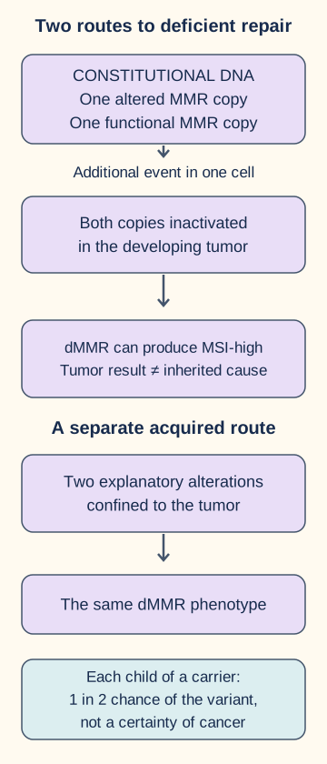
Try separating the inherited variant from the later tumor event in the first diagram. If a healthy parent transmits the variant, must the child already have dMMR throughout the body? No. The usual heterozygous carrier retains another functional copy; tumor formation involves additional events. Transfer that distinction to a cancer-free sibling: genetic testing can identify risk before any tumor exists.
Distinguish the inherited predisposition from the tumor phenotype by tracing both routes. [1]
MLH1 and MSH2 generally confer higher and earlier colorectal risk than MSH6 or PMS2. Later colorectal onset with MSH6 does not make its endometrial risk trivial. PMS2 usually has lower penetrance. The gene, family history, organs present and prior cancers all matter; a single lifetime percentage would conceal important differences. [1]
Does an abnormal tumor screen establish the inherited diagnosis?
Screen the tumor broadly, then explain the abnormal result. Universal evaluation of newly diagnosed colorectal and endometrial cancers identifies people missed by age or pedigree criteria. Colorectal pathways may use MMR immunohistochemistry (IHC), MSI analysis or a validated sequencing equivalent. NICE recommends IHC as the initial endometrial pathway. A small family, adoption or an apparently negative family history should not prevent tumor screening. [1][2]
Gross colon carcinoma photographed by. Image: Netha Hussain, 17 January 2012. The specimen's Lynch status is not documented. Its appearance cannot establish dMMR or a germline diagnosis. image, CC BY-SA 3.0. Netha Hussain; original source; CC BY-SA 3.0 Unported. [1].
IHC asks whether tumor nuclei show the four MMR proteins. MSI testing asks whether repetitive DNA lengths are unstable. These assays examine different consequences of defective repair. A retained but dysfunctional protein can stain, and technical or biologic discordance occurs. Review tissue quality, internal controls and discordant results with pathology and genetics rather than letting one reassuring assay erase the other. [1]
Establish the tumor's MMR or MSI result.
Use the loss pattern to choose reflex testing.
With MLH1 loss, assess tumor MLH1 promoter methylation; BRAF V600E is an additional sporadic-pathway marker in colorectal cancer.
Refer unexplained dMMR for informed genetics evaluation and constitutional testing.
Use an established familial pathogenic variant for targeted testing of relatives.
Consider an older adult with MLH1/PMS2-deficient colorectal cancer, tumor MLH1 methylation and BRAF V600E. This combination strongly favors acquired MLH1 silencing. In an endometrial tumor with the same protein loss, use MLH1 methylation, not a colorectal BRAF shortcut. Very young onset, multiple primaries or a compelling pedigree can justify further genetics assessment even with tumor methylation, including assessment for rare constitutional MLH1 methylation. BRAF wild type does not itself prove Lynch syndrome. [1][2]
Now sort three reports by what they establish: tumor MSH2/MSH6 loss; a pathogenic MSH2 variant in blood; two acquired MSH2 alterations confined to tumor with negative adequate germline testing. The first requires explanation, the second establishes constitutional predisposition, and the third supports a double-somatic explanation. A negative germline panel alone does not explain an abnormal tumor: paired tumor-normal analysis may help. If results remain unresolved, personal and family history still guide risk management. [1]
For a new situation, imagine a relative tests negative before anyone has identified the family's cause. That is not the same as a true negative for a known familial variant. Likewise, a variant of uncertain significance is not a pathogenic result and should not, by itself, trigger predictive testing or irreversible risk-reducing surgery. [1]
Why do proteins disappear in pairs?
Partners explain the pattern, but do not replace germline testing. MSH2 pairs with MSH6 in mismatch recognition; MLH1 pairs with PMS2 in the repair response. Loss of MSH2 often destabilizes MSH6. Loss of MLH1 often destabilizes PMS2. In the reverse direction, MSH2 and MLH1 can remain detectable when their usual partners are lost. EPCAM is not one of the four stained repair proteins: certain EPCAM deletions cause MSH2 silencing. [1]
Read as a testing guide, not a germline answer key
Absent tumor proteins
Leading evaluation
Absent tumor proteinsMLH1 and PMS2
Leading evaluationMLH1 methylation; colorectal BRAF testing as appropriate; germline evaluation if unexplained
Absent tumor proteinsMSH2 and MSH6
Leading evaluationGenetics-guided panel including MSH2 and EPCAM deletion analysis
Absent tumor proteinsMSH6 alone
Leading evaluationGermline evaluation with attention to MSH6
Absent tumor proteinsPMS2 alone
Leading evaluationPMS2 testing with methods that address pseudogenes; consider MLH1 if unresolved
Absent tumor proteinsAll retained, but MSI-high
Leading evaluationReconcile assays and consider dysfunctional retained protein or technical issues
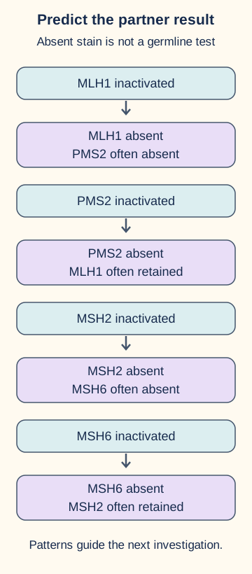
The second diagram is a protein-dependency comparison. Cover its outcome column with your hand, trace loss of MLH1 to its partner, and predict the result. Then repeat with loss of PMS2 alone. The visible comparison shows why paired loss points upstream toward MLH1, while isolated PMS2 loss primarily points toward PMS2. Some MLH1 variants also produce isolated PMS2 loss, so the pattern is a starting hypothesis rather than an exclusive gene assignment. [1]
Cover the outcome area, predict the staining pattern, then compare directional dependency. These are typical patterns, not exclusive germline diagnoses. [1]Prediction check: if MSH6 is lost alone, must MSH2 also disappear?
No. MSH2 can remain detectable because it has other binding partners. Isolated MSH6 loss still warrants inherited evaluation when found in the appropriate tumor pathway. [1][2]
Transfer check: paired MSH2/MSH6 loss, but MSH2 sequencing is negative. What testing capability matters?
Confirm that deletion/duplication analysis and relevant EPCAM deletions were assessed. A sequence-only result can miss a structural explanation. If germline evaluation remains negative, tumor analysis may identify acquired biallelic inactivation. [1]
Visible worked result: MLH1 absent commonly means PMS2 absent; PMS2 absent does not require MLH1 absence. The same directional logic applies to MSH2 and MSH6. Non-neoplastic cells must provide appropriate positive controls before absent tumor staining is interpreted as biologic loss. [1]
Does every organ on the cancer list need annual imaging?
Risk association and effective screening are different claims. Complete high-quality colonoscopy is the central surveillance intervention. It examines proximal as well as distal colon and permits polypectomy. Relatively rapid progression and interval lesions justify short follow-up, even after a reassuring examination. Stool testing or distal-only sigmoidoscopy does not provide an equivalent Lynch surveillance strategy. An examination with inadequately visualized mucosa cannot justify the usual normal-examination interval; obtain an adequate assessment before assigning that interval. [1][8]
For the endometrium, teach prompt reporting of abnormal uterine bleeding, especially postmenopausal bleeding. Evaluate symptoms diagnostically rather than waiting for the surveillance date. For postmenopausal bleeding, the ACOG 2026 update generally favors ultrasound plus endometrial tissue sampling; a thin stripe alone should not reassure a symptomatic high-risk carrier. [9] Endometrial biopsy every 1 to 2 years from age 30 to 35 can be considered if the uterus remains, but a survival benefit from screening has not been demonstrated. Routine ovarian screening with ultrasound or CA-125 has not established comparable protection to risk-reducing surgery. Persistent bloating, pelvic pain or early satiety warrants assessment, even after a normal screening result. [1][5]
For the stomach and duodenum, upper endoscopy decisions depend on the guideline, gene, family history and risk context, including ancestry from a high-incidence region. Assess for H. pylori and treat infection when identified. ACG's older risk-selected approach considers a baseline examination at 30 to 35 with 3- to 5-year follow-up in selected families; the NCI summary of NCCN 2024 describes a broader 30- to 40-year start with 2- to 4-year intervals. Do not merge those into a supposedly universal schedule. [1][6]
For urothelial cancer, evidence for screening benefit is limited. Annual urinalysis from age 30 to 35 may be considered with a family history or particularly an MSH2-associated risk profile. Urine cytology has low sensitivity and false-positive concerns; it is not a proven interchangeable alternative. New visible hematuria requires diagnostic evaluation rather than the next screening urinalysis. A renal-pelvis abnormality is an upper-tract finding, not a renal cortical mass; a normal bladder examination does not settle it. CT urography and, when needed, diagnostic ureteroscopy address the upper tract. [1][7]
For selected MLH1, MSH2 or MSH6 carriers with pancreatic cancer in the same family lineage, discuss MRI/MRCP and/or endoscopic ultrasound at an experienced center. The NCI-described approach starts at 50 or 10 years before the youngest family diagnosis. Discuss uncertain benefit and incidental findings. Routine capsule endoscopy, brain MRI or whole-body imaging is not a default for all asymptomatic carriers. Assess concerning neurologic symptoms and suspicious sebaceous lesions promptly; skin assessment can be part of individualized care. [1][6]
Try assigning a purpose to each plan: colonoscopy can detect and excise precursors; biopsy investigates endometrial tissue; symptom education prompts diagnosis; surgery prevents selected organ cancers. A normal pelvic ultrasound does not compensate for a missed colonoscopy. Transfer the comparison to an MSH2 carrier with hematuria: the finding calls for urologic evaluation, not simply more intensive colon surveillance.
When should surveillance begin, and what changes the date?
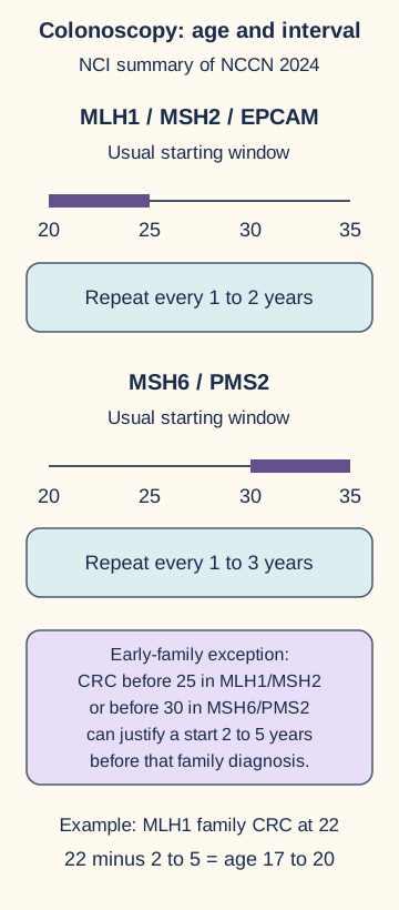
Use the gene first, then check for unusually early family cancer. The third diagram separates starting age from repeat interval. The following practical framework is the NCCN 2024 row reproduced in the NCI PDQ table, not a claim to reproduce every current guideline. Local specialist guidance may differ. [1]
Calculate an earlier family-based start, then distinguish that age from the interval between later examinations. Schedule is the NCI table's NCCN 2024 summary. [1]
High-quality colonoscopy for confirmed carriers
Gene
Usual start
Repeat interval
GeneMLH1, MSH2; EPCAM managed as MSH2
Usual startAge 20 to 25
Repeat intervalEvery 1 to 2 years
GeneMSH6, PMS2
Usual startAge 30 to 35
Repeat intervalEvery 1 to 3 years
If colorectal cancer occurred before 25 in an MLH1/MSH2 family, consider starting 2 to 5 years before that diagnosis. For MSH6/PMS2, the corresponding unusually early family threshold is before 30. Findings, examination quality, previous colorectal cancer and individual risk may justify shorter intervals. A normal colonoscopy does not reset a carrier to average-risk timing. [1]
Calculate before checking: an MLH1 carrier's parent developed colorectal cancer at 22. Subtracting 2 to 5 gives a possible start window of 17 to 20, requiring genetics and gastroenterology planning rather than waiting automatically until 25. Contrast a 24-year-old PMS2 carrier whose family's earliest colorectal cancer was at 63: that family history does not itself invoke the unusually early-onset exception.
For transfer, change only the gene from PMS2 to MLH1 while keeping age 24 and the late family diagnosis. The person is now in the usual earlier starting window. These are ages and intervals, not additive scores; summing surveillance ages has no clinical meaning. Selected upper-GI and urinary surveillance ages answer separate organ-specific questions and cannot delay colonoscopy.
Which prevention decisions remain after a cancer is treated?
Organ-directed treatment does not erase constitutional risk. After segmental colectomy, remaining colon is still susceptible to a new primary cancer, termed metachronous cancer. At initial surgery, compare segmental and extended colectomy using tumor site and stage, age, gene, comorbidity, bowel function and preferences. Extended surgery reduces the amount of colon at risk but can worsen bowel function and does not eliminate rectal risk if rectum remains. An unaffected carrier with a manageable colon is not automatically a candidate for prophylactic colectomy. [1]
Separate tumor biology, anatomic stage and treatment intent. dMMR/MSI-high may affect systemic treatment selection, including eligibility for immune-directed therapy in appropriate cancer settings, whether the cause is inherited or acquired. It does not tell you the depth of invasion or whether metastases exist. A germline result informs prevention for the family; it is not a substitute for cancer staging. [1]
Discuss risk-reducing hysterectomy after completion of childbearing, with timing individualized. Ovarian risk reduction requires a separate discussion of gene, age, menopause and priorities. A matched retrospective study found no endometrial cancers after prophylactic hysterectomy versus 33% in controls, and no ovarian cancers after prophylactic bilateral salpingo-oophorectomy versus about 5% in controls. This supports incidence reduction, not a randomized proof of mortality benefit or a guarantee for every individual. [4]
As one explicit gene-specific example, NICE permits consideration of hysterectomy alone from age 45 for a heterozygous PMS2 carrier without ovarian cancer family history; adding salpingo-oophorectomy depends on verified family history, age and menopausal status. This should not be generalized to all MSH2 carriers. Premenopausal ovarian surgery requires counseling about surgical menopause and its management. Colon surveillance continues after gynecologic surgery. [5]
Aspirin is an additional prevention discussion, not an endoscopy replacement. CAPP2 assigned 861 participants to 600 mg aspirin daily or placebo and found a colorectal cancer hazard ratio of 0.65 (95% confidence interval 0.43 to 0.97) in long-term intention-to-treat analysis. Benefit was delayed. The trial dose is evidence about that trial, not an instruction for every carrier to self-start 600 mg. Choose a regimen with the treating clinician, considering bleeding or ulcer history, concurrent medicines and current guidance. [3]
Finally, offer genetics-guided testing to at-risk relatives for the confirmed familial variant. A true negative usually returns to population screening unless independent history or findings indicate another risk. An uninformative negative or uncertain variant does not justify that reassurance. Consent includes implications for relatives and reproductive decisions. [1][2]
Try subtracting one intervention from an example plan: a 42-year-old MSH2 carrier has a hysterectomy and bilateral salpingo-oophorectomy. Which prevention disappears? Surveillance of organs no longer present is no longer the same issue; surveillance of the remaining colon is unchanged. For a new application, the carrier's sibling tests negative for the known variant and has no other risk factors. That sibling does not inherit the carrier's entire surveillance plan simply because they share a family.
Apply the distinctions
Case 1
Show answer and explanations for case 1
A. MLH1/PMS2 deletion and duplication analysis (Why this does not fit)
Structural MLH1/PMS2 defects could matter with a different protein pattern, but this tumor retains that pair.
Reasoning steps for option A
What does retained MLH1/PMS2 imply for MLH1/PMS2 deletion testing?
This intact pair makes an MLH1/PMS2 structural defect a poor match for the tumor's MSH2/MSH6 loss.
Which missing testing capability would this option supply?
It would assess structural changes, but in the wrong protein pair for this pattern.
Why does the prior negative coding panel not justify switching to MLH1/PMS2?
Coding results leave structural variants unresolved; the absent MSH2/MSH6 pair still directs the structural search toward MSH2 and EPCAM.
B. MSH2 structural analysis and relevant EPCAM deletion testing (Best answer)
Paired MSH2/MSH6 loss directs attention to MSH2 and EPCAM-related silencing. Structural testing addresses the limitation of the prior broad sequencing panel.
Reasoning steps for option B
How can an EPCAM deletion produce paired MSH2/MSH6 loss?
A relevant EPCAM deletion can silence adjacent MSH2; loss of MSH2 destabilizes its partner MSH6.
Which limitation of the prior blood panel does MSH2 structural analysis address?
The panel checked coding sequence but did not assess deletions or duplications, so a constitutional MSH2 structural variant remains possible.
Does paired tumor protein loss itself establish inherited MSH2 or EPCAM disease?
No. It prioritizes appropriate constitutional structural testing, which is needed to establish an inherited cause.
C. MSH6-only deletion and duplication analysis (Why this does not fit)
MSH6 is absent, but paired MSH2 loss points upstream; isolated MSH6 structural testing is too narrow.
Reasoning steps for option C
Why is isolated MSH6 structural testing too narrow when both MSH2 and MSH6 are absent?
Loss of upstream MSH2 can secondarily eliminate MSH6 staining; an MSH2 or relevant EPCAM lesion remains unexamined.
Which retained proteins distinguish this pattern from an MLH1-driven defect?
MLH1 and PMS2 are retained, focusing the inherited investigation on the MSH2/MSH6 axis.
What structural blind spot would MSH6-only analysis leave?
It would not assess MSH2 deletions or relevant EPCAM deletions, despite the paired loss.
D. Repeat coding-sequence analysis of the four MMR genes (Why this does not fit)
Repeating the already performed test category does not fill the unassessed structural-variant gap.
Reasoning steps for option D
What did the earlier blood panel already test?
It assessed coding-sequence variants in all four mismatch-repair genes and found none.
What untested variant class makes repeating that panel insufficient?
Structural variants were not assessed, including MSH2 copy-number changes and relevant EPCAM deletions.
How does the IHC pattern prioritize the missing assay?
Absent MSH2/MSH6 with retained MLH1/PMS2 directs structural evaluation to the MSH2/EPCAM pathway rather than another coding-only round.
Takeaway: Use paired protein loss to select genetics evaluation, without treating the stain as a constitutional diagnosis.
A. Mother: germline evaluation; daughter: target a presumed MSH6 variant now (Why this does not fit)
The maternal investigation fits, but the protein stain has not identified a specific pathogenic familial variant for targeting.
Reasoning steps for option A
What does isolated MSH6 loss establish about the mother's evaluation?
With valid controls, it warrants constitutional assessment of the MSH6-associated pathway, especially with her sister's colorectal cancer.
Does an MSH6-negative stain identify the daughter's exact familial allele?
No. Protein loss is a tumor phenotype, not identification of a specific inherited pathogenic variant.
What must precede informative targeted testing of the daughter?
Establish a pathogenic familial variant through evaluation of the affected mother; a presumed MSH6 target is not yet known.
B. Mother: MLH1 methylation; daughter: target only an established pathogenic familial variant (Why this does not fit)
Deferring targeted testing until a familial variant exists is correct, but MLH1 methylation does not explain isolated MSH6 loss.
Reasoning steps for option B
Why is MLH1 methylation a mismatch for isolated MSH6 loss?
Methylation of MLH1 explains MLH1/PMS2 loss, not a tumor retaining those proteins and losing only MSH6.
Which part of this proposed daughter plan is appropriately conditional?
Targeted predictive testing becomes informative only after a pathogenic familial variant has been established.
What should replace methylation in the mother's branch?
Germline evaluation prompted by valid isolated MSH6 loss and the relevant family history.
C. Mother: MLH1 methylation; daughter: target a presumed MSH6 variant now (Why this does not fit)
Neither the MLH1 reflex pathway nor a presumed familial target follows from the isolated MSH6 result.
Reasoning steps for option C
Which two assumptions fail in the combined methylation and presumptive-target plan?
The isolated MSH6 stain does not suggest MLH1 silencing and does not reveal a specific familial pathogenic allele.
What IHC pattern would instead raise an MLH1 methylation reflex?
Paired MLH1/PMS2 loss, rather than isolated MSH6 loss with intact controls.
Why is targeting a presumed MSH6 variant in the healthy daughter premature?
Without identification of a pathogenic variant in the family, there is no established allele for an informative targeted test.
D. Mother: germline evaluation; daughter: target only an established pathogenic familial variant (Best answer)
The isolated-loss pathway requires constitutional evaluation; informative targeted testing for the adult daughter follows identification of a familial pathogenic variant.
Reasoning steps for option D
What finding directs the affected mother toward germline evaluation?
Her endometrial tumor has valid isolated MSH6 loss, with a sister who had colorectal cancer at 52.
Why should the daughter's predictive test await a molecular diagnosis?
The stain identifies lost protein, not an inherited pathogenic variant; targeted testing requires an established familial allele.
How does testing the affected person improve interpretation for her daughter?
If a pathogenic familial variant is found, the daughter can be tested specifically for it; otherwise a presumed target cannot yield an informative familial result.
Takeaway: Isolated MSH6 loss in endometrial cancer deserves inherited evaluation.
A. Treat the defect as acquired and assess relatives by their own clinical history (Best answer)
A tumor alteration plus loss of the other allele can explain biallelic inactivation without a constitutional cause; relatives do not have an established familial variant to target.
Reasoning steps for option A
How do the two tumor MSH2 events explain paired MSH2/MSH6 absence?
A pathogenic tumor MSH2 alteration and loss of the other allele produce biallelic MSH2 inactivation, with secondary MSH6 loss.
What does matched normal testing indicate about transmission?
Neither alteration was detected constitutionally, so no transmissible familial MSH2 variant is established.
On what basis should relatives be assessed instead?
Use each relative's own clinical and family history rather than predictive testing for a tumor-only alteration.
B. Offer relatives predictive testing for the tumor MSH2 variant as a familial target (Why this does not fit)
A tumor-only alteration is not an established transmissible familial variant.
Reasoning steps for option B
Why is the tumor MSH2 alteration not an established familial target?
It was detected in the tumor but not in the matched normal sample.
What additional tumor event explains the observed protein loss?
Loss of the other MSH2 allele completes biallelic inactivation in the tumor.
What would relatives need before targeted predictive testing could be informative?
A demonstrated constitutional pathogenic familial variant, which the supplied testing has not found.
C. Assign each child a 50% variant risk and institute carrier surveillance (Why this does not fit)
The 50% rule applies to constitutional heterozygosity, which has not been demonstrated here.
Reasoning steps for option C
When does a 50% transmission risk apply to children?
It applies to a heterozygous constitutional pathogenic variant in a parent, not automatically to a tumor-only alteration.
What do the paired tumor and normal specimens show here?
MSH2 alteration and loss of the other allele are tumor-confined; matched normal testing does not show them.
Why does this distinction change surveillance advice?
Children cannot be assigned carrier status from acquired tumor events; assess their independent clinical and family risks.
D. Repeat MLH1 methylation testing to distinguish inherited from acquired MSH2 loss (Why this does not fit)
MLH1 methylation does not explain this MSH2-associated pattern or resolve the already localized alterations.
Reasoning steps for option D
Which protein pair is absent in this endometrial tumor?
MSH2 and MSH6, not MLH1 and PMS2.
What does MLH1 methylation normally help explain?
Tumor MLH1 silencing and consequent MLH1/PMS2 loss, which is not the observed mechanism.
What existing result already explains the MSH2 loss?
Tumor-confined pathogenic MSH2 alteration plus loss of the other allele supplies an acquired biallelic explanation.
Takeaway: An abnormal tumor and a negative germline result can be reconciled by double-somatic inactivation.
A. Test relatives predictively for the uncertain MSH6 variant (Why this does not fit)
Uncertain significance does not establish a disease-causing familial variant.
Reasoning steps for option A
What does uncertain significance say about the MSH6 variant's pathogenicity?
Its disease-causing role is not established, so it cannot serve as a predictive familial pathogenic target.
Why would testing relatives for that variant not determine carrier risk?
A positive result would inherit the same uncertainty rather than prove Lynch syndrome.
Whose testing has greater potential to clarify the family's cause?
The father with colorectal cancer at 44 is alive and willing to undergo testing.
B. Apply pathogenic-MSH6 surveillance and surgical planning to the daughter (Why this does not fit)
An uncertain variant is not equivalent to an established pathogenic MSH6 result for gene-specific prevention.
Reasoning steps for option B
What evidence is missing before applying pathogenic-MSH6 preventive measures?
The daughter has only an MSH6 variant of uncertain significance, not a confirmed pathogenic familial variant.
Why does the father's cancer not make the daughter's VUS pathogenic by itself?
Early-onset family history raises risk but does not classify a particular uncertain allele as causal.
How should her current screening risk be assessed?
Use her family history while pursuing informative evaluation of the affected father, not automatic gene-specific surgery or surveillance based on the VUS.
C. Test the affected father and retain history-based screening for the daughter (Best answer)
Testing the affected father may identify a familial cause. Until then, the daughter's uncertain result does not erase her early-onset family history.
Reasoning steps for option C
Why test the father rather than infer causation from the daughter's panel?
He is affected and available, so testing him may identify a pathogenic familial cause that the unaffected daughter's uncertain result cannot establish.
Does the daughter's VUS provide a true negative result for the family?
No. No familial pathogenic variant is known, and a VUS is neither confirmation nor exclusion.
What supports continued history-based screening for her?
Her father's colorectal cancer at 44 remains relevant regardless of her uninformative panel finding.
D. Use population screening for the daughter because her panel found no pathogenic variant (Why this does not fit)
An uninformative panel in this setting is not a true negative for a known familial cause.
Reasoning steps for option D
What makes a negative predictive test a true familial negative?
A known familial pathogenic variant must be specifically excluded by an adequate test.
Why does this daughter's broad panel fail that condition?
No pathogenic familial cause has been identified, and her only finding is an uncertain MSH6 variant.
Which fact prevents defaulting to population-risk screening now?
Her father developed colorectal cancer at 44, so her risk assessment still depends on that family history.
Takeaway: Do not treat uncertainty as either pathogenicity or reassurance.
A. Keep the order because the preceding colonoscopy was normal (Why this does not fit)
The inherited result changes the appropriate interval despite the prior normal examination.
Reasoning steps for option A
Does a normal colonoscopy 12 months ago justify retaining the nine-year population order after PMS2 confirmation?
No. The adequate examination counts, but the new pathogenic PMS2 result changes subsequent surveillance to a carrier interval.
What interval replaces the scheduled nine-year wait?
NCCN 2024 as summarized by NCI uses colonoscopy every 1 to 3 years for PMS2 carriers.
What changed since the population-risk order was placed?
A confirmed pathogenic PMS2 result now governs follow-up despite the normal colonoscopy.
B. Repeat at 40, then use 5-year intervals because family onset was late (Why this does not fit)
Late family onset does not justify a population-like interval in a confirmed PMS2 carrier.
Reasoning steps for option B
Does the family's youngest colorectal cancer at 64 postpone this carrier's next colonoscopy until 40?
No. Late family onset does not override the confirmed PMS2-associated surveillance schedule.
Why are five-year repeats insufficient after age 40?
A confirmed PMS2 carrier needs the cited 1-to 3-year interval, even after a normal examination.
Does a late youngest family diagnosis cancel gene-based surveillance?
No. Colorectal cancer at 64 does not justify delaying a known carrier to age 40.
C. Repeat within 1 to 3 years of the prior examination, then individualize (Best answer)
Her prior examination occurred in the PMS2 starting window; she needs the 1-to 3-year carrier interval, not another 9-year wait.
Reasoning steps for option C
Can the high-quality colonoscopy at age 30 serve as the baseline examination?
Yes. It occurred within the PMS2 starting window and need not be discarded merely because carrier status was confirmed later.
When should surveillance resume after that normal examination?
Repeat within 1 to 3 years of the examination, then individualize carrier surveillance rather than waiting nine years.
Why is a fresh initiation date unnecessary?
She already underwent a high-quality colonoscopy at 30, so the next decision concerns its carrier-based repeat interval.
D. Restart at 35, then use 5-year intervals because PMS2 has lower penetrance (Why this does not fit)
PMS2 has later and lower colorectal risk than MLH1, but the cited repeat interval remains 1 to 3 years and a prior examination does not justify this prolonged gap.
Reasoning steps for option D
Does lower PMS2 penetrance require restarting screening at 35 despite an adequate examination at 30?
No. The prior high-quality examination remains useful within the PMS2 starting window.
Does a five-year interval follow from PMS2's later risk profile?
No. The NCCN 2024 framework still calls for a 1-to 3-year repeat interval.
How does the proposed age-35 restart affect the prior examination?
It imposes an unnecessary gap after a valid examination rather than scheduling its next carrier-based repeat.
Takeaway: A later starting window is still an intensive surveillance plan.
A. Start at 18, using the usual 10-year offset from the maternal diagnosis (Why this does not fit)
This imports a generic 10-year family-history offset rather than the cited gene-specific early-onset rule.
Reasoning steps for option A
Is ten years before the mother's colorectal cancer at 28 the MSH6 early-onset rule?
No. The cited gene-specific exception considers a start 2 to 5 years before the earliest family diagnosis when it occurred before 30.
What age would the proposed ten-year subtraction produce?
It produces 18, earlier than the 23-to 26 window derived from the applicable 2-to 5-year rule.
Which relationship makes the age-28 diagnosis informative?
Her mother carries the same MSH6 variant, directly linking the early cancer to this carrier family.
B. Plan initiation in the 23-to 26 window because of unusually early family onset (Best answer)
Maternal CRC before 30 triggers consideration of a start 2 to 5 years earlier:28 minus 5 to 28 minus 2 gives 23 to 26. At 24 she is within that planning window.
Reasoning steps for option B
Does maternal colorectal cancer at 28 meet the early-family-onset threshold?
Yes. It occurred before 30 in a relative carrying the same MSH6 variant.
What initiation window follows from that diagnosis for this 24-year-old?
Subtracting 5 to 2 years from 28 gives ages 23 to 26; she is already in that window.
What practical consequence follows at her current age of 24?
She should plan initiation now within the calculated 23-to 26-year window rather than defer to routine ages.
C. Wait until 30 to 35 because MSH6 always uses the later starting window (Why this does not fit)
The usual later starting window can be modified by unusually early cancer in the family.
Reasoning steps for option C
Can the routine MSH6 starting window of 30 to 35 be used without modification here?
No. A same-variant maternal colorectal cancer at 28 activates consideration of an earlier start.
What does applying the family-history adjustment instead yield?
A 2-to 5-year lead on age 28 yields an initiation window of 23 to 26, which includes her current age.
Why is the age-30 threshold not absolute?
The early-onset family exception modifies the usual MSH6 start when colorectal cancer occurs before 30.
D. Wait until 28 and repeat every 5 years because the family has only one cancer (Why this does not fit)
The family-based start precedes the diagnosis, and a 5-year interval is longer than the cited carrier schedule.
Reasoning steps for option D
Should the carrier wait until the exact age of her mother's diagnosis?
No. The early-onset exception considers colonoscopy 2 to 5 years before the maternal diagnosis at 28.
Does having only one affected relative justify five-year colonoscopy intervals?
No. The proposed five-year spacing is longer than the cited MSH6 carrier surveillance schedule.
What does the proposed five-year interval confuse?
A family-based adjustment to starting age does not convert carrier follow-up to five-year spacing.
Takeaway: Recognize when family timing modifies the usual gene-specific starting age.
A. Repeat complete colonoscopy promptly with improved preparation, then use population intervals (Why this does not fit)
The early repeat addresses inadequate visualization, but subsequent population intervals disregard MSH2-associated risk.
Reasoning steps for option A
What does prompt repeat with better preparation fix in today's examination?
It allows assessment of the ascending colon obscured by solid stool despite washing.
What is still wrong with population intervals after an adequate repeat?
They ignore continuing MSH2-associated colorectal risk; short carrier intervals remain necessary.
Does the high-quality examination 18 months earlier make today's blind spot acceptable?
No. The adequate examination 18 months earlier does not establish what is present in the poorly visualized ascending colon today.
B. Accept this examination and wait another full 2-year carrier interval (Why this does not fit)
The current examination is not adequate to assign a normal surveillance interval; the unseen proximal colon remains unevaluated.
Reasoning steps for option B
Can lesion-free visualized segments establish a negative whole-colon examination?
No. Solid stool left the ascending colon inadequately examined.
Why not wait another full two-year carrier cycle?
An incomplete examination cannot reset the routine surveillance clock while proximal bowel remains unevaluated.
Which immediate defect must be corrected before assigning a new interval?
The solid stool obscuring the ascending colon requires a prompt adequate whole-colon repeat.
C. Repeat only the distal examination, then resume short-interval colonoscopy (Why this does not fit)
Repeating distal visualization does not address the obscured ascending colon.
Reasoning steps for option C
Which segment was not adequately visualized today?
The ascending colon, not the distal bowel, remained obscured despite washing.
Why does repeating only the distal examination fail?
It leaves the actual blind spot in the ascending colon unresolved; a complete colonoscopy with improved preparation is needed.
Why is improved bowel preparation relevant to the repeat?
It targets the solid-stool obstruction that prevented inspection of the ascending colon.
D. Repeat complete colonoscopy promptly with improved preparation, then retain carrier intervals (Best answer)
First obtain an adequate whole-colon examination rather than waiting another routine cycle. Then use the short carrier interval; stool testing is not an equivalent replacement.
Reasoning steps for option D
What immediate action addresses the obscured ascending colon?
Promptly repeat a complete colonoscopy with improved preparation instead of accepting the apparent negative examination.
After an adequate examination, can annual stool testing or population spacing replace MSH2 surveillance?
No. Maintain short MSH2 carrier colonoscopy intervals; stool testing is not an equivalent replacement.
What does a negative view of the segments seen today actually establish?
Only those visualized segments had no lesions; the ascending colon still requires adequate examination.
Takeaway: A reassuring examination changes findings, not the inherited predisposition.
A. Less colon at risk; no rectal surveillance; possible increased stool frequency (Why this does not fit)
Retaining rectum preserves a site at risk, so surveillance remains necessary despite the reduced colonic burden.
Reasoning steps for option A
Does ileorectal anastomosis remove the rectum?
No. Subtotal colectomy leaves rectal tissue susceptible to subsequent cancer.
Which prediction fails despite reduced colon and possibly more frequent stools?
Eliminating rectal surveillance is incorrect because retained rectum still needs surveillance.
Does removing more colon remove every colorectal cancer surveillance target?
No. The rectum retained for ileorectal anastomosis remains a surveillance target.
B. Less colon at risk; continued rectal surveillance; possible increased stool frequency (Best answer)
The operation reduces susceptible colon but leaves rectum and can increase stool frequency; these separate outcomes inform the shared decision.
Reasoning steps for option B
How does removing most of the colon affect future colonic cancer risk?
Less susceptible colon remains, reducing future colonic risk relative to right hemicolectomy.
What two consequences remain after ileorectal anastomosis?
The retained rectum requires surveillance, and diminished colonic reservoir can increase stool frequency.
Why is this a functional tradeoff rather than an unqualified benefit?
Reducing colon at risk can come with more frequent stools after extensive resection.
C. Less colon at risk; continued rectal surveillance; predictably reduced stool frequency (Why this does not fit)
Retained rectal surveillance and colonic risk reduction fit, but subtotal colectomy is not expected to predictably reduce stool frequency.
Reasoning steps for option C
Which proposed effects of subtotal colectomy are accurate?
It reduces colon at risk but retains a rectum requiring surveillance.
Is stool frequency predictably lower after removal of most of the colon?
No. Loss of colonic capacity may increase rather than reliably decrease stool frequency.
What functional prediction better fits reduced colonic length?
Possible increased stool frequency, not a predictable reduction.
D. Comparable colonic risk; continued rectal surveillance; possible increased stool frequency (Why this does not fit)
Function and rectal surveillance fit, but leaving less colon reduces future colonic cancer risk rather than preserving an equivalent metachronous risk.
Reasoning steps for option D
Are stool-frequency increase and rectal surveillance plausible after subtotal colectomy?
Yes. Bowel function may change, and the retained rectum remains at risk.
Is future colonic risk comparable when far less colon remains?
No. Removing more susceptible colon reduces metachronous colonic cancer risk relative to segmental resection.
What surgical comparison makes the risk claim wrong?
Subtotal colectomy leaves less colon at risk than a right hemicolectomy.
Takeaway: Cancer surgery balances future organ risk against current functional costs.
A. A: Lynch-specific remnant-colon surveillance;B: Lynch-specific remnant-colon surveillance (Why this does not fit)
A needs inherited-risk surveillance, but B's well-supported acquired pathway does not establish the same inherited indication.
Reasoning steps for option A
Why does patient A need Lynch-specific surveillance of remaining colon?
A has a constitutional pathogenic MLH1 variant and retains colon after segmental surgery.
Does patient B's MLH1/PMS2 loss alone establish the same inherited indication?
No. Tumor methylation, BRAF V600E, negative genetics evaluation and an unremarkable pedigree support acquired silencing; B still needs standard post-cancer endoscopy.
Which finding makes the patients unlike despite identical deficient IHC proteins?
Only A has a confirmed constitutional MLH1 variant; B has evidence for acquired tumor MLH1 silencing.
B. A: standard post-cancer surveillance alone;B: Lynch-specific remnant-colon surveillance (Why this does not fit)
This reverses the constitutional and acquired implications of the supplied results.
Reasoning steps for option B
Which patient has a confirmed constitutional MLH1 predisposition?
Patient A, not B, has a pathogenic germline MLH1 variant.
Why is assigning Lynch-specific follow-up to B but standard follow-up to A reversed?
B's reflex results support acquired MLH1 silencing, while A's inherited risk persists in the remaining colon.
Does B’s prior cancer remove the need for endoscopic follow-up?
No. The correct distinction is standard post-cancer surveillance rather than no surveillance.
C. A: Lynch-specific remnant-colon surveillance;B: standard post-cancer surveillance (Best answer)
A's constitutional predisposition determines intensive retained-colon surveillance. B still needs post-cancer endoscopy but does not have an established Lynch-specific indication.
Reasoning steps for option C
What distinguishes A's retained-colon risk after segmental colectomy?
A's constitutional MLH1 variant warrants Lynch-specific remnant-colon surveillance despite normal current examinations.
What endoscopy does B still need without established Lynch syndrome?
Standard post-cancer surveillance remains indicated; supported acquired MLH1 silencing does not itself impose Lynch-specific intervals.
Do normal remnant-colon examinations remove either patient’s future follow-up?
No. A retains inherited-risk surveillance and B retains standard post-cancer surveillance.
D. A: standard post-cancer surveillance alone;B: standard post-cancer surveillance (Why this does not fit)
B's standard post-cancer plan fits, but A's confirmed MLH1 predisposition requires its additional inherited-risk framework.
Reasoning steps for option D
Is standard post-cancer surveillance suitable for B?
Yes. B still requires post-cancer endoscopy despite evidence favoring an acquired tumor pathway.
Why is that standard schedule alone insufficient for A?
A has confirmed constitutional MLH1-associated risk in the retained colon and needs Lynch-specific surveillance.
Does A’s age of 68 alone negate inherited-risk follow-up?
No. The stem specifies adequate life expectancy and fitness for ongoing endoscopy.
Takeaway: Constitutional risk modifies retained-colon surveillance; an acquired tumor pathway does not erase post-cancer follow-up.
A. A: hysterectomy alone under the PMS2 exception;B: consider hysterectomy alone (Why this does not fit)
B's option fits, but the PMS2-specific exception cannot simply be assigned to A's MSH2 risk profile.
Reasoning steps for option A
Can A's MSH2 variant be managed using the PMS2-only hysterectomy exception?
No. The NICE exception for hysterectomy alone in this setting concerns the heterozygous PMS2 profile, not A's MSH2 profile.
Which part of this pairing remains appropriate for B?
At 46 with heterozygous PMS2 and no ovarian cancer family history, B can consider hysterectomy alone after childbearing.
What gene difference prevents applying B’s exception to A?
A carries MSH2, whereas the hysterectomy-alone exception described here concerns heterozygous PMS2.
B. A: defer endometrial prevention and discuss ovarian surgery alone;B: consider hysterectomy alone (Why this does not fit)
Ovarian surgery alone does not address A's endometrial risk-reduction goal after childbearing.
Reasoning steps for option B
Does ovarian surgery alone address A's endometrial risk-reduction goal?
No. A's uterus would remain; MSH2 supports discussing hysterectomy with bilateral salpingo-oophorectomy.
Is the proposed hysterectomy-alone discussion for B the error?
No. B meets the PMS2-specific conditions for considering that option; the mismatch is A's ovarian-only plan.
Which organ remains exposed if A has ovarian surgery without hysterectomy?
Her uterus remains, leaving the endometrial risk that the proposed risk-reduction discussion must address.
C. A: discuss hysterectomy with bilateral salpingo-oophorectomy;B: require the identical combined operation (Why this does not fit)
The discussion for A fits the named framework, but it does not make combined surgery mandatory for B.
Reasoning steps for option C
Is combined hysterectomy and bilateral salpingo-oophorectomy appropriate to discuss with A?
Yes. A's MSH2 status and completed childbearing make the combined risk-reduction discussion pertinent.
Must B undergo the identical combined operation?
No. At 46 with heterozygous PMS2 and no ovarian cancer family history, NICE permits consideration of hysterectomy alone.
Which patient’s ovarian operation must not be made mandatory by analogy?
B’s: heterozygous PMS2 status without ovarian cancer family history permits hysterectomy-alone consideration.
D. A: discuss hysterectomy with bilateral salpingo-oophorectomy;B: consider hysterectomy alone (Best answer)
NICE distinguishes MSH2-associated combined risk-reduction discussion from consideration of hysterectomy alone in this PMS2 setting; individual counseling remains essential.
Reasoning steps for option D
What operation should be discussed for A's MSH2-associated gynecologic risks?
Discuss hysterectomy with bilateral salpingo-oophorectomy, including the surgical and menopause consequences.
What distinction allows B a different discussion at age 46?
For a heterozygous PMS2 carrier without ovarian cancer family history, NICE allows consideration of hysterectomy alone; ovarian surgery is individualized.
Why do the distinct discussions not eliminate shared counseling?
Both patients still need consent, surgical-risk and menopause counseling before individualized decisions.
Takeaway: A shared syndrome label does not make uterine and ovarian surgical decisions identical across genes.
A. Consider hysterectomy alone now; individualize additional ovarian surgery (Best answer)
NICE permits hysterectomy alone from 45 in this PMS2 context; adding ovarian surgery depends on history, age and menopause.
Reasoning steps for option A
Does this carrier meet the age and gene conditions for the NICE hysterectomy-alone option?
Yes. She is 46, heterozygous for PMS2, has completed childbearing and has no ovarian cancer family history.
Is removal of the ovaries automatically required with hysterectomy?
No. Hysterectomy alone can be considered from 45 in this PMS2 setting; ovarian surgery requires individualized counseling about risk and menopause.
What is the purpose of the hysterectomy discussion in this question?
To reduce endometrial risk after childbearing, without automatically committing her to ovarian removal.
B. Offer combined hysterectomy and ovarian surgery as the only gene-appropriate option (Why this does not fit)
The named NICE framework permits consideration of hysterectomy alone in this PMS2 context; combined surgery is not the only gene-appropriate option.
Reasoning steps for option B
What makes combined surgery an unnecessarily exclusive recommendation here?
NICE permits consideration of hysterectomy alone from age 45 for this heterozygous PMS2 carrier without ovarian cancer family history.
What remains to discuss if ovarian surgery is contemplated?
Ovarian risk, family history, age and the consequences of premenopausal oophorectomy should inform that separate decision.
Which missing risk factor weakens the claim that ovarian surgery is obligatory?
She reports no ovarian cancer family history, supporting individualized rather than compulsory ovarian surgery.
C. Require deferring hysterectomy until natural menopause (Why this does not fit)
At 46 she is within the NICE age framework for considering hysterectomy alone. Waiting for natural menopause is not a prerequisite for that discussion; ovarian surgery is a separate decision.
Reasoning steps for option C
Must a 46-year-old PMS2 carrier await natural menopause before discussing hysterectomy?
No. She has completed childbearing and already meets the NICE age threshold of 45 for considering hysterectomy alone.
Why might menopause still matter to surgical counseling?
Additional ovarian surgery could cause surgical menopause, but menopause is not a prerequisite to discussing uterine risk reduction.
Which timing threshold does the proposed delay ignore?
NICE permits considering hysterectomy alone from age 45 in this PMS2 setting; she is 46.
D. Offer ovarian surgery alone now and rely on surveillance for endometrial prevention (Why this does not fit)
The stated aim is endometrial prevention. Ovarian surgery alone does not accomplish it, and endometrial screening has no established equivalent preventive benefit.
Reasoning steps for option D
Would ovarian surgery alone remove the source of endometrial risk?
No. The uterus remains, so it fails her stated endometrial prevention objective.
Can endometrial surveillance substitute for a proven preventive operation in this proposal?
No. Screening has no established equivalent preventive benefit, while hysterectomy alone is a permissible discussion for her PMS2 profile.
Why does the option misalign the operation with her stated goal?
It removes ovaries but leaves the endometrium in place despite her endometrial risk-reduction aim.
Takeaway: Use gene-specific ovarian counseling rather than a single operation for every carrier.
A. Repeat ultrasound in 6 weeks and sample only if the stripe thickens (Why this does not fit)
A thin stripe does not provide sufficient reassurance to postpone tissue assessment in this symptomatic high-risk patient.
Reasoning steps for option A
Does a 3 mm stripe exclude the need for tissue evaluation of new bleeding in this MSH6 carrier?
No. A thin ultrasound stripe alone is insufficient reassurance for this symptomatic high-risk patient.
Why not postpone sampling until a thicker stripe appears in six weeks?
The new postmenopausal bleeding poses a current diagnostic question that requires tissue assessment rather than conditional ultrasound follow-up.
What feature changes this from routine surveillance to diagnostic assessment?
She now has postmenopausal bleeding, a new symptom arising after her prior benign sample.
B. Obtain endometrial tissue as part of the current diagnostic evaluation (Best answer)
Her prior surveillance sample predates the new symptom. ACOG's 2026 update favors ultrasound plus tissue sampling for most postmenopausal bleeding evaluations, especially when risk makes ultrasound-only reassurance inappropriate.
Reasoning steps for option B
Does the benign biopsy eight months before bleeding assess this episode?
No. It predates the new symptom and cannot rule out pathology causing current postmenopausal bleeding.
What should be obtained despite an adequately measured 3 mm stripe?
Endometrial tissue during the current diagnostic evaluation; ultrasound alone does not settle this high-risk presentation.
What specifically is absent from the current episode’s workup?
No endometrial tissue has been obtained since this bleeding began.
C. Treat presumed atrophic bleeding first and sample only if treatment fails (Why this does not fit)
Atrophy can cause bleeding, but presuming it before appropriate evaluation risks missing endometrial pathology in this carrier.
Reasoning steps for option C
Can atrophy be assumed as the cause before assessing the endometrium?
No. Although atrophy can cause bleeding, MSH6-associated risk makes endometrial pathology important to exclude.
What is the danger of sampling only if empiric treatment fails?
It delays tissue diagnosis of a potentially consequential cause of new postmenopausal bleeding.
Does an ultrasound finding establish atrophy as the cause?
No. A 3 mm stripe does not diagnose the source of bleeding or exclude a lesion needing tissue assessment in this setting.
D. Wait for the next scheduled surveillance biopsy because the stripe is thin (Why this does not fit)
A surveillance calendar and a thin stripe do not settle a new diagnostic problem; tissue from eight months earlier does not assess this episode.
Reasoning steps for option D
Does the next surveillance biopsy replace evaluation of this new symptom?
No. Scheduled surveillance is different from diagnostic assessment of current postmenopausal bleeding.
Why do the prior benign biopsy and thin stripe not justify waiting?
The biopsy preceded the episode and a 3 mm stripe alone does not adequately resolve the present high-risk diagnostic question.
What temporal limitation applies to the benign biopsy?
It was taken eight months before the new bleeding and cannot evaluate pathology arising or presenting afterward.
Takeaway: New postmenopausal bleeding requires diagnostic evaluation; neither an earlier surveillance biopsy nor a thin stripe alone settles the current episode.
A. Treat the infection and consider repeat upper endoscopy in 3 to 5 years (Best answer)
ACG supports treatment of documented H. pylori and consideration of 3-to 5-year follow-up with a gastric or duodenal family history. Other guidance differs.
Reasoning steps for option A
What action follows the positive gastric H. pylori sample?
Treat the documented infection even though baseline upper endoscopy found no neoplasia.
Which finding supports considering repeat upper endoscopy in 3 to 5 years under ACG 2015?
Her mother's gastric cancer supplies the gastric family-history criterion in the risk-selected upper-GI framework.
Does residence in a high-incidence region replace the family-history discriminator?
No. Her maternal gastric cancer specifically supports considering risk-selected follow-up in the named framework.
B. Treat the infection and use the 1-to 2-year colonoscopy interval for the stomach (Why this does not fit)
Treating infection is appropriate, but the colonoscopy interval is not automatically the upper-endoscopy interval.
Reasoning steps for option B
Is treating biopsy-proven H. pylori appropriate?
Yes. The infection is actionable despite a normal neoplasia assessment.
Can the 1-to 2-year colonoscopy interval be copied to upper endoscopy?
No. Under the specified ACG 2015 risk-selected framework, gastric family history supports considering a 3-to 5-year upper-GI interval.
Which organ-specific interval is being confused?
The 1-to 2-year schedule belongs to Lynch colonoscopy, not automatically to gastric endoscopy.
C. Treat the infection and stop upper endoscopic follow-up after this normal examination (Why this does not fit)
Normal current mucosa does not erase the family-history context supporting consideration of follow-up.
Reasoning steps for option C
Does no neoplasia at baseline erase future upper-GI risk considerations?
No. Her mother's gastric cancer remains relevant to risk-selected follow-up.
What is missing from a treat-and-stop plan?
It omits consideration of repeat upper endoscopy in 3 to 5 years under the specified ACG 2015 framework.
Why does baseline normality not settle the future surveillance choice?
No neoplasia today does not remove the maternal gastric cancer history relevant to subsequent endoscopy.
D. Defer infection treatment and consider repeat upper endoscopy in 3 to 5 years (Why this does not fit)
The selected interval is plausible under this framework, but documented H. pylori should be treated rather than deferred.
Reasoning steps for option D
Is a 3-to 5-year upper-endoscopy discussion reasonable with maternal gastric cancer?
Yes. That family history supports considering this interval under ACG 2015.
Can treatment of documented H. pylori be deferred simply because endoscopy found no neoplasia?
No. The positive gastric sampling calls for treatment now, independently of surveillance scheduling.
Are infection management and endoscopy scheduling interchangeable decisions?
No. The 3-to 5-year endoscopy discussion does not excuse deferring treatment of confirmed H. pylori.
Takeaway: Treat an actionable infection while keeping organ-specific surveillance decisions distinct.
A. Renal cortical carcinoma; proceed to percutaneous cortical mass sampling (Why this does not fit)
The described defect is in the collecting system, not a cortical mass suitable for this proposed target.
Reasoning steps for option A
Does the renal-pelvis filling defect identify a cortical biopsy target?
No. The persistent defect lies in the urothelial collecting system, and CT shows no parenchymal mass to sample.
Why would cortical sampling miss the suspected source of hematuria?
A cortical specimen would not directly assess the renal-pelvis lesion in this MSH2 carrier.
What CT feature contradicts a renal cortical carcinoma target?
The scan explicitly reports no renal parenchymal mass.
B. Bladder urothelial carcinoma; repeat bladder-only cystoscopy to explain the renal finding (Why this does not fit)
The normal bladder assessment does not settle a persistent renal-pelvis defect; repeating only it misses the relevant compartment.
Reasoning steps for option B
Does a negative bladder cystoscopy inspect the renal pelvis?
No. Cystoscopy found no bladder lesion but does not evaluate the persistent upper-tract filling defect.
What is missed by repeating only bladder cystoscopy?
The abnormality is in the renal pelvis, so bladder-only reassessment would leave its cause unresolved.
Where is the persistent finding relative to the normal bladder exam?
It is in the renal pelvis, upstream of the bladder inspected by cystoscopy.
C. Upper-tract urothelial lesion; pursue diagnostic ureteroscopy and directed assessment (Best answer)
The collecting-system lesion raises concern for upper-tract urothelial disease. When imaging and voided cytology are insufficient, ureteroscopy can inspect and sample the upper tract; carcinoma is not yet proved.
Reasoning steps for option C
What diagnosis does the renal-pelvis defect raise in an MSH2 carrier?
It raises concern for an upper-tract urothelial lesion, although imaging alone does not prove carcinoma.
Why proceed to diagnostic ureteroscopy despite negative voided cytology?
Voided cytology can miss upper-tract disease; ureteroscopy permits directed inspection and sampling of the persistent pelvic defect.
Does the proposed assessment assume carcinoma is already proved?
No. Directed ureteroscopic assessment seeks a diagnosis because imaging does not establish one.
D. Benign upper-tract bleeding; return to annual urinalysis because cytology is negative (Why this does not fit)
Negative voided cytology is insufficient reassurance with symptoms and a persistent upper-tract abnormality.
Reasoning steps for option D
Does negative voided cytology establish benign bleeding here?
No. Negative cytology cannot exclude an upper-tract lesion in a symptomatic carrier with a persistent renal-pelvis defect.
Why is annual urinalysis insufficient follow-up?
Visible hematuria and a structural collecting-system finding call for diagnostic assessment rather than routine screening.
Which two findings make routine urinalysis an inadequate response?
Visible hematuria and a persistent renal-pelvis filling defect require investigation despite negative cytology.
Takeaway: A low-sensitivity screen does not exclude disease in a symptomatic high-risk patient.
Does retaining colonoscopy resolve this option's safety problem?
No. Continuing surveillance is appropriate, but aspirin risk still needs assessment before starting 600 mg.
Which co-medication raises bleeding concerns with 600 mg aspirin?
His anticoagulant for atrial fibrillation adds bleeding risk to his history of ulcer bleeding.
B. Start low-dose aspirin now and continue the established colonoscopy schedule (Why this does not fit)
A lower dose does not eliminate the need to evaluate the stated bleeding risks before starting.
Reasoning steps for option B
Does a low dose remove the need for bleeding-risk assessment?
No. Anticoagulation and prior ulcer bleeding remain relevant even if the contemplated aspirin dose is lower.
What should happen before starting aspirin in this patient?
Assess individual bleeding risk and the benefit-harm balance rather than prescribe immediately on the basis of dose alone.
Which prior event remains relevant even with low-dose aspirin?
A previous ulcer bleed is still a material harm consideration before adding aspirin to anticoagulation.
C. Continue colonoscopy; assess bleeding risk before prescribing aspirin (Best answer)
Delayed trial benefit supports discussion, but anticoagulation and prior ulcer bleeding materially affect the individual balance.
Reasoning steps for option C
What did CAPP2 support, and what remains patient-specific?
Long-term colorectal prevention benefit supports discussing aspirin; anticoagulation and past ulcer bleeding make net benefit and regimen individual decisions.
Should aspirin deliberation interrupt his colonoscopy schedule?
No. Continue established colonoscopy while assessing bleeding risk before any aspirin prescription.
What trial conclusion remains applicable during individualized prescribing?
CAPP2 supports delayed colorectal prevention benefit, but it does not remove the need to weigh bleeding harms.
D. Continue colonoscopy; permanently exclude aspirin because it lacks efficacy (Why this does not fit)
CAPP2 supports efficacy over long follow-up; the uncertainty here is individual net benefit and regimen, not an absence of colorectal benefit.
Reasoning steps for option D
Does CAPP2 show aspirin has no colorectal preventive efficacy?
No. Its long-term results support benefit, so a categorical efficacy-based exclusion misstates the evidence.
What justifies caution without a permanent efficacy-based ban?
Anticoagulation and previous ulcer bleeding require individualized harm assessment, while colonoscopy continues.
Is the obstacle lack of efficacy or individual safety?
Individual safety: CAPP2 supports efficacy, while anticoagulation and past ulcer bleeding complicate prescribing.
Takeaway: Prevention evidence must be integrated with competing harms, not used to cancel proven surveillance.
A. About 4 points crude absolute difference; assess his bleeding risk before choosing a regimen (Best answer)
The proportions are about 9.4% and 13.4%, a crude difference near 4 points. A relative hazard reduction is not a personal absolute benefit; anticoagulation and prior major bleeding require individualized assessment.
Reasoning steps for option A
What crude absolute difference follows from 40/427 versus 58/434?
The proportions are about 9.4% and 13.4%, giving roughly 4 percentage points, not 35 points.
Why not prescribe the trial dose automatically despite that difference?
The hazard ratio is a group relative measure; his anticoagulation and major ulcer bleed require individualized harm assessment while colonoscopy continues.
What does the hazard ratio of 0.65 measure instead of personal absolute benefit?
It is a relative time-to-event hazard estimate, not a 35-percentage-point change in this patient’s risk.
B. About 4 points crude absolute difference; initiate the trial dose without further harm assessment (Why this does not fit)
The calculation is correct, but group benefit does not establish net benefit for this higher-bleeding-risk individual or justify automatic trial-dose prescribing.
Reasoning steps for option B
Is the proposed four-point calculation accurate?
Yes. The trial proportions differ by approximately four crude percentage points.
What clinical fact defeats immediate trial-dose prescribing?
His anticoagulant use and prior major ulcer bleed mean group efficacy does not establish his personal net benefit.
Which two patient-specific facts remain after the correct arithmetic?
He takes anticoagulation after venous thromboembolism and has had a major ulcer bleed.
C. About 35 points absolute difference; assess his bleeding risk before choosing a regimen (Why this does not fit)
Harm assessment is appropriate, but 0.65 denotes a relative hazard measure, not a 35-point absolute difference.
Reasoning steps for option C
Does a hazard ratio of 0.65 mean a 35-point absolute benefit?
No. It expresses a relative time-to-event hazard comparison, while observed event proportions differ by about four percentage points.
Which part of this plan remains appropriate?
Assessing his bleeding risk before selecting an aspirin regimen is appropriate given anticoagulation and major ulcer bleeding.
How is the observed absolute difference obtained?
Subtract about 9.4% on aspirin from about 13.4% on placebo to obtain roughly 4 percentage points.
D. About 35 points absolute difference; initiate the trial dose without further harm assessment (Why this does not fit)
Both the effect-size interpretation and automatic prescribing inference are unsupported.
Reasoning steps for option D
What is wrong with the claimed 35-point difference?
40/427 and 58/434 yield roughly 9.4% and 13.4%; a hazard ratio of 0.65 is not a 35-point absolute difference.
Why is immediate trial-dose initiation additionally unsupported?
Anticoagulation after venous thromboembolism and a major ulcer bleed require a personal bleeding-risk assessment first.
Does retaining colonoscopy fix either incorrect inference?
No. Continued colonoscopy is appropriate but cannot turn a relative hazard into a 35-point absolute benefit or negate bleeding risk.
Takeaway: Distinguish observed absolute risk differences from time-to-event relative measures.
A. Use lifelong MSH2-carrier intervals solely because her brother carries the variant (Why this does not fit)
An adequate true negative does not establish the familial MSH2 predisposition in her.
Reasoning steps for option A
What does her adequately negative familial MSH2 test establish?
The known variant in her brother was detectable by this assay but is absent in her, so it does not assign her MSH2-carrier status.
Why are carrier-specific intervals not justified solely by her brother's result?
His variant is not hers on this informative test; her own colorectal cancer supplies a separate surveillance indication.
What confirms this is an informative true negative for the family variant?
The laboratory confirms that the brother’s pathogenic MSH2 variant was detectable by her targeted assay.
B. Return directly to population screening because the familial test is negative (Why this does not fit)
The negative familial test does not erase her personal history of colorectal cancer and retained colon.
Reasoning steps for option B
Does a true negative familial test erase a previous colon cancer?
No. It addresses inherited familial-variant status, not the follow-up need after curative segmental colectomy.
Why is population screening alone too little?
She retains colon and has a personal colorectal cancer history requiring post-cancer surveillance.
What anatomic history preserves her need for surveillance?
She underwent segmental rather than total colectomy and retains colon after colorectal cancer.
C. Suspend endoscopy until a second germline panel identifies another variant (Why this does not fit)
Her cancer history already requires follow-up; endoscopy should not depend on identifying another inherited cause.
Reasoning steps for option C
Must another germline variant be found before endoscopy resumes?
No. Her prior colorectal cancer already establishes the follow-up indication independently of further germline testing.
What do the completed tumor and broader genetics assessments change?
They reveal no additional suspicious finding, but do not justify suspending post-cancer endoscopic surveillance.
Why is a second panel not a prerequisite here?
Broader genetics and tumor assessment are already completed without another suspicious finding, and cancer follow-up is independent of a molecular explanation.
D. Use appropriate post-cancer surveillance without assigning MSH2-carrier status (Best answer)
She is negative for the known familial cause but still has an independent post-cancer indication for surveillance. Other concerning findings would deserve separate assessment.
Reasoning steps for option D
How does her negative result differ from an uninformative negative panel?
It specifically excludes her brother's detectable pathogenic MSH2 variant on an adequate targeted test.
Which indication still calls for colonoscopy?
Her personal colorectal cancer treated by segmental colectomy requires appropriate post-cancer surveillance without labeling her an MSH2 carrier.
Does the familial result exclude every reason for follow-up?
No. It excludes the known inherited MSH2 variant, not the post-cancer surveillance indication.
Takeaway: An informative familial negative is different from an unexplained negative panel.
A. A:25% chance for each child;B:50% chance for each child (Why this does not fit)
A has an autosomal dominant constitutional predisposition, not a 25% transmission rule; B's demonstrated events are acquired.
Reasoning steps for option A
What inheritance probability applies to A's blood-confirmed heterozygous MSH6 variant?
Each child has a 50% chance of inheriting that constitutional variant, not 25%.
Can B's tumor-confined MSH2 alterations be assigned 50% transmission?
No. Both explanatory events are absent from matched normal tissue, and adequate germline testing is negative.
Does a 25% figure describe transmission from a single heterozygous autosomal dominant parent?
No. Each child independently has a one-in-two chance of inheriting A’s identified MSH6 variant.
B. A:50% chance for each child;B:no transmissible variant established (Best answer)
A's constitutional heterozygous variant has a 1 in 2 transmission probability. B's tumor events do not establish a familial variant; neither tumor phenotype makes cancer inevitable in children.
Reasoning steps for option B
Why does A have a one-in-two transmission probability?
A pathogenic heterozygous MSH6 variant confirmed in blood is constitutional and follows autosomal dominant transmission.
What has B's matched tumor-normal testing established for offspring?
The MSH2 events are tumor-only, so no transmissible variant is established; inheriting a predisposition is not the same as inevitable cancer.
What makes B’s alterations somatic rather than demonstrated constitutional variants?
Both MSH2 events are absent from matched normal tissue, alongside adequate negative germline testing.
C. A:50% chance for each child;B:50% chance for each child (Why this does not fit)
The probability for A is correct, but the same dMMR phenotype does not make B's tumor-only events constitutional.
Reasoning steps for option C
Which half of the two 50% claims is supported?
A's blood-confirmed heterozygous MSH6 variant has a 50% chance of transmission to each child.
Why does B's dMMR tumor not establish the same risk?
B's two MSH2 events are absent from matched normal tissue, with adequate negative germline testing; tumor phenotype is not a constitutional result.
Does dMMR alone imply a 50% risk of inheriting cancer?
No. A’s 50% applies to variant transmission, not certainty of cancer; B has no established inherited variant.
D. A:no transmissible variant established;B:no transmissible variant established (Why this does not fit)
A's blood result establishes a constitutional variant even though B's does not.
Reasoning steps for option D
Is A's pathogenic MSH6 variant restricted to the tumor?
No. Its confirmation in blood demonstrates a constitutional variant capable of transmission.
Which adult actually lacks an established transmissible variant?
B does: both identified MSH2 events are tumor-specific and adequate germline testing is negative.
Which sample distinguishes A from B for reproductive counseling?
A’s blood contains the pathogenic MSH6 variant, whereas B’s identified MSH2 events are absent from normal tissue.
Takeaway: Transmission of predisposition and development of cancer are distinct events.
A. End inherited evaluation because tumor methylation fully explains both primary cancers (Why this does not fit)
Methylation favors a sporadic mechanism in many settings but is not sufficient reassurance with this striking early multiple-primary history.
Reasoning steps for option A
Does tumor MLH1 methylation explain away both exceptionally early primaries?
No. It often favors acquired silencing but does not settle constitutional risk in a patient with endometrial cancer at 25 and colorectal cancer at 28.
Which pedigree detail prevents ending inherited evaluation?
Her mother's colorectal cancer at 34 reinforces suspicion despite the tumor methylation result.
What does MLH1/PMS2 loss plus tumor methylation localize without proving?
It points to MLH1 silencing in this tumor, but does not prove both primary cancers have exclusively sporadic origins.
B. Limit inherited evaluation to MLH1 coding sequence and ignore methylation outside the tumor (Why this does not fit)
Coding sequence analysis alone would not address the constitutional methylation mechanism suggested by this unusually early presentation.
Reasoning steps for option B
Why is MLH1 coding analysis alone incomplete?
A coding assay would not detect constitutional MLH1 methylation, a rare mechanism relevant to this unusually early presentation.
What history supports assessment beyond coding sequence?
Two primary cancers by age 28 and maternal colorectal cancer at 34 sustain inherited-risk concern despite tumor methylation.
Why consider methylation beyond the colorectal tumor?
Rare constitutional MLH1 methylation would not be resolved by checking only the coding sequence of MLH1.
C. Evaluate inherited causes, including constitutional MLH1 methylation (Best answer)
Very early cancers and a strong pedigree justify further assessment despite tumor methylation, including constitutional MLH1 methylation where appropriate.
Reasoning steps for option C
What does methylation in the tumor usually suggest?
It often supports acquired MLH1 silencing, but is not conclusive evidence against inherited causes in this extreme clinical context.
Which additional constitutional mechanism should evaluation consider?
Constitutional MLH1 methylation is a rare possibility alongside other inherited causes given multiple very early primaries and the maternal history.
Which ages make this unlike a routine late-onset methylated tumor?
Endometrial cancer occurred at 25, colorectal cancer at 28, and her mother had colorectal cancer at 34.
D. Use tumor-only MMR sequencing as the sole inherited-risk assessment (Why this does not fit)
Tumor sequencing may help, but tumor-only results cannot settle whether a constitutional cause explains the striking personal and family history.
Reasoning steps for option D
Can tumor-only sequencing distinguish a constitutional cause here?
No. Findings restricted to tumor cannot by themselves establish or exclude an alteration present outside the tumor.
Why does this history demand more than tumor-only assessment?
Endometrial cancer at 25, colorectal cancer at 28 and maternal colorectal cancer at 34 sustain suspicion requiring constitutional evaluation.
What compartment must be evaluated to test constitutional suspicion?
Tissue outside the tumor is needed to investigate a constitutional mechanism such as MLH1 methylation.
Takeaway: Clinical context can prevent an overly absolute interpretation of a sporadic-pathway marker.