
Choose an answer, then open any option to work its reasoning.
MSK · Endocrinology
Diabetic Foot and Charcot Arthropathy: The Hot Red Foot and the Amputation Clock
Fifteen percent of people with diabetes develop a foot ulcer, and 85 percent of amputations are preceded by one. The clock runs on three failures at once: neuropathy (the foot cannot feel the pressure that starts the ulcer), peripheral arterial disease (the foot cannot heal it), and impaired healing (infection climbs while hyperglycemia smothers repair). Learn the discriminator that stops the clock: a hot, red, swollen foot with intact skin that does not hurt is Charcot arthropathy, not cellulitis; an ulcer that probes to bone is osteomyelitis until proven otherwise; and the annual monofilament, the daily inspection, and the total contact cast are the moves that keep the patient out of the operating room.
What this page makes you able to do
- Examine the swollen diabetic foot and run the four-way discriminator: Charcot, cellulitis, osteomyelitis, gout
- Grade the ulcer on the Wagner ladder and the University of Texas depth-infection-ischemia frame
- Work up the infected foot in order: probe to bone, markers, plain film, MRI, bone biopsy, and 4 to 6 weeks of therapy
- Read the ischemic foot: claudication, dependent rubor, the ABI, and the calcified-vessel trap that forces toe pressures
- Dr. Fatima Ali, DOPsychiatry residentPrimary reviewer
Last reviewed
The pearl
A hot, red, swollen foot in a neuropathic diabetic is four diseases wearing the same costume. Painless plus intact skin plus bounding pulses plus normal markers is Charcot: offload with a total contact cast, never antibiotic. Ulcer plus probe to bone plus climbing ESR and CRP is osteomyelitis: debride and treat 4 to 6 weeks. Tender plus fever plus erythema is cellulitis: antibiotics. The discriminator is three sentences long, and it decides between a cast, a cure, and a collapse.
Prove it
Opening question
Answer before you read anything, then carry the discriminator through every section.
A 58-year-old man with type 2 diabetes and established peripheral neuropathy presents with a hot, red, swollen right foot enlarging over 4 days. He denies injury and says the foot does not really hurt despite the swelling. Temperature is 37.0 C. Examination shows a diffusely swollen, erythematous midfoot with intact skin and no ulcer and bounding dorsalis pedis and posterior tibial pulses. Laboratory studies show a WBC count of 7,200, ESR 22, and CRP 3.Which of the following is the most likely diagnosis?
- Why this is rightA hot, red, swollen foot that is remarkably PAINLESS with intact skin, bounding pulses, and normal white count and inflammatory markers in a neuropathic diabetic is acute Charcot arthropathy: the midfoot is breaking under unperceived load. The treatment is mechanical, not antimicrobial: non-weight-bearing plus a total contact cast, with serial exams until the foot cools. Rule: hot plus red plus swollen plus painless plus intact skin plus bounding pulses is Charcot until proven otherwise.
- Why this failsCellulitis is tender, erythematous, febrile, and leukocytotic, and it enters through a break in the skin. This foot has no entry wound, does not hurt, and carries a normal WBC, ESR, and CRP. Rule: tender plus fever plus erythema is infection; this foot has none of the three.
- Why this failsOsteomyelitis needs the ulcer doorway: an ulcer or sinus tract that probes to bone, with climbing ESR and CRP. The skin here is intact, so there is nothing to probe, and the markers are normal. Rule: no ulcer, no probe, no bone infection; do not probe through intact skin.
- Why this failsGout is exquisitely tender, monoarticular, and classically hits the first metatarsophalangeal joint (podagra) with an elevated uric acid. This foot is painless, midfoot-centered and diffuse, and normouricemic. Rule: gout is the most tender foot in the room; Charcot is the least.
- Why this failsA DVT is a venous occlusion: unilateral leg swelling and pain, often after immobility, with a positive Wells score, and it does not produce the midfoot-centered, painless, red-hot bony foot of acute Charcot. Rule: venous swelling is not positionally spared; the painless hot midfoot is not a clot.
Work the reasoning
Loss of protective sensation plus autonomic hyperemia lets unperceived stress fracture and remodel the midfoot bones. The patient cannot feel the destruction, so the collapse proceeds silently while the foot stays hot and red.
Tenderness, fever, leukocytosis, and climbing ESR and CRP name infection. Charcot keeps the skin intact, the pulses bounding, and the markers normal; MRI or a WBC-labeled scan separates the two when the exam is ambiguous.
Mechanical: non-weight-bearing plus a total contact cast through the active Eichenholtz stages, with serial skin temperature checks until the foot cools. Antibiotics do nothing for a sterile collapsing midfoot.
The answer is A: acute Charcot arthropathy. The trap is treating the Charcot foot with antibiotics while the midfoot quietly collapses: offload with a total contact cast.
THE TRIPLE THREAT
The Triple Threat: Why the Diabetic Foot Fails
Every diabetic foot ulcer is the product of three failures working at once: neuropathy removes the alarm, peripheral arterial disease removes the blood supply, and impaired healing removes the defense. Kill any one limb of the triad and the amputation clock slows.
Fifteen percent of people with diabetes develop a foot ulcer at some point, and 85 percent of diabetes-related amputations are preceded by one. The ulcer is never a single failure. Neuropathy removes the alarm: distal symmetric polyneuropathy kills protective sensation in a stocking-glove pattern, so the pressure that starts the ulcer is never felt and the patient keeps walking on a wound. Peripheral arterial disease removes the repair crew: atherosclerotic narrowing in the tibial vessels cuts the blood flow that builds granulation tissue, so the wound does not close. Impaired healing removes the defense: hyperglycemia weakens neutrophil function and collagen repair, so the ulcer colonizes and the infection climbs the Wagner ladder.
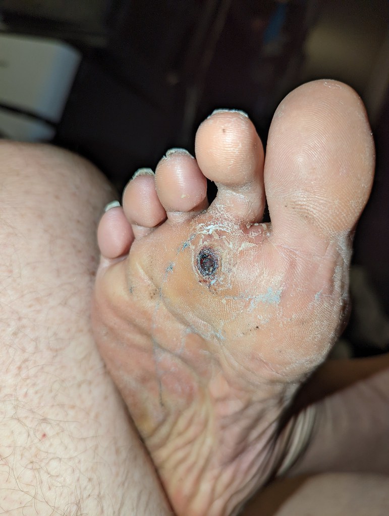
The triad shows up on boards as a contrast pair. The neuropathic ulcer is plantar over the metatarsal heads or heel, painless, on a warm dry foot with bounding pulses and claw toes: the problem is pressure the patient cannot feel. The ischemic ulcer is distal and lateral, painful with rest pain relieved by dependency, on a cool hairless foot with absent pulses and dependent rubor: the problem is flow the wound cannot get. The plan flips too: the neuropathic ulcer is offloaded, the ischemic ulcer is revascularized first. Hold the pair and half the foot questions on boards answer themselves.
Flip the switch between the two ulcers and hold the contrast.
THE HOT RED FOOT
The Hot Red Foot: Charcot Versus Cellulitis Versus Osteomyelitis Versus Gout
A hot, red, swollen foot in a neuropathic diabetic is four diseases wearing the same costume. The discriminator is three sentences long, and the wrong call costs the midfoot or the limb.
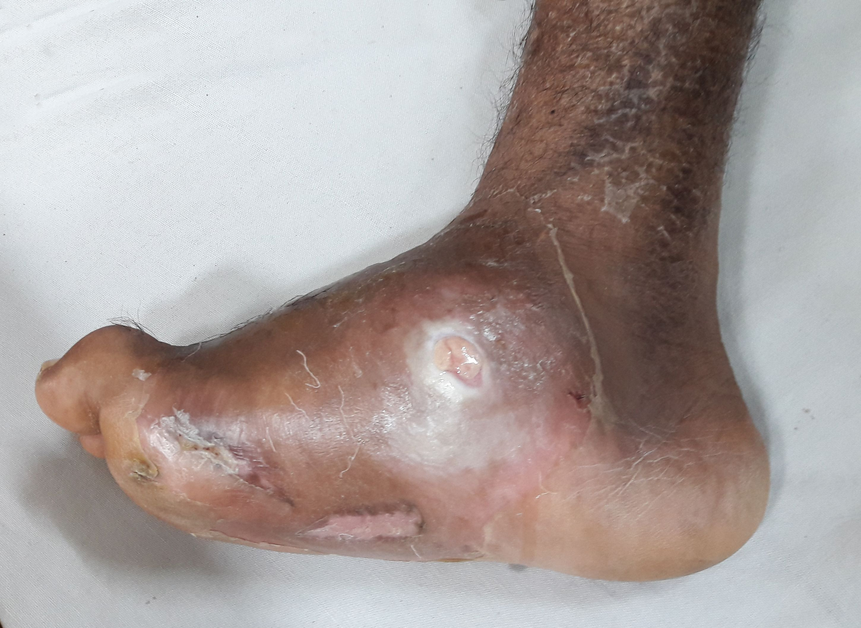
Acute Charcot arthropathy is the one that fools everyone: a neuropathic osteoarthropathy that is remarkably PAINLESS despite the swelling, with intact skin and bounding pulses, in a patient who is afebrile with normal white count and inflammatory markers. It is classically unilateral, centered on the midfoot at the Lisfranc area, and the affected foot runs more than 2 C warmer than the other side by skin temperature. The mechanism is unperceived stress fracture: loss of protective sensation plus autonomic hyperemia lets the bones break and remodel while the patient keeps walking. The treatment is offloading with a total contact cast and non-weight-bearing, never antibiotics.
Cellulitis is the tender red spread: erythema, warmth, swelling, and tenderness with fever and leukocytosis, entering through a fissure, web-space crack, or ulcer. Osteomyelitis is the bone infection behind an ulcer that probes to bone, with climbing ESR and CRP and marrow edema on MRI. Gout is the exquisitely tender monoarticular flare, classically podagra, with crystals in the joint. When the exam is ambiguous, MRI or a WBC-labeled scan separates Charcot from infection, and remember the rule that protects both: do not probe through intact skin, because a sterile Charcot foot does not need an iatrogenic doorway.
Answer the opening challenge in module form before the reveal.
A 58-year-old man with type 2 diabetes and established peripheral neuropathy has a hot, red, swollen right midfoot that has enlarged over 4 days. He denies injury and says the foot does not really hurt. Temperature is 37.0 C. The skin is intact with no ulcer, and the dorsalis pedis and posterior tibial pulses are bounding. WBC is 7,200, ESR 22, CRP 3. Which of the following is the most appropriate next step?
A. Offload with a total contact cast. Hot plus painless plus intact skin plus bounding pulses plus normal markers is acute Charcot arthropathy: the midfoot is actively breaking under unperceived load, and the treatment is mechanical, non-weight-bearing plus a total contact cast through the active stages. B. IV antibiotics. There is no infection: afebrile, normal WBC and inflammatory markers, intact skin; treating the Charcot foot with antibiotics lets the midfoot collapse while the patient keeps walking. C. Colchicine. Gout is exquisitely tender and monoarticular; this foot is painless and midfoot-centered. D. Debridement. There is no necrosis or abscess to debride; surgery in the acute stage is reserved for instability that fails offloading. Rule: the hot, painless, intact-skin foot is offloaded, never antibioticed.
DIABETIC NEUROPATHY
Diabetic Neuropathy: The Loss of Protection
Distal symmetric polyneuropathy is the engine of the diabetic foot: it silences pain in a stocking-glove pattern, dries the skin through autonomic failure, and claws the toes through motor loss. The 10 g monofilament is the board test for loss of protective sensation.
Distal symmetric polyneuropathy is length-dependent: the longest axons die first, so numbness starts in the toes and forefeet, climbs past the ankles in a stocking distribution, and reaches the hands (the glove) only when the feet are profoundly numb. Screening starts at diagnosis of type 2 diabetes and 5 years after onset of type 1 diabetes, then annually, because the patient who cannot feel the 10 g monofilament cannot feel the pressure that starts a painless ulcer. Tight glycemic control prevents or slows the neuropathy; it is the one intervention with real disease-modifying evidence.
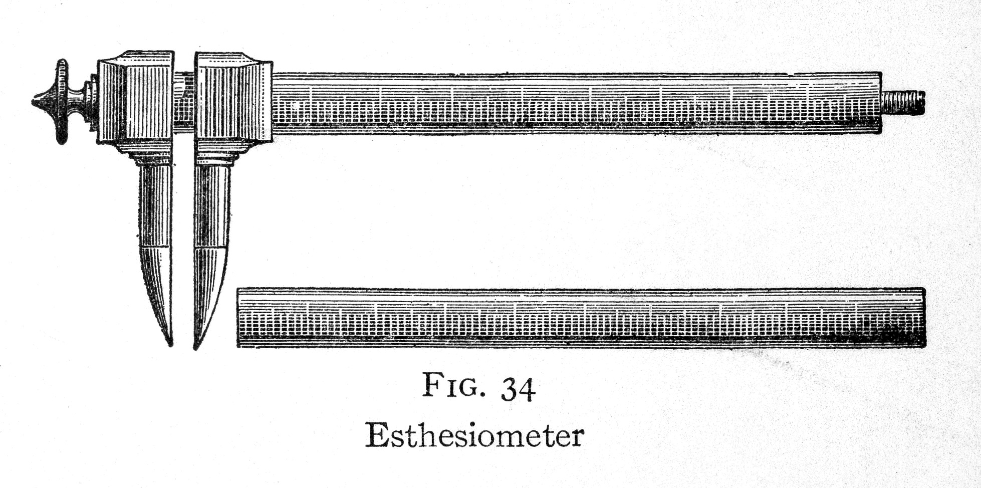
Three domains fail together. Sensory loss removes the warning: the 10 g Semmes-Weinstein monofilament pressed to the plantar great toe and metatarsal heads until it buckles is not felt, and a 128 Hz tuning fork on the great toe loses its buzz. Autonomic failure dries the skin: anhidrosis leaves the foot scaly and fissured, and the cracks between the toes are the entry wounds of cellulitis. Motor weakness claws the toes: intrinsic muscle loss unbalances the flexors and extensors, the metatarsal heads become prominent, and the fat pad migrates, concentrating weight on bone under skin. Tap the mapper below to watch the loss climb, and remember that stocking-glove loss in a diabetic is DSPN until proven otherwise.
Tap each body zone and read what has gone silent at that level.
Tap a zone to read what has gone silent there.
THE PREVENTION LOOP
The Prevention Loop: Six Moves That Keep the Amputation Clock Stopped
The annual monofilament, the daily inspection, the right shoe, the target HbA1c, the quit date, and the same-day call: each move is cheap, and each missed move is expensive.
The amputation clock starts long before the operating room: it starts with a pebble in the shoe that the patient cannot feel. Prevention is a loop, not a list. Every day, the patient inspects the feet, including between the toes and the soles, because a break caught at grade 0 or 1 is a wound managed in clinic, not an admission. Every year, the clinician repeats the 10 g monofilament exam to detect loss of protective sensation before the first ulcer forms. Always, the foot is protected: footwear with a wide toe box, cushioned insoles, and offloading that redistributes pressure off the metatarsal heads and deformities.
Two metabolic moves complete the loop. Glycemic control keeps the HbA1c at target so the wound can heal and the nerves can survive; smoking cessation protects the microvascular and macrovascular supply that the ischemic limb cannot replace. And the loop closes with the rule that saves limbs: the moment skin breaks, the patient seeks care the same day. Fifteen percent of people with diabetes develop a foot ulcer, and 85 percent of amputations are preceded by one; the loop works because every ulcer that ever amputated a foot started as a small, catchable event.
Tap the six moves in the order the prevention loop runs.
Tap the six moves in the order the prevention loop runs.
THE ISCHEMIC FOOT
The Ischemic Foot: PAD and the Amputation Second Hand
Neuropathy starts the ulcer; peripheral arterial disease keeps it from healing. Claudication is the early voice, dependent rubor with elevation pallor is the bedside sign of critical ischemia, and the ABI carries one diabetic trap: calcified arteries can make the number lie high.
Claudication is the first voice of PAD: reproducible calf or thigh cramping on walking that resolves within minutes of standing still, because the muscles outrun the blood supply. The exam adds the pair boards love: elevation pallor (the foot blanches when raised) with dependent rubor (it flushes red when dangled), on a cool, hairless foot with weak or absent pedal pulses. The ankle brachial index reads the flow: below 0.9 confirms PAD, 0.9 to 1.3 is the healthy window, and above 1.3 is the calcified-vessel trap. Monckeberg medial calcification makes the tibial arteries stiff and noncompressible, so the number reads falsely high while the limb is dying; when the ABI is falsely elevated or the vessel is noncompressible, order toe pressures, and a toe brachial index below about 0.7 confirms ischemia. Screen for PAD annually with pulse exam and ABI.
Critical limb ischemia is the amputation second hand: rest pain in the forefoot at night relieved by hanging the leg out of bed, a nonhealing ulcer (no progress after 2 weeks of appropriate care), or gangrene. The plan is urgent vascular referral for revascularization, because an ischemic limb cannot heal any wound; debridement and antibiotics support the healing but never replace the flow.
Flag the ABI readings that demand action, then read the cumulative verdict.
Flagged 0
THE WAGNER LADDER AND THE INFECTION WORKUP
The Wagner Ladder and the Infection Workup
Grade the wound, then run the infection workup in order: probe to bone, ESR and CRP, plain film, MRI with contrast, bone biopsy for culture, and 4 to 6 weeks of therapy.
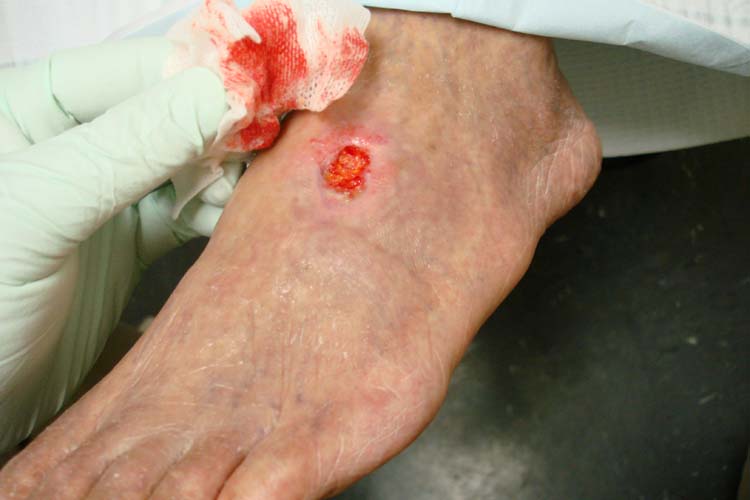
The Wagner classification turns the diabetic foot into a numbered ladder. Grade 0 is intact skin with a pre-ulcerative lesion or healed ulcer; 1 is a superficial ulcer; 2 is a deep ulcer to tendon or capsule; 3 is a deep ulcer with abscess or osteomyelitis; 4 is forefoot gangrene; 5 is whole-foot gangrene. The University of Texas classification adds the second axis boards now reward: each depth grade (0 to 3) is modified by infection and by ischemia, so a shallow ulcer on an ischemic limb is recognized as the high-risk wound it is.
Infection workup runs a fixed order. Probe to bone comes first: a sterile probe gently passed into the ulcer that touches bone is osteomyelitis until proven otherwise, with a positive likelihood ratio around 87 percent, and it outranks a normal plain film. Never probe through intact skin: the probe is for ulcers and sinus tracts, and a sterile Charcot foot does not need an iatrogenic doorway. ESR and CRP both rise in osteomyelitis; normal markers make it unlikely, and acute Charcot keeps them normal. Plain film comes next: osteopenia, periosteal reaction, and lucency, remembering that osteomyelitis needs about 50 percent bone loss before the film turns, plus gas, foreign bodies, and Charcot fragmentation. MRI with contrast is the gold standard: marrow edema, abscess, and the sinus tract that reaches bone, and it separates osteomyelitis from Charcot marrow edema. Bone biopsy is the definitive culture, taken through clean skin or at surgery, and guides the final regimen.
Therapy is polymicrobial by design: Staph aureus plus anaerobes plus gram negatives. Severe infection gets IV piperacillin-tazobactam (add vancomycin when MRSA is suspected) with surgical debridement; mild infection gets oral amoxicillin-clavulanate, or clindamycin plus a fluoroquinolone; osteomyelitis runs 4 to 6 weeks of IV or highly bioavailable oral therapy guided by bone culture, and amputation is reserved for extensive, unreconstructable disease.
Commit to the fork in the infection workup, then read the branch.
A 64-year-old man with diabetes has a draining plantar ulcer and a hot foot. Where does the workup fork?
THE EICHENHOLTZ CLOCK
The Eichenholtz Clock and the Four-Way Discriminator
The Charcot foot runs a clock of its own: stage 0 acute inflammation, stage 1 fragmentation, stage 2 coalescence, stage 3 reconstruction into the rocker-bottom. The treatment is mechanical, and the discriminator table is the one screen that stops the wrong antibiotic.
The Eichenholtz classification stages the Charcot foot as it evolves. Stage 0 is the acute inflammatory foot: hot, swollen, red, with intact skin and normal plain films, the stage the boards show as the hot painless foot, where offloading must start NOW. Stage 1 (development) is fragmentation: bone debris, fracture, and joint subluxation, the shattered-glass look at the midfoot. Stage 2 (coalescence) is new bone formation with sclerosis and fusion. Stage 3 (reconstruction) is remodeling into the stable rocker-bottom deformity, where the plantar skin sits over bone and ulcerates. Treatment across the active stages is non-weight-bearing plus a total contact cast for 8 to 12 or more weeks, with serial skin temperature checks until the foot cools; bisphosphonates are not the board answer (controversial, without strong evidence), and surgery is reserved for deformity, instability, or ulceration that fails offloading.
Run the four-way discriminator in order. Charcot: hot, painless, intact skin, bounding pulses, normal markers; offload. Cellulitis: tender, erythematous, febrile, leukocytotic, with an entry wound; antibiotics. Osteomyelitis: ulcer that probes to bone, climbing ESR and CRP, marrow edema with a sinus tract; debride and treat 4 to 6 weeks. Gout: exquisitely tender monoarthritis with crystals; treat the flare. The table below is the one screen to hold on the wards and on boards.
Tap each letter of the Wagner ladder mnemonic.
SSDD FF
SSkin intact: grade 0, the pre-ulcerative or healed foot, offloading footwear and screening
SSuperficial ulcer: grade 1, full-thickness but not to tendon, offloading and wound care
DDeep to tendon or capsule: grade 2, no abscess, no osteomyelitis, debridement and offloading
DDeep with bone: grade 3, abscess or osteomyelitis, probe to bone, MRI, debridement and 4 to 6 weeks
FForefoot gangrene: grade 4, revascularize first, then amputate the nonviable tissue
FFull foot gangrene: grade 5, below-knee amputation to save the patient
Open each stage and regimen on the Eichenholtz clock.
Hot, swollen, red, with intact skin and normal plain films: the stage the boards show as the hot painless foot. Offloading must start NOW, before the films ever turn; the clinical diagnosis outranks the normal radiograph.
Plain films show bone debris, fracture, and joint subluxation: the shattered-glass look at the midfoot, classically at the Lisfranc area. The foot is still actively collapsing under unperceived load.
New bone formation with sclerosis and fusion of fragments as the foot begins to consolidate. Offloading continues; the foot is still vulnerable to recurrence of activity.
Remodeling into the stable rocker-bottom deformity: the arch is collapsed, weight concentrates on the plantar apex, and the insensate skin over the prominence ulcerates, opening the road to osteomyelitis.
Non-weight-bearing plus a total contact cast for 8 to 12 or more weeks across the active stages, with serial skin temperature checks until the foot cools. Bisphosphonates are controversial, not the board answer. Surgery (arthrodesis) is reserved for deformity, instability, or ulceration over the prominence that fails offloading.
The flora is polymicrobial: Staph aureus plus anaerobes plus gram negatives. Severe infection: IV piperacillin-tazobactam, adding vancomycin when MRSA is suspected, with surgical debridement. Mild infection: oral amoxicillin-clavulanate, or clindamycin plus a fluoroquinolone. Osteomyelitis: 4 to 6 weeks of IV or highly bioavailable oral therapy guided by bone culture; the bone must be debrided, not just soaked.
FeatureCharcotCellulitisOsteomyelitisGout
TendernessPainlessTenderTender at the ulcerExquisite
SkinIntactErythema, entry woundUlcer or sinus tractIntact
PulsesBoundingVariableVariableVariable
WBC, ESR, CRPNormalElevatedElevatedVariable
FeverAbsentOften presentVariableAbsent
The testPlain film: fragmentation, subluxation; skin temp asymmetry over 2 CClinical plus WBCProbe to bone, MRI marrow edema with sinus tractCrystals on synovial fluid
The planTotal contact cast, offloadAntibioticsDebride, 4 to 6 weeks of antibioticsNSAID, colchicine, or steroid
Prove it
Walkthrough: the amputation clock in seven cases
Original practice scenarios, one at a time. Choose an answer, then open any option to work its reasoning.
Clinical walkthrough
