
Choose an answer, then open any option to work its reasoning.
MSK · Rheumatology
Gout vs Pseudogout: The Crystal Split
One drop of joint fluid, two crossed polarizers, and the whole crystal question resolves: needle-shaped crystals that flash yellow when parallel are monosodium urate, rhomboid bricks that flash blue are calcium pyrophosphate, and the destroyed shoulder with nothing under the scope is basic calcium phosphate. Learn to read the polarized field, treat the flare within hours, and sequence urate lowering so the crystal never comes back.
What this page makes you able to do
- Read a compensated polarized-light readout: shape plus birefringence sign names the crystal
- Treat the acute flare within hours: NSAID, colchicine, or glucocorticoid, dosed to the kidney
- Sequence urate lowering: after the flare settles, with colchicine prophylaxis, to a target below 6 mg/dL
- Rule sepsis out of every hot joint before the crystal wins the argument
- Dr. Fatima Ali, DOPsychiatry residentPrimary reviewer
Last reviewed
The pearl
One drop of joint fluid, two crossed polarizers: needle-shaped and yellow when parallel is monosodium urate (gout); rhomboid and blue when parallel is calcium pyrophosphate (pseudogout). The aspirate names the crystal, and the crystal names the plan: anti-inflammatory firepower within hours for the flare, urate lowering after it settles, and a gram stain on every tap because crystals never clear a septic joint.
Prove it
Opening question
Answer before you read anything, then hold the polarized-field logic through every section.
A 64-year-old man comes to the office because of 12 hours of excruciating pain and swelling of the left big toe that woke him from sleep. He drank beer and ate steak the night before, and he has hypertension treated with hydrochlorothiazide. Temperature is 37.6 C. Examination shows a hot, red, swollen first metatarsophalangeal joint. Aspiration returns cloudy fluid, and compensated polarized microscopy shows needle-shaped crystals that are yellow when parallel to the compensator axis and blue when perpendicular.Which of the following is the most likely diagnosis?
- Why this is rightNeedle-shaped crystals that are yellow when parallel and blue when perpendicular are monosodium urate with strong negative birefringence: that is gout. The first MTP joint with acute podagra, the beer, and the thiazide diuretic complete the classic underexcretion package. Rule: podagra plus negatively birefringent needles equals gout, and the thiazide explains the underexcretion.
- Why this failsCPPD crystals are rhomboid and weakly positively birefringent: blue when parallel, yellow when perpendicular. The colors here are reversed, and CPPD flares classically in the knee or wrist of an older adult, not the first MTP. Rule: rhomboid and positive belongs to CPPD; needles and negative belong to gout.
- Why this failsA hot swollen joint can be septic, which is exactly why the fluid was aspirated and sent for gram stain and culture. The crystals here are diagnostic of gout, and the temperature is only mildly elevated; still, the culture is the safety net. Rule: rule out sepsis in every hot joint, but the crystals name the disease.
- Why this failsCellulitis lives in the skin and soft tissue, not inside the joint: joint aspiration would not return cloudy, crystal-laden fluid. The effusion and marked tenderness localize to the MTP joint itself. Rule: cellulitis does not tap out joint fluid with urate needles.
Work the reasoning
Serum urate is frequently normal during an acute flare and elevated in people who never flare. The crystal in the fluid is the diagnosis; the serum level is a chronic-disease number.
Both push urate up. Thiazides reduce renal urate clearance (underexcretion, the 90 percent bucket), while beer adds purines and its ethanol generates lactate that competes with urate for tubular secretion.
A hot, red first MTP can be gout, pseudogout, or infection. The aspirate rules infection in or out and names the crystal; gram stain and culture ride along on every tap.
The answer is A: acute gout. Needle-shaped crystals with negative birefringence (yellow parallel, blue perpendicular) are monosodium urate, and podagra plus a thiazide is the classic underexcretion package.
THE CRYSTAL SPLIT
The Crystal Split: Shape and Birefringence
One drop of joint fluid, two crossed polarizers, and the whole question resolves. Shape names the crystal family; the color swap under the compensator names the disease.
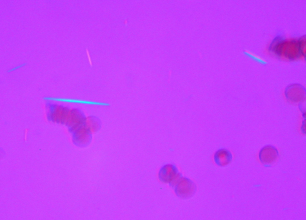
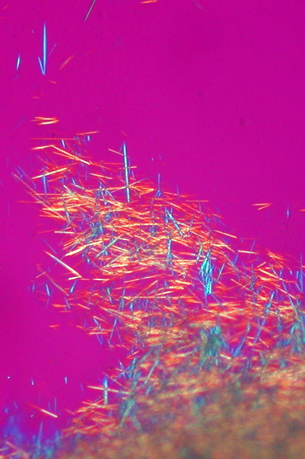
Board stems hand you the readout in one sentence, so learn to read it the way they write it. Needle-shaped crystals that are yellow when parallel to the compensator axis and blue when perpendicular are monosodium urate (MSU): strong negative birefringence, and that is gout. Rhomboid crystals that are blue when parallel and yellow when perpendicular are calcium pyrophosphate dihydrate (CPPD): weak positive birefringence, and that is pseudogout. The two crystals answer the compensator in opposite directions, and so do the two diseases. Shape plus sign is the entire ID.
The compensator is a first-order red plate that adds a known retardation to the field. A crystal aligned with its slow axis shifts one color; a crystal perpendicular shifts the other. That is why the swap, not the brightness, is the test. The memory hooks boards reward: Negative equals Needle for MSU, and the blue P's for CPPD: blue when parallel, positive birefringence, pyrophosphate, pseudogout. A third door waits for the empty field: basic calcium phosphate (BCP) clumps are too small to polarize, so the destroyed shoulder reads crystal-poor under routine light.
And one number never decides. Serum urate is frequently normal during an acute gout flare, and most people with an elevated urate never flare. The crystal is the diagnosis; the serum level is the chronic-disease number that guides prevention, not the flare call.
Flip between the two crystals and lock the split.
THE JOINT PATTERN
The Joint Pattern: Podagra vs the Knee and Wrist
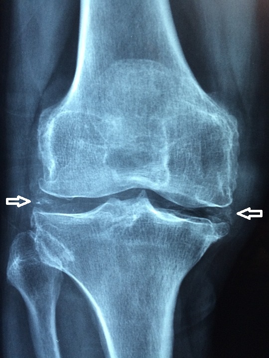
Gout owns the first metatarsophalangeal joint; pseudogout owns the knee and wrist of the older adult. The joint points, the fluid proves, and the film backs the call.
Gout picks the first metatarsophalangeal joint: podagra, the sudden hot red toe that wakes a middle-aged man from sleep, is the classic first attack, and roughly half of first flares land there. Pseudogout picks the knee and wrist of the older adult, often triggered by an illness, surgery, or trauma that shifts calcium and phosphate. The joint pattern is a strong clue, but the fluid is the answer: the same needle-shaped negative crystals can flare in the knee, and CPPD can rarely hit the first MTP, so the polarized field always settles the argument.
Chondrocalcinosis is the pseudogout film: linear calcification of cartilage, classically the knee menisci, the wrist triangular fibrocartilage, and the symphysis pubis. CPPD also wears chronic costumes. The pseudo-osteoarthritis pattern shows bilateral knees, wrists, and MCPs with osteophytes and joint space loss that keep flaring acutely; the pseudo-rheumatoid pattern shows symmetric small-joint morning stiffness with negative rheumatoid factor and anti-CCP. Hemochromatosis arthropathy targets the MCPs and wrists and usually carries CPPD with it, which is one reason every new CPPD diagnosis triggers a metabolic screen.
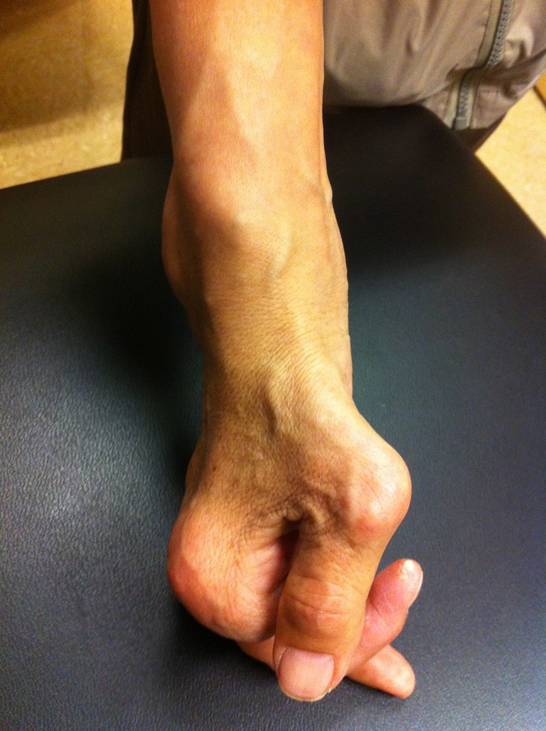
Gout has its own chronic film: erosions with overhanging edges in long-standing disease, tophi at the helix of the ear and olecranon bursa, and uric acid nephrolithiasis. The takeaway for the boards: location plus film narrows the differential, and the aspirate closes it.
Commit to the joint pattern before the crystal readout arrives.
A 71-year-old woman has had three episodes of acute right knee swelling over the past year, each resolving within days. A radiograph shows linear calcification along the knee menisci. Which of the following findings would most strongly support pseudogout rather than gout?
A. Rhomboid, blue when parallel. Rhomboid shape plus weak positive birefringence (blue parallel, yellow perpendicular) is the CPPD signature, and the knee with meniscal chondrocalcinosis in an older adult is the classic frame. B. Needle-shaped negative crystals. That is monosodium urate: gout, not pseudogout. C. A normal urate during a flare. Serum urate is frequently normal during an acute gout flare, so it neither rules gout in nor out and says nothing about CPPD. D. Colchicine response. Colchicine suppresses neutrophil-driven crystal inflammation for MSU and CPPD alike; response to therapy does not discriminate between the crystals. Rule: the crystal shape and the color swap are the only true discriminators.
THE POLARIZED-FIELD SIMULATOR
The Polarized-Field Simulator: Yellow Parallel, Blue Parallel
Rotate the mental compensator on three fields: MSU needles, CPPD rhomboids, and the empty field that still means crystal disease.
Compensated polarized light adds a known retardation through a first-order red plate, so the background reads magenta and the crystal colors carry the information. Negative birefringence turns the crystal yellow when its long axis is parallel to the slow axis and blue when perpendicular: that is monosodium urate. Positive birefringence does the reverse, blue parallel and yellow perpendicular: that is CPPD. The sign, not the presence of color, is the ID, and the board stem almost always quotes the colors verbatim.
The third field is the trap. The elderly woman with a swollen, destroyed shoulder and a rotator cuff tear who taps out bloody, low-cell-count fluid with no visible crystals still has crystal disease: basic calcium phosphate (Milwaukee shoulder). BCP clumps are sub-microscopic and non-birefringent, so the fluid reads crystal-poor under routine light; alizarin red staining or electron microscopy finds them. BCP activates synovial lining cells to release collagenase, which is how the cuff and cartilage get digested.
Whatever the field shows, the safety net never moves: crystals and infection can coexist, so every hot joint gets a gram stain and culture, and fever plus a hot joint is treated as septic until proven otherwise.
Tap each region of the field and read what the optics mean.
Tap a region of the field.
THE FLARE TIMELINE
The Flare Timeline: Supersaturation to Self-Limited Fire
The flare is a neutrophil response to crystals, not to the urate number. Learn why it ignites, why it burns out, and why colchicine only works early.
When serum urate stays above the solubility point, monosodium urate crystallizes and deposits in cartilage and synovium. Most of the time the deposits sit quietly. Then a trigger arrives: cold, trauma, alcohol, illness, surgery, or a thiazide-driven rise in urate, and the deposits shed crystals into the joint space. Neutrophils chase and phagocytose them, and the phagocytosis itself releases IL-1 and chemoattractants that recruit more neutrophils: an amplification loop that is the flare.
The flare is self-limited because shedding stops and anti-inflammatory mediators rise even without treatment, which is why an attack of podagra burns for days and then quiets. But the course is compressed by early therapy: colchicine binds tubulin and blocks microtubule polymerization, so neutrophils cannot polarize, migrate, or efficiently take up crystals. Given within the first 24 to 48 hours it cuts the amplification loop before it commits; given late, the fire is already built. That timing is why the acute treatment rule says hours, not days.
The same physics explains the initiation flare of chronic therapy: as serum urate falls, surface crystals dissolve and can re-seed the joint. Every urate-lowering start therefore rides with flare prophylaxis for the first months, and repeated untreated flares accumulate tophi: MSU mats wrapped in giant cells and fibrosis at the ear, olecranon, and joints.
Put the flare timeline in order.
THE FLARE TREATMENT
The Flare Treatment: NSAID, Colchicine, or Steroid Within Hours
The acute flare is treated with anti-inflammatory firepower, never with urate lowering. The drug, the dose, and the timing all change with the kidney and the pill list.
An acute crystal flare is treated with an NSAID, colchicine, or a glucocorticoid, started within 24 to 36 hours of onset. The NSAID (indomethacin is the classic) is first line in the healthy adult. Colchicine is the neutrophil weapon: 1.2 mg followed by 0.6 mg one hour later for an acute attack, and it works best early. A glucocorticoid, oral or intra-articular, is the answer when both NSAIDs and colchicine are out. What you do not do is start urate lowering mid-flare without cover: the falling urate mobilizes crystals and hands the patient a second attack.
The kidney changes every choice. NSAIDs are avoided in chronic kidney disease, especially with an eGFR below 30, because they cut renal perfusion. Colchicine is dose-reduced in CKD and whenever a CYP3A4 or P-glycoprotein inhibitor is on board: clarithromycin, ketoconazole, cyclosporine, and grapefruit juice can push colchicine into diarrhea, marrow suppression, and myopathy. When neither NSAID nor colchicine is safe, the glucocorticoid route takes over.
And the aspiration is part of the treatment plan, not a formality: crystals and infection can coexist, so the same tap that names the crystal sends the gram stain and culture, and fever plus a hot joint is managed as septic until proven otherwise.
Tick every situation that forces a regimen change off the healthy-adult NSAID default.
Checked 0
THE URATE-LOWERING CHAPTER
The Urate-Lowering Chapter: After the Flare Settles
Urate lowering is a chronic plan with a hard sequence: treat the flare, start after it settles, cover the first months, and screen the patient who can crash.
The indications are disease, not numbers: recurrent flares (generally two or more per year), tophi, or urate nephrolithiasis. Asymptomatic hyperuricemia, however high, is counseled about lifestyle and reassessed, not medicated. When the door opens, allopurinol is first line: a xanthine oxidase inhibitor started low at 100 mg daily, titrated every two to four weeks to a target serum urate below 6 mg/dL, and lower with tophi so the deposits dissolve.
The timing is the classic board trap. Urate lowering is started after the flare settles, never as a first move in an untreated attack, and always with low-dose colchicine prophylaxis for the first three to six months because falling urate mobilizes crystals and triggers initiation flares. Modern guidance permits starting during a flare when the flare is already being treated and prophylaxis is on board; the default answer on boards remains: cool the fire first, then drain the swamp, with a fire extinguisher in hand.
Before the first tablet, screen the patient who can crash: HLA-B*5801 testing before allopurinol in high-risk populations (Han Chinese, Thai, Korean, and people of African descent), because the allele predicts allopurinol hypersensitivity syndrome and Stevens-Johnson syndrome. The alternative after hypersensitivity is febuxostat, a non-purine xanthine oxidase inhibitor, which is also the answer after allopurinol DRESS. Probenecid is the uricosuric alternative that needs functioning tubules and no uric acid stones; pegloticase is the PEGylated uricase reserved for severe refractory tophaceous disease; rasburicase belongs to tumor lysis syndrome alone, and only after a G6PD check.
One interaction completes the chapter: xanthine oxidase inhibitors block the enzyme that inactivates azathioprine and 6-mercaptopurine. Allopurinol with azathioprine requires cutting the azathioprine dose by roughly 50 to 75 percent with close CBC monitoring; febuxostat is contraindicated with azathioprine, 6-mercaptopurine, or thioguanine outright.
Send each hot joint down the right door.
Which door fits which hot joint?
Open each cause and hold the metabolic story.
The kidney is the bottleneck: thiazide and loop diuretics, cyclosporine, niacin, pyrazinamide, and low-dose aspirin reduce urate clearance; chronic kidney disease and lead nephropathy (saturnine gout) do the same. Alcohol fits here too, because its lactate competes with urate for tubular secretion. The board clue is the drug list in the first sentence.
Purine metabolism runs hot: Lesch-Nyhan syndrome (X-linked HGPRT deficiency, the self-mutilating boy with orange diaper crystals), tumor lysis syndrome, myeloproliferative disease, hemolytic anemias, and psoriasis. A 24-hour urine uric acid above 800 mg flags the overproduction bucket.
Calcium pyrophosphate deposition follows a metabolic address: hemochromatosis (ferritin and transferrin saturation), hyperparathyroidism (flares can follow parathyroidectomy as calcium drops), hypothyroidism (TSH), hypomagnesemia (cisplatin, diuretics, proton pump inhibitors), and hypophosphatasia. Find and treat the association and the recurrent flares may settle.
Uric acid stones form in concentrated, acidic urine. The plan is allopurinol to cut urate production, fluids to dilute, and alkalinization (citrate or bicarbonate) to keep urate soluble. Probenecid is forbidden in a uric acid stone former because uricosuria feeds the stone machine.
Xanthine oxidase inactivates azathioprine and 6-mercaptopurine. Allopurinol with azathioprine is the studied protocol: cut the azathioprine by about 50 to 75 percent and monitor the CBC. Febuxostat is contraindicated with azathioprine, 6-mercaptopurine, or thioguanine; switching to it does not dodge the interaction, it steps into a contraindication.
THE ONE-SCREEN DISCRIMINATOR
The One-Screen Discriminator: Three Hot Joints, Three Doors
Every hot joint on boards is one of three doors: gout, pseudogout, or septic arthritis. Run the doors in order and the fluid makes the call.
Run the doors in order. Hot first MTP with needle-shaped negative crystals: gout; treat the flare now and sequence urate lowering later. Hot knee or wrist in an older adult with rhomboid positive crystals and chondrocalcinosis: pseudogout; treat the flare and screen the metabolic guest list. Any hot joint with fever, a prosthetic joint, or a host who cannot fight infection: septic until proven otherwise; aspirate, stain, culture, and start empiric antibiotics. The table below is the whole page on one screen: the crystals, the joints, the films, and the first move.
| Feature | Gout (MSU) | Pseudogout (CPPD) | Septic arthritis |
|---|---|---|---|
| Crystal ID | Needle, strong negative: yellow parallel | Rhomboid, weak positive: blue parallel | None; gram stain and culture carry the diagnosis |
| Classic joint | First MTP (podagra) | Knee and wrist | Knee; any hot joint |
| Film | Erosions with overhanging edges; tophi | Chondrocalcinosis of menisci and triangular fibrocartilage | Joint space loss late; MRI marrow edema early |
| Fever | Usually absent | Usually absent | Common and often high |
| First move | NSAID, colchicine, or steroid within 24 to 36 hours | Same anti-inflammatory firepower; no urate lowering | Aspirate, gram stain, culture, empiric IV antibiotics |
The elimination rule closes the page: crystals diagnose crystal disease, organisms diagnose infection, and both can be true in one joint. A positive crystal reading never cancels the culture, and fever plus a hot joint is sepsis until the fluid says otherwise.
Tap each letter of the blue P's, then the two N's that balance them.
BPPP NN
BBlue when parallel to the compensator axis
PPositive birefringence (weak)
PPyrophosphate: calcium pyrophosphate dihydrate
PPseudogout: the flare the rhomboids cause
NNegative equals Needle: MSU is needle-shaped with strong negative birefringence (yellow parallel)
NNormal urate never rules out gout: the crystal is the diagnosis
Prove it
Walkthrough: the crystal ID under pressure
Original practice scenarios, one at a time. Choose an answer, then open any option to work its reasoning.
Clinical walkthrough
