
Choose an answer, then open any option to work its reasoning.
MSK · Trauma
Fractures and Trauma: Eponyms, Growth Plates, and the Salvage Clock
Every fracture is a mechanism plus a pattern plus a complication risk. The dinner fork tells you the fall, the snuffbox tells you the scaphoid, the child who will not move the arm tells you the supracondylar, and the leg that hurts too much tells you the compartment. Name the fracture, then name the complication before it names you: AVN, nonunion, fat embolism, and the contracture that follows the missed compartment.
What this page makes you able to do
- Name the eponym from the mechanism and the lateral film: Colles, Smith, Barton, Monteggia, Galeazzi
- Run the scaphoid rule and the clavicle triggers: thumb spica, repeat imaging, sling first
- Sort the growth plate injuries and the fractures that accuse: Salter-Harris, supracondylar, buckle, abuse patterns
- Race the salvage clock: open fracture antibiotics and tetanus, compartment, fat embolism, and the union outcomes
- Dr. Fatima Ali, DOPsychiatry residentPrimary reviewer
Last reviewed
The pearl
Snuffbox tenderness after a fall on an outstretched hand plus a normal three-view radiograph is a scaphoid fracture until proven otherwise: the wrist goes into a thumb spica cast and the radiograph is repeated in 7 to 14 days, never reassurance. The blood enters the scaphoid distally, so a high fracture can kill the proximal pole while the first film still looks clean.
Prove it
Opening question
Answer before you read anything, then carry the mechanism-first rule through every section.
A 24-year-old man comes to the emergency department after falling onto his outstretched left hand while playing basketball. He reports wrist pain and tenderness directly over the anatomic snuffbox but no deformity. Temperature is 37.1 C. Examination shows snuffbox tenderness with no swelling or deformity. A three-view radiograph of the wrist shows no fracture.Which of the following is the most appropriate next step?
- Why this is rightSnuffbox tenderness after a fall on an outstretched hand is scaphoid until proven otherwise, and the first radiograph misses many scaphoid fractures. The thumb spica immobilizes the thumb, and the repeat radiograph in 7 to 14 days catches what the first film hid. Rule: snuffbox tenderness plus a negative film equals thumb spica plus repeat imaging, never reassurance.
- Why this failsThe scaphoid hides on the first film, and reassurance hands a missed fracture weeks to nonunion. The trap is trusting the normal radiograph. Rule: a negative film never clears snuffbox tenderness.
- Why this failsA soft wrap does not immobilize the thumb, and the scaphoid fracture, if present, moves and fails to heal. Analgesia is not treatment for a possibly broken carpal bone. Rule: the thumb must be immobilized, which a wrap cannot do.
- Why this failsAdvanced imaging can be used when the diagnosis matters acutely, but the standard first move is immobilization plus a repeat radiograph. A bone scan does not replace the thumb spica. Rule: immobilize first; imaging decides, it does not treat.
- Why this failsA scaphoid fracture must be immobilized with the thumb included, because the thumb moves the scaphoid through its ligaments. A short arm cast that leaves the thumb free fails the one job the cast has. Rule: no thumb, no treatment.
Work the reasoning
The scaphoid is the most commonly fractured carpal bone, and its fracture line can be invisible on the initial radiograph. Snuffbox tenderness is the clinical diagnosis; the film only confirms it later.
The scaphoid spans two rows of carpal bones, and thumb motion pulls it through ligamentous attachments. Only a thumb spica holds it still enough to heal.
Blood enters the scaphoid distally, so the proximal pole is fed retrograde. A missed fracture can nonunion, and the proximal fragment can die (AVN). The repeat radiograph at 7 to 14 days is the safety net.
The answer is A: immobilize in a thumb spica and repeat the radiograph in 7 to 14 days. A missed scaphoid can nonunion and the proximal fragment can die; never trust the normal first film.
THE EPONYM GALLERY
The Eponym Gallery: Mechanism Names the Fracture
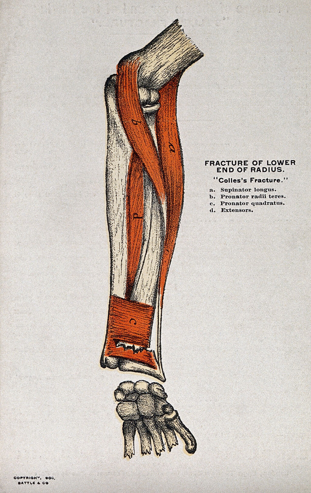
The board gives you the mechanism and the film; the eponym is the shortcut. The lateral view of the wrist is the whole answer: dorsal tilt is Colles, volar tilt is Smith, a rim with a slipped carpus is Barton. Then the forearm pairs: proximal ulna plus radial head is Monteggia, distal radius plus DRUJ is Galeazzi.
The direction of displacement on the lateral wrist film is the discriminator, and the reduction follows the direction. A Colles fracture is the classic fragility injury: a fall on an outstretched hand with the wrist extended gives dorsal angulation of the distal radius fragment, the dinner fork deformity, with the carpus in place. It reduces in flexion with ulnar deviation; surgery is reserved for comminution, articular step-off, or loss of reduction. A Smith fracture is the mirror: volar angulation after a fall on the dorsum of a flexed wrist, the garden spade, and it reduces in extension, never flexion, because flexing a Smith reproduces the deformity; unstable patterns need volar plate fixation. A Barton fracture is intra-articular: a triangular rim fragment with the carpus subluxating with it, unstable by definition and fixed surgically, because a cast cannot hold a joint surface.
The forearm pairs work the same way, one joint higher or lower. Monteggia is a proximal ulna fracture with the radial head dislocated, classically anteriorly; the dislocation is the part that gets missed, so every ulna fracture gets a radiocapitellar line check on the lateral film. Galeazzi is the reverse: a distal radius shaft fracture with a distal radioulnar joint dislocation; fix the radius and the length holds the DRUJ reduced. The mnemonic that sticks: M for the middle of the arm, G for the far side at the wrist.
Flip between the three lateral wrist films.
THE SNUFFBOX AND THE CLAVICLE
The Snuffbox and the Clavicle: Small Bones, Big Complications
The scaphoid is the carpal bone that hides fractures on the first film and pays for the delay with a dead proximal pole. The clavicle is the most fractured bone in the body, and its middle third is the classic fall-on-the-shoulder injury.
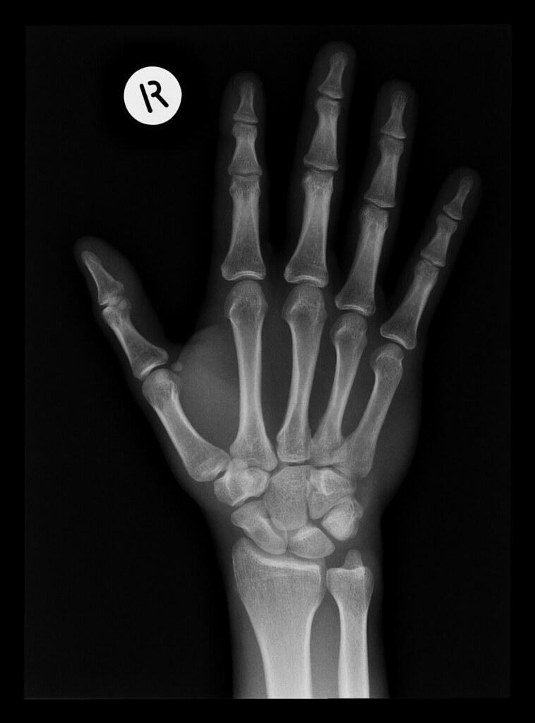
The scaphoid is the most commonly fractured carpal bone, and its blood supply is the whole story: branches of the radial artery enter distally, so the proximal pole is fed retrograde. A fracture across the waist, and more so across the proximal pole, cuts that retrograde supply and the proximal fragment can die. The exam clue is snuffbox tenderness, and the rule is absolute: tenderness plus a normal three-view radiograph equals thumb spica immobilization and a repeat radiograph in 7 to 14 days, never reassurance. The delayed film can show the fracture line, and a dense sclerotic proximal pole is the mark of dead bone, the endpoint of the missed diagnosis. The clavicle, by contrast, heals: middle-third fractures from a fall onto the shoulder are managed with a sling, and surgery waits for the named triggers (open fracture, skin tenting, neurovascular compromise, major shortening). The neonate with a clavicle fracture after a difficult delivery shows arm pseudoparalysis and a callus lump within days; it heals and reassures.
Commit to the scaphoid rule.
A 26-year-old man comes to the emergency department after falling onto his outstretched hand while playing basketball. He reports wrist pain and tenderness over the anatomic snuffbox. Temperature is 37.1 C. Examination shows snuffbox tenderness with no deformity. A three-view radiograph of the wrist shows no fracture. Which of the following is the most appropriate next step?
A. Thumb spica and repeat imaging. Snuffbox tenderness after a fall on an outstretched hand is scaphoid until proven otherwise, and the initial radiograph misses many scaphoid fractures. The thumb spica immobilizes the thumb, and the radiograph is repeated in 7 to 14 days. B. Reassurance. A negative first film never clears snuffbox tenderness; a missed scaphoid can nonunion and the proximal pole can die. C. NSAID and wrap. A soft wrap does not immobilize the thumb, and the fracture, if present, moves and fails to heal. D. Short arm cast. The thumb must be immobilized, because thumb motion pulls the scaphoid through its ligaments. Rule: snuffbox tenderness plus a negative film equals thumb spica plus repeat imaging, never reassurance.
Tap each zone of the scaphoid and read the AVN risk.
Tap a zone.
KIDS’ BONES
Kids’ Bones: Growth Plates, Supracondylar, and the Fractures That Accuse
Children heal fast and break differently: greenstick and buckle instead of complete breaks, growth plate injuries that can stop a bone from growing, a supracondylar humerus that threatens the forearm, and fracture patterns that name the abuser.
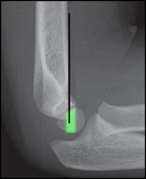
The child’s bone bends before it breaks. A buckle (torus) fracture is a crumpled cortex from compression; a greenstick fracture breaks one cortex and bends the other, like a fresh branch; both are incomplete and heal quickly. Growth plate injuries are graded by the Salter-Harris system, and the mnemonic writes itself: Straight through the physis, Above (into the metaphysis), Lower (into the epiphysis), Through both, ERasure (crush). The crush injury is the one that silently stops growth, because it destroys the germinal layer of the physis. The supracondylar humerus fracture is the elbow emergency of childhood: the classic test is the anterior humeral line, which must pass through the middle third of the capitellum on the lateral film. The anterior interosseous nerve (a branch of the median nerve) is the classic victim: the child cannot make an OK sign, because flexor pollicis longus and the index flexor digitorum profundus are weak. The same fracture threatens the brachial artery and the forearm compartment, so the pulse and the hand are checked with the nerve. Then the fractures that accuse: a metaphyseal corner (bucket-handle) fracture, posterior rib fractures, and a spiral femur fracture in a non-walker are abuse until proven otherwise, and the workup is a skeletal survey, retinal exam, and social work, not the family’s story.
Tap the five growth plate injuries into order, type I through type V.
THE OPEN FRACTURE
The Open Fracture: Antibiotics and Tetanus Before the OR
An open fracture is a contaminated fracture: the bone is exposed to the world, and the clock runs toward infection. The immediate package is antibiotics, tetanus, irrigation, and debridement, in that order, before the operating room.
Every open fracture gets cefazolin as the baseline, because a first-generation cephalosporin covers the skin organisms, chiefly Staphylococcus aureus. Gross contamination pulls in gram-negative and environmental organisms, so the regimen adds gram-negative coverage, typically an aminoglycoside or a third-generation cephalosporin. Farm or fecal contamination adds penicillin for anaerobes and enteric organisms. Empiric antibiotics start immediately and never wait for cultures, and antibiotics never replace irrigation and debridement, which is the definitive move. Tetanus prophylaxis runs on its own clock: a clean wound needs a booster only if the last dose was more than 10 years ago; a dirty wound needs one after 5 years; and a dirty wound with an unknown history gets Tdap plus tetanus immune globulin now, the toxoid for active immunity and the globulin for immediate cover.
Tick every factor present and read the regimen that follows.
Checked 0
HIP AND AXIAL FALLS
Hip and Axial Falls: The Femoral Head Blood Supply Decides
The hip fracture question is a blood supply question: intracapsular femoral neck fractures cut off the head and displace toward AVN, extracapsular intertrochanteric fractures keep the head alive. The same mechanism-first thinking names the calcaneus, the plateau, the Weber ankle, the pelvic ring, and the Chance fracture.
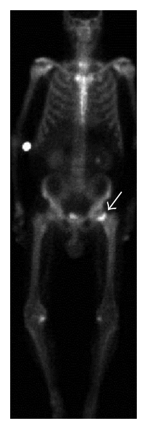
The femoral head is fed by the retinacular vessels that run along the femoral neck inside the capsule. A femoral neck fracture is intracapsular, so displacement cuts the supply and the head can die: the Garden classification tracks the displacement, and a displaced neck fracture gets a hemiarthroplasty because the head is already dead, while a nondisplaced one gets cannulated screws to preserve it. An intertrochanteric fracture sits outside the capsule, the head keeps its blood supply, and the bone heals: sliding hip screw fixation, never replacement. The same blood supply logic explains the rest of the axial falls. A calcaneus fracture after a fall from height transmits force up the axial skeleton, so the lumbar spine is examined and imaged first. The Chance fracture is the lap belt injury: a horizontal split through the vertebral body and posterior elements from flexion-distraction, unstable and surgically stabilized. A tibial plateau fracture from a dashboard knee is an intra-articular weight-bearing injury: depression of the joint surface is a surgical conversation. The Weber ankle system names stability by the fibula fracture level relative to the syndesmosis (A below, B at, C above), and the pelvic ring after high energy is a hemodynamic emergency before it is an orthopaedic one.
Four hip-region doors: which treatment follows which fracture?
A 74-year-old woman fell at home and cannot bear weight. Which door matches which management?
THE SALVAGE CLOCK
The Salvage Clock: Compartment, Fat Embolism, and the Complications That Name Themselves
The tibia breaks, the compartment swells, and the clock starts: muscle and nerve die within hours unless the fasciotomy beats the pressure. The same clock ticks for fat embolism at 24 to 72 hours, for DVT prophylaxis from day one, and for the union problems that show up weeks later.
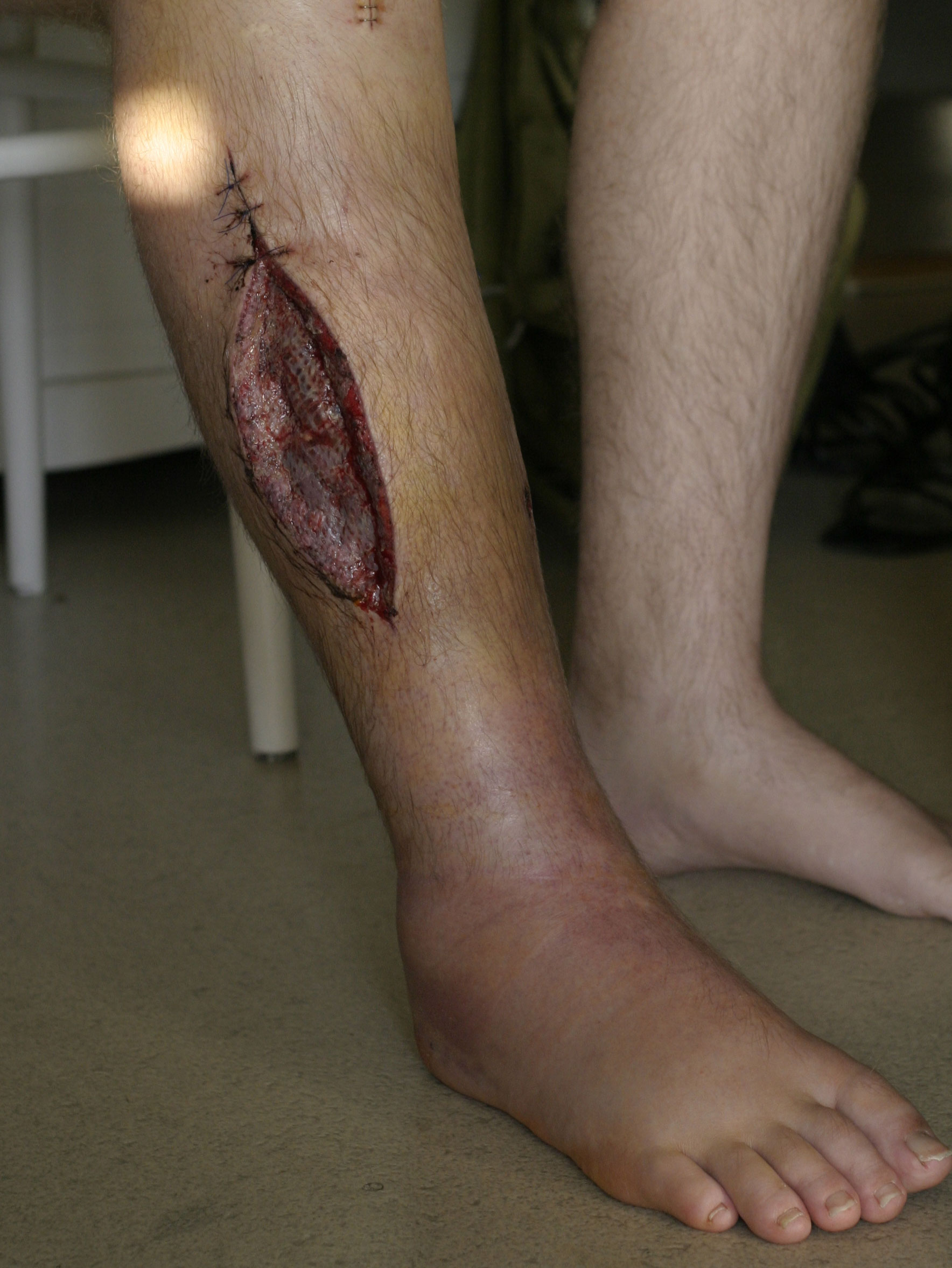
Compartment syndrome is the emergency where the pulse lies. The 5 Ps run in a strict order: pain out of proportion (the earliest sign, worse with passive stretch), paresthesias, pallor or poikilothermia, paralysis, and pulselessness last, too late, because compartment pressure collapses veins and capillaries long before it exceeds arterial pressure, so a palpable pulse does not clear a tense compartment. After a casted fracture, the first move is to remove the cast and bivalve the dressing, because the constriction is part of the pressure; persistent signs mean urgent fasciotomy before Volkmann ischemic contracture. The fat embolism syndrome has its own clock: a long bone or pelvic fracture plus petechiae, hypoxia, and confusion at 24 to 72 hours is fat until proven otherwise, with thrombocytopenia completing the picture; treatment is supportive with oxygen, and early fracture stabilization stops the marrow from leaking more. The chronic complications name themselves by time and alignment: delayed union is slow but progressing, nonunion has stopped (the sequestrum must be debrided), and malunion healed crooked. Post-fracture burning pain with skin changes and patchy osteopenia is CRPS type I, treated with early physical therapy and pain control. And every lower-extremity fracture patient gets DVT prophylaxis from day one.
Tap each letter of the 5 Ps in the order they appear.
PPPPP
PPain out of proportion: the earliest sign, worse with passive stretch
PParesthesias: numbness and tingling as nerves lose perfusion
PPallor or poikilothermia: the skin pales and cools
PParalysis: motor loss, late and ominous
PPulselessness: the last sign, too late to save the muscle, because pressure exceeds venous outflow long before arterial inflow
THE HEALING TIMELINE
The Healing Timeline: From Hematoma to Remodeling
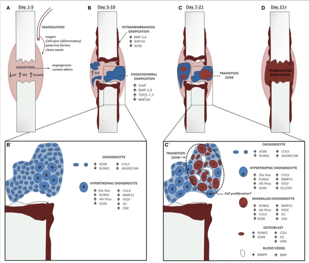
A fracture heals in four acts: hematoma, granulation tissue, callus, remodeling. The board wants the order, the timing, and the factors that stall it: smoking, NSAIDs, steroids, infection, diabetes.
The fracture hematoma is the first act and the most important: it delivers the inflammatory cells and growth factors that start the repair, which is why the inflammation is not the enemy and why prolonged high-dose NSAIDs are avoided, since they suppress the prostaglandins that drive the inflammatory and osteogenic phases. Granulation tissue and a fibrocartilaginous soft callus bridge the gap within weeks, the hard callus of woven bone follows, and remodeling restores the cortex along stress lines over months to years. The impairing factors are the testable list: smoking (vasoconstriction and hypoxia), high-dose NSAIDs, steroids, infection, and diabetes. The outcomes are three words the boards separate: delayed union is slow but progressing, nonunion has stopped and needs intervention (an infected nonunion heals only after the sequestrum is debrided), and malunion healed in an unacceptable position.
Open each act and hold the timing.
The first 24 to 48 hours: bleeding forms the fracture hematoma, the scaffold that delivers inflammatory cells, cytokines, and growth factors. This is why the inflammation is the first act of the healing play, not a complication.
Days: capillaries and fibroblasts invade the hematoma and build granulation tissue, the soft tissue bridge that stabilizes the gap before bone arrives.
Weeks: cartilage and woven bone form the fibrocartilaginous (soft) callus that splints the fracture from the outside, visible on film as early periosteal new bone.
Weeks to months: the soft callus ossifies into the hard callus of woven bone, and the fracture becomes clinically and radiographically bridged.
Months to years: woven bone is resorbed and lamellar bone is laid down along stress lines, restoring the cortex and the medullary canal. The bone returns toward its original shape and strength.
Delayed union is slow but progressing; nonunion has stopped and needs intervention, and an infected nonunion heals only after the sequestrum is debrided; malunion healed crooked. Smoking, high-dose NSAIDs, steroids, infection, and diabetes all slow the clock.
Prove it
Walkthrough: race the salvage clock
Original practice scenarios, one at a time. Choose an answer, then open any option to work its reasoning.
Clinical walkthrough
