/>

Cardiology · Heart Sounds

Wide S2 Split

Why inspiration makes the second heart sound split wider, and what a wide split tells you about the right side of the heart.

clinical medicine High Yield

Before We Dive In

A patient takes a deep breath in. On auscultation, you hear the second heart sound split into two distinct components with a w i d e gap between them. The split widens even further with inspiration and narrows (but does not disappear) on expiration.

What does this tell you?

Yes. Wide split = P2 delayed. Something is slowing the right ventricleThe RV pumps blood to the lungs. It has to empty fully before the pulmonic valve closes, generating P2. from emptying. The A2 (aortic valve closure) fires on time; the P2 (pulmonic valve closure) is late. Inspiration makes it worse because more venous blood rushes into the right side, giving the RV even more to pump through.

The gap between A2 and P2 is the split. Wide gap = slow RV.
Loading images...

Hear the Split

Watch A2 and P2 move apart as you change the pattern and breathing phase.

Select pattern

Breathing phase

S1 A2 P2 gap
A2 = Aortic valve closes
P2 = Pulmonic valve closes
S1 = Mitral/tricuspid close (systole starts)
Normal inspiration: Inspiration pulls extra venous blood into the right heart. The RV has more blood to pump, so it takes slightly longer to empty. P2 is mildly delayed. A2 fires early (LV gets less return). Result: slight split, heard on inspiration, gone on expiration.

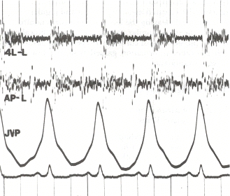
The second heart sound has two components. A2Aortic valve closure. The left ventricle finishes ejecting and pressure drops, snapping the aortic valve shut. This is the louder component because aortic pressure is higher. is the aortic valve slamming shut. P2Pulmonic valve closure. Same idea on the right side. Normally softer than A2 because pulmonary pressures are lower. If P2 becomes loud, think pulmonary hypertension. is the pulmonic valve shutting. Normally they're so close together they sound like one "dub."

The split gap depends on how long each ventricle takes to finish emptying. Longer = later valve closure = later sound component. Wide split = the right ventricle is slow.

Why Is P2 Late?

Tap each cause to see the mechanism. Every one slows the right ventricle.

RBBB
Right Bundle Branch Block
The electrical signal to the right ventricle travels down a side road instead of the express lane. The RV depolarizes late and finishes contracting late. P2 is delayed.

Board pearl: RBBB is the classic textbook cause of wide splitting. Wide, mobile split on exam = think RBBB first.
🚫
Pulmonic Stenosis
Outflow obstruction
The pulmonic valve is narrowed. The RV has to squeeze harder against resistance to push blood through. It takes longer, so it finishes late. P2 is delayed AND often soft (the stenotic valve barely moves).

Clue: Harsh crescendo-decrescendo murmur at upper left sternal border.
💪
Right Heart Failure
Pressure overload / dysfunction
A weak or overloaded right ventricle empties slowly. The pulmonic valve closes late. P2 is delayed.

Context: Look for JVD, peripheral edema, hepatomegaly (right-sided congestion signs).
🩺
Mitral Regurgitation
Volume overload of right side
Blood leaks backward through the mitral valve. More blood eventually floods the right side. The RV deals with increased volume load and closes P2 late.

Note: MR can also make A2 come earlier because the LV empties fast into the low-pressure LA. Both effects widen the split.
🧠
All causes of wide S2 split share one thing: the right ventricle takes longer to finish ejecting. The mechanism differs; the result is identical. P2 is late.

Four Patterns, One Test💡Memory: "Wide FAP" = Wide, Fixed, Absent on insp (paradox), Physiologic. Four patterns. Fixed = ASD. Absent = paradoxical. Wide mobile = RV slow. Physiologic = normal.

Tap any card to flip it and see the full clinical breakdown.

N
Normal Split
Physiologic
Heard onInspiration only
On expirationSingle "dub"
A2 vs P2A2 early, P2 slight delay
▶ Disappears on expiration
tap to flip →
Normal Physiologic Split
Why It Happens
Inspiration increases venous return to the RV. More volume = RV takes slightly longer to empty = P2 is mildly delayed. A2 fires slightly early (less LV return). Gap appears. On expiration, venous return normalizes, P2 returns close to A2, split disappears.
The Rule
Heard on inspiration. Gone on expiration. Any split that persists on expiration is pathologic.
Board Punchline
If it disappears on expiration, it is normal. If it does NOT disappear, figure out why.
W
Wide Split
Mobile
On inspirationWider
On expirationNarrower but persists
Which delayedP2 late
⚠ Persists on expiration
tap to flip →
Wide Mobile Split
Trace It
P2 is delayed. The right ventricle takes longer than normal to finish ejecting. The pulmonic valve closes late. A2 fires on time. A2...P2 gap is wide.
Classic Causes
RBBB (electrical detour to RV) · Pulmonic Stenosis (outflow obstruction) · Right Heart Failure · Severe Mitral Regurgitation
Board Punchline
Wide on inspiration, narrows on expiration, never disappears. Mobile = it responds to breathing. Wide + mobile = RV slow.
F
Fixed Wide Split
ASD Signature
On inspirationUnchanged
On expirationUnchanged
DiagnosisASD until proven otherwise
★ Same gap always = ASD
tap to flip →
Fixed Wide Split = ASD
Why It is Fixed
ASD (hole in atrial septum) shunts blood left-to-right constantly. On inspiration, extra venous blood enters RA, but LA-to-RA shunting decreases by the same amount. Net RV preload: identical. On expiration: same compensation in reverse. The RV is equally overloaded at all times.
Memory Hook
ASD = Always Same Distance. The split gap does not move.
Board Punchline
The one word that seals it: FIXED. Fixed split that does not change with breathing = ASD. Write it. Move on.
P
Paradoxical Split
Reversed Order
On expirationSplit heard
On inspirationDisappears
Which delayedA2 late (past P2)
↻ Opposite of everything
tap to flip →
Paradoxical Split
Trace It
A2 is delayed past P2. Order reverses: S1 → P2 → A2. On expiration, P2 is in its normal spot, A2 is late: you hear the reversed split. On inspiration, normal physiology delays P2 toward the already-delayed A2. The gap closes. Split disappears.
Classic Causes
LBBB (left electrical detour delays LV) · Aortic Stenosis (LV squeezes through tiny orifice) · Severe hypertension (high afterload on LV)
Board Punchline
Present on expiration, gone on inspiration. Opposite of normal. Left-side problems. LBBB and AS are the big two.

Fixed Wide Split = ASD

This is the exam answer that never changes. Literally.

Atrial Septal Defect (ASD)

Auscultation

In a normal heart, breathing changes how much blood enters the right side. On inspiration, more venous blood rushes in, making P2 slightly later. On expiration, less blood, P2 returns toward A2. The split varies with breathing.

In ASDAtrial Septal Defect: a hole in the wall between the left and right atria. Blood shunts left-to-right because left atrial pressure is higher. The right heart is always volume overloaded., there is a hole between the atria. Blood shunts left-to-right all the time. On inspiration, yes, more venous blood comes in from the veins, but simultaneously more blood shunts from LA to RA to compensate. The net result: the RV is always equally overloaded, breath to breath. The split does not change.

Board Trap: Fixed Split

Fixed wide split that does NOT vary with breathing = ASD until proven otherwise.

Every other cause of wide splitting (RBBB, PS, RHF, MR) produces a mobile split that gets wider on inspiration and narrows on expiration. The fixed split is ASD's signature. If the stem mentions the split stays the same width regardless of breathing phase, write ASD.

Phonocardiogram showing S1 and S2 heart sound components
Phonocardiogram trace: S1 and S2 with their components Wikimedia Commons. The split between A2 and P2 within S2 is the gap you are learning to interpret.

S2 Split Diagnosis Tree

Walk the branches. Every node is a board question.

You auscultate the second heart sound. Is S2 split into A2 and P2?
Yes, I hear two components
No, single sound
Single S2. That is normal on expiration.
A single S2 on expiration is physiologic. On inspiration you should hear a faint split. If S2 is persistently single on both phases, consider aortic stenosis with calcified immobile valve (absent A2) or severe pulmonary hypertension (early loud P2 fusing with A2). But in most clinical medicine, single S2 on expiration = normal.
Split present. Does it change with breathing?
Toggle breathing phase. Does the gap change width between inspiration and expiration?
No change with breathing (fixed)
Yes, changes with breathing (mobile)
★ Fixed split. This is ASD until proven otherwise.
The respiratory cycle does not change the A2-P2 gap. Left-to-right shunting through the ASD equalizes RV preload breath-to-breath. Both inspiration and expiration show the same wide split. Classic secundum ASD. Look for: systolic ejection murmur at upper left sternal border, right heart enlargement on CXR, right axis deviation on EKG.
Mobile split confirmed. When is it wider?
Does the split widen on inspiration or expiration?
Wider on inspiration (closes on expiration)
Present on expiration, disappears on inspiration
Wide mobile split. P2 is delayed. RV is slow.
Wide on inspiration, narrower on expiration (but persists). This is the standard wide split. P2 is late. The right ventricle takes longer than normal to empty. Classic causes: RBBB (delayed RV depolarization) · Pulmonic Stenosis (outflow obstruction, also soft P2) · Right Heart Failure · Severe Mitral Regurgitation. Check if P2 is soft (PS) or normal/loud (others).
Paradoxical split. A2 is delayed past P2.
Split heard on expiration, disappears on inspiration. The order has reversed: P2 fires before A2. On inspiration, normal physiology delays P2 toward the already-late A2, closing the gap. Classic causes: LBBB (left bundle delays LV depolarization, delays A2) and Aortic Stenosis (LV squeezes blood through a tiny valve, takes longer, A2 late). Also severe hypertension.

Paradoxical Split: Know the Difference

When the split disappears on inspiration, something is wrong on the left.

Wide split = P2 is late. Simple. Now here is the exam trap: what if A2 is late instead?

In paradoxical splitAlso called reversed split. The split is present on expiration and narrows or disappears on inspiration. This is the opposite of normal physiology., A2 is delayed so much that the order reverses: P2 fires first, A2 fires second. You still hear a split on expiration. On inspiration, the normal physiologic delay of P2 makes P2 catch up to the delayed A2, and the split disappears.

Feature Wide Split Fixed Wide (ASD) Paradoxical Normal
Which component is late? P2 delayed P2 always delayed A2 delayed Neither (slight P2 delay on insp)
On inspiration Split widens Split unchanged Split narrows / disappears Split heard (S1 -- A2 -- P2)
On expiration Split narrows (persists) Split unchanged Split heard (S1 -- P2 -- A2) No split (single "dub")
Classic causes RBBB, PS, RHF, MR ASD🧠ASD: Always Same Distance. The split gap is the same on inspiration AND expiration. LBBB, Aortic Stenosis🧠Left-side problems delay A2 paradoxically: LBBB (left bundle slows LV), AS (LV squeezes through a tiny aortic valve). None (healthy)
P2 softer than normal? Only in PS No (often loud) No No

Decision Tree: Causes of Wide S2 Splitting

Pick the direction of the abnormality. The cause falls out automatically.

What is the direction of the abnormality causing the wide split?

Clinical Vignettes

Five questions. S2 is judging you.

clinical Questions

Two questions. One concept. Can you nail the distinguisher?

A 58-year-old man with no cardiac history presents for a routine physical. On auscultation, S2 splits on inspiration and comes back together on expiration. The gap between A2 and P2 is larger than expected. His chest X-ray and ECG are pending. Which condition is most consistent with this finding?
On a busy cardiology rotation, two patients each have S2 splitting. Patient A has wide splitting: heard on inspiration, nearly gone on expiration. Patient B has wide splitting on inspiration AND expiration, and the width does not change with breathing. Which pairing correctly identifies both causes?
Medically reviewed by Kaitlyn Cocuzzo, MD and Fatima Ali, DO · Last reviewed June 2026
Bone Wizardry is an independent educational resource for visual learning in the medical sciences. It is not affiliated with, endorsed by, or sponsored by any licensing or examination board, contains no real or recalled examination questions, and does not guarantee any educational or examination outcome.