Gram Stain Chart: From Appearance to Clinical Decisions
Read bacterial stains in context, compare major organisms, test processing errors, and practice safe diagnostic and treatment decisions with original cases.
Start with what the specimen actually shows. Then ask whether the preparation is reliable, which organisms fit the site, and what decision the patient needs. This chart connects appearance to clinical reasoning without turning a stain into an automatic antibiotic prescription.
Read the stain before naming the organism
A report says pink cocci, but the known Gram-positive control is pink too. Did the patient suddenly acquire a different organism? First question the preparation. A stain describes the specimen after processing; it does not bypass quality control. [1]
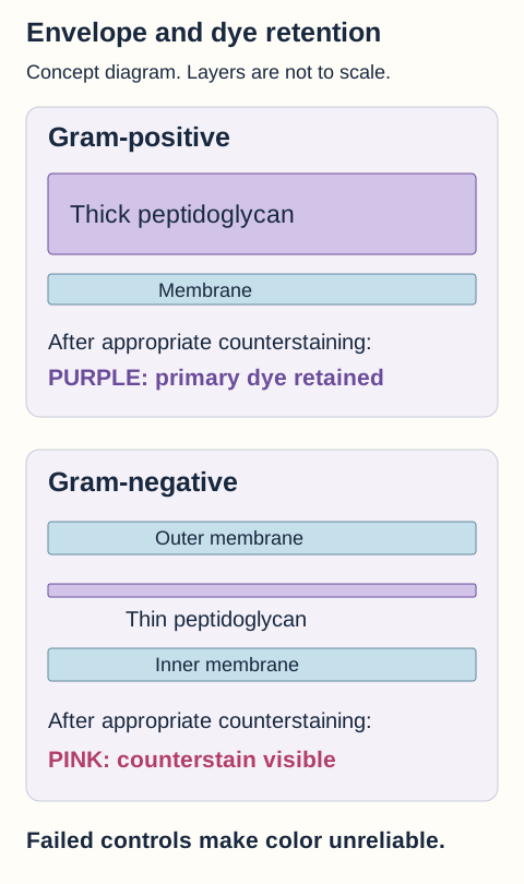
Compare the envelope layers before comparing the final colors. Purple retention depends on processing as well as the envelope.
Crystal violet initially colors both groups. Iodine forms a larger dye complex. During appropriate alcohol or acetone decolorization, the thick peptidoglycan of most Gram-positive organisms retains that complex. In Gram-negative organisms, disruption of the outer membrane and the thin peptidoglycan layer permit dye loss. Safranin then makes those decolorized cells pink. Decolorizer does not simply dissolve peptidoglycan. [1]
Change the processing, not the organism
Predict what will happen to both reference organisms when decolorization is excessive. Then compare excessive and insufficient decolorization. Compare how each known control responds to the change in decolorization.
Known Gram-positive control
Purple. The crystal violet and iodine complex remains.
Known Gram-negative control
Pink. Decolorized cells take up the counterstain.
With appropriate processing, the final control colors differ. Color is interpretable only when controls and the specimen are satisfactory.
Help is available without making a selection. Read the complete comparison immediately below. Reset restores the final stage with appropriate processing. No colors in this demonstration identify a bacterial species or its antibiotic susceptibility.
Complete worked comparison after counterstaining
Processing
Known positive
Known negative
Interpretation
ProcessingAppropriate
Known positivePurple
Known negativePink
InterpretationThe controls separate as expected.
ProcessingExcessive decolorization
Known positiveMay appear pink
Known negativePink
InterpretationDye loss can falsely suggest a Gram-negative organism.
ProcessingInsufficient decolorization
Known positivePurple
Known negativeMay remain purple
InterpretationRetained primary dye can falsely suggest a Gram-positive organism.
The comparison exaggerates control behavior to make the error visible. Real smears can be patchy. Thick preparations, damaged cells, prior antibiotics, and older cultures can produce variable appearances. Recheck technique and fresh controls; do not report an organism solely because the demonstration gives it a color. [1]
Color, shape, arrangement, then specimen
Cocci are round; bacilli are rods; coccobacilli are short rods. Staphylococci commonly form irregular clusters, streptococci often form linear groups, and pneumococci often occur as lancet-shaped pairs. These are useful starting patterns, not exclusive species signatures. A rod inside a neutrophil is still a rod. Intracellular location cannot turn it into a diplococcus. [1][3][4]
Now ask where the material came from and whether inflammation accompanies it. Saliva with many squamous epithelial cells does not provide the same evidence as a high-quality lower respiratory specimen. Organisms on a colonized surface can be bystanders, whereas growth from a normally sterile site carries different weight. A negative stain can reflect low organism burden, prior therapy, sampling problems, or a poorly visualized organism; it does not rule out infection. [1][2]
Apply the distinction to a new specimen. If a final smear contains purple rods and both controls are also purple, insufficient decolorization is possible. If the controls separate correctly but the specimen alone is variable, examine specimen quality and organism biology instead. The useful report separates observation from inference. [1]
Optional organism comparison index: all 37
Filter this duplicate quick-reference index, then follow an organism link into the complete lesson. Filtering does not hide any teaching section.
Showing 37 of 37 organisms.
Use the distinguishing feature to choose the next question, not an automatic antibiotic.
A serogroup-limited urinary assay cannot exclude all Legionella infections.
Try it here · Checkpoint 1 of 3
Make your prediction before reading the choices. A first attempt is just a starting point.
Case 1
Show answer and explanations for case 1
A. Perform a coagulase test as the sole corrective step (Why this does not fit)
Coagulase can help identify a staphylococcal isolate. It does not repair or validate the failed Gram-stain control.
Reasoning steps for option A
Would coagulase testing establish whether the over-decolorized blood-culture cocci are truly Gram-positive?
Coagulase can help identify staphylococci among the cultured cocci, but it does not establish the Gram reaction after the reference control turns pink.
Why is coagulase testing alone insufficient after the positive Gram-stain control turns pink?
Coagulase may help identify the isolate, but it cannot repair or validate the failed Gram stain. Repeat staining with satisfactory controls is needed.
B. Report a Gram-negative coccus from the final pink color (Why this does not fit)
Pink cocci can represent a genuinely Gram-negative organism. The failed positive control shows that processing can explain this color without a different bacterial envelope.
Reasoning steps for option B
When could a pink coccus legitimately be reported as Gram-negative?
Pink cocci can truly be Gram-negative if valid controls retain their expected colors; here the positive control did not.
What does the pink Gram-positive reference control reveal about this smear?
The failed positive control shows that processing can explain this color without a different bacterial envelope.
C. Repeat staining using proper processing and fresh controls (Best answer)
Control failure can make a Gram reaction uninterpretable. The positive control also lost its primary dye, so the specimen cannot yet be classified reliably.
Reasoning steps for option C
What does the pink known Gram-positive control after prolonged decolorization demonstrate?
The processing can strip primary dye from a known Gram-positive organism.
Can the simultaneously pink patient cocci be classified by this run?
No. The same processing error could explain its pink appearance.
How should the laboratory obtain an interpretable Gram reaction?
Repeat the stain under appropriate conditions with satisfactory controls before interpreting its Gram reaction.
D. Discard the culture as a mixed skin-flora contaminant (Why this does not fit)
Skin flora can contaminate blood cultures. A shared staining failure is not evidence of contamination, particularly with several positive bottles.
Reasoning steps for option D
Could several positive blood-culture bottles be dismissed as skin contamination on the basis of this stain?
Skin flora can contaminate blood cultures, but multiple positive bottles and an invalid stain cannot by themselves establish contamination.
Why does the control failure not establish a mixed contaminant?
A shared staining failure is not evidence of contamination, particularly with several positive bottles.
Takeaway: Repeat the stain under appropriate conditions with satisfactory controls before interpreting its Gram reaction.
Separate Gram-positive cocci by behavior and setting
Two patients have purple cocci in blood. One has an infected prosthetic valve; the other has an acutely destructive infection after influenza. Before selecting a species, compare the device history, time course, culture pattern, and confirmatory tests. [3][40]
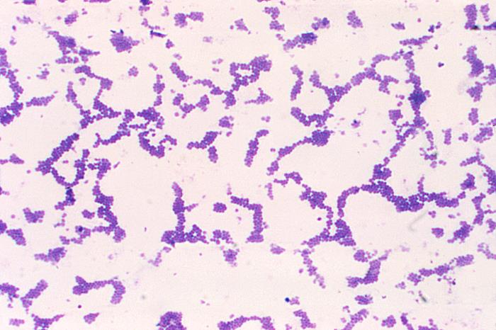
Describe round cells and irregular groupings first. The photographed organism is S. aureus; use additional testing to determine methicillin susceptibility. Image. Image: CDC / Dr. Richard Facklam. Public domain. CDC original and reuse record. Original downloaded image.
Staphylococci
Staphylococcus aureus is usually catalase-positive and coagulase-positive. Catalase separates the familiar staphylococcal branch from streptococci; coagulase supports S. aureus but is not an exclusive species test. Protein A binds the Fc portion of IgG, disrupting effective opsonization. Purulent skin infection, bacteremia, endocarditis, and post-influenza necrotizing pneumonia fit its invasive behavior. Injection-associated tricuspid infection can send infected emboli into the lungs. [3][2][40]
Separate the toxins by what they injure. Exfoliative toxins disrupt superficial epidermal adhesion. TSST-1 acts as a superantigen, linking MHC II and T-cell receptors outside ordinary antigen recognition and provoking cytokine release. Preformed enterotoxin can cause abrupt vomiting after food exposure. Leukocidins can injure leukocytes, but neither a toxin association nor a severe presentation proves the strain carries a particular toxin gene. [2]
Methicillin resistance commonly reflects mecA-encoded PBP2a, an altered binding target rather than ordinary penicillinase alone. Most beta-lactams then fail, although selected anti-MRSA cephalosporins are exceptions. Drain an abscess when indicated. Invasive susceptible S. aureus usually favors cefazolin or an antistaphylococcal penicillin; severe MRSA disease needs an active agent selected for the infection site, often intravenous vancomycin. A Gram stain cannot choose that regimen. [39][40]
S. epidermidis is coagulase-negative and classically novobiocin-susceptible. It is skin flora, but prosthetic material and concordant positive blood cultures can indicate genuine infection. Surface attachment and biofilm physiology support persistence. A fixed claim that every biofilm is a thousand times more resistant is not justified; source control depends on the infected device and clinical circumstances. [3][40]
S. saprophyticus is coagulase-negative, classically novobiocin-resistant, and urease-positive. It can cause cystitis in a sexually active young adult. That urinary setting separates it from the device-associated pattern above. Nitrofurantoin or trimethoprim-sulfamethoxazole may suit susceptible uncomplicated cystitis, but a lower-tract drug choice must not be transferred automatically to pyelonephritis or bacteremia. [3][2]
Streptococci and enterococci
S. pyogenes, group A streptococcus, is typically beta-hemolytic and PYR-positive. Bacitracin susceptibility is a historical screening distinction, not definitive identification. M protein helps resist phagocytosis. Phage-associated pyrogenic exotoxins can produce scarlet fever with a sandpaper-like rash, Pastia lines, and strawberry tongue. Streptolysins O and S damage cells; antibodies against streptolysin O or DNase B can support a preceding infection. [3][5]
Compare the delayed complications. Acute rheumatic fever classically follows pharyngitis and can cause migratory arthritis, carditis, or chorea through immune cross-reactivity. Timely treatment of streptococcal pharyngitis reduces that risk. Post-streptococcal glomerulonephritis can follow throat or skin infection and causes nephritic urine, low complement, and edema. Antibiotics treat remaining infection and transmission risk but have not been shown reliably to prevent that renal complication. Anti-DNase B is useful after skin infection when an ASO response may be less informative. [5][6][2]
S. agalactiae, group B streptococcus, commonly shows beta hemolysis, hippurate hydrolysis, and CAMP enhancement of staphylococcal hemolysis. Genital colonization matters because newborns can develop sepsis or meningitis. Routine pregnancy screening is at 36 0/7 through 37 6/7 weeks. Intrapartum prophylaxis reduces early-onset disease; it does not guarantee protection against late-onset infection. GBS bacteriuria at any time in the current pregnancy is an indication for intrapartum prophylaxis even if a later screening culture is negative. [3][7]
Do not confuse maternal prophylaxis with treatment of a sick newborn. Empiric neonatal meningitis therapy must cover the likely Gram-negative organisms and Listeria as well as GBS. NICE uses intravenous amoxicillin plus cefotaxime when the pathogen is unknown, then narrows confirmed GBS to a penicillin-based regimen. Ampicillin is used in corresponding local protocols. Cefotaxime and ceftriaxone are not interchangeable in neonates; bilirubin and calcium-related ceftriaxone risks matter. [8]
S. pneumoniae often appears as lancet-shaped diplococci with alpha hemolysis, optochin susceptibility, and bile solubility. Its capsule impedes phagocytosis; the Quellung reaction demonstrates capsule swelling. IgA protease aids mucosal colonization. Pneumonia, otitis, sinusitis, and meningitis are familiar syndromes. Loss of splenic clearance increases invasive encapsulated infection risk. Vaccination follows current age and risk recommendations, not a single vaccine for every patient. Suspected pneumococcal meningitis commonly requires ceftriaxone plus vancomycin pending susceptibility. [3][2]
Viridans streptococci are usually alpha-hemolytic, optochin-resistant, and bile-insoluble. Oral species such as S. mutans contribute to caries and produce adhesive extracellular polysaccharides. Oral bacteremia can seed a previously damaged valve, producing a more indolent endocarditis pattern. Susceptibility guides penicillin or alternative treatment; a recent dental procedure is not required. [3][40]
Enterococcus faecalis and E. faecium can be PYR-positive, hydrolyze esculin in bile, and tolerate salt in classic identification schemes. They cause urinary, bloodstream, and endocardial infections. E. faecium is often more resistant. Cephalosporin monotherapy is intrinsically unreliable, but ampicillin plus ceftriaxone can act synergistically against appropriate E. faecalis endocarditis. High-level aminoglycoside resistance defeats the usual gentamicin synergy. VanA or VanB resistance commonly replaces D-Ala-D-Ala with D-Ala-D-Lac. VRE therapy is site- and susceptibility-dependent; daptomycin is not a pneumonia treatment because pulmonary surfactant inactivates it. [3][40]
Return to the prosthetic-valve example. Coagulase-negative cocci in several independently drawn cultures plus a valve abnormality should not be dismissed as skin contamination. Transfer the same reasoning to a single positive bottle without illness or prosthetic material: contamination becomes more plausible, but the whole clinical record still decides. [40]
Use spores, toxin targets, and invasion to separate rods
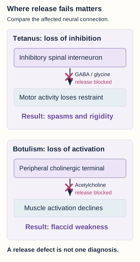
Stiffness and flaccid weakness can both follow exposure to a spore-forming rod. The important difference is where neurotransmitter release fails. Predict the result of blocking an inhibitory interneuron before comparing it with blocking the neuromuscular junction. [2]
Loss of inhibition permits spasms. Loss of peripheral acetylcholine release produces flaccid weakness. Similar molecular tools can have opposite clinical results.
Aerobic spore-formers
Bacillus anthracis classically forms broad rods, is nonmotile and nonhemolytic, and has a poly-D-glutamate capsule rather than a polysaccharide capsule. Protective antigen enables toxin entry. Edema factor raises cyclic AMP through adenylate cyclase activity; lethal factor is a zinc-dependent protease that cleaves MAP kinase kinases. A painless eschar with substantial edema or systemic illness after a relevant exposure requires urgent specialist and public-health coordination. Alert the laboratory rather than requesting routine bench handling. Inhalational disease can produce mediastinal widening and pleural effusions; ingestion can cause gastrointestinal disease. Neither form is excluded by the absence of a skin lesion. [2][10]
Systemic anthrax is not treated as a routine outpatient skin infection. Current guidance uses appropriate antimicrobial combinations and consideration of antitoxin, with evaluation for meningitis. A prolonged postexposure prophylaxis course addresses possible delayed spore germination; its duration must not be copied as a universal treatment duration for active disease. Protective antigen is also the principal antigen in established anthrax vaccines. [10]
B. cereus can produce two foodborne syndromes. Vomiting soon after a meal, classically cooked rice, fits preformed heat-stable cereulide. A later diarrheal illness follows enterotoxin production in the intestine, including pore-forming toxins. The diarrheal mechanism is not the same cyclic-AMP mechanism as cholera. Uncomplicated food poisoning is usually managed supportively; bacteremia, ocular infection, and other invasive disease require a different evaluation. [3][2]
Anaerobic spore-formers
Clostridium perfringens can produce rapidly progressive myonecrosis with severe pain, tissue gas, and systemic toxicity. Its alpha toxin is phospholipase C, also called lecithinase, which damages cell membranes and can cause hemolysis. Tissue gas reflects metabolism in damaged tissue; it is not a species-specific finding. Suspected necrotizing infection requires immediate surgical evaluation and broad antibiotics. Confirmed clostridial myonecrosis is treated with debridement and penicillin plus clindamycin. Do not delay surgery for imaging or hyperbaric treatment. [2][39]
C. perfringens also causes a separate enterotoxin-mediated diarrheal syndrome after contaminated food, often with abdominal cramps and relatively little vomiting. Keep that intestinal presentation separate from destructive muscle invasion. The same species name does not imply the same toxin, site, or urgency. [2]
C. tetani classically has terminal spores. Tetanospasmin reaches inhibitory interneurons and cleaves a vesicle-release protein, preventing release of GABA and glycine. The motor system loses inhibition, causing trismus, risus sardonicus, painful spasms, and sometimes opisthotonus. Tetanus immune globulin neutralizes unbound toxin, not toxin already fixed to nerve endings. Treatment also requires wound care, antibiotics, respiratory and spasm support, and active immunization because disease does not confer reliable immunity. [2][15]
Wound prevention depends on both vaccination history and wound type. After a complete primary series, the usual booster threshold is ten years for a clean minor wound and five years for a dirty or major wound. An unknown or incomplete series changes the vaccine plan; immune globulin is added for qualifying dirty or major wounds, not for clean minor wounds. HIV or severe immunodeficiency also changes immune globulin eligibility. Antibiotics do not replace vaccine and immune globulin decisions for wound prophylaxis. [47][15]
C. botulinum toxins disrupt vesicle-release proteins at peripheral cholinergic terminals, reducing acetylcholine release. Cranial nerve findings, autonomic dysfunction, and symmetric descending flaccid weakness contrast with tetanus. Foodborne disease generally follows ingestion of preformed toxin; infant disease follows intestinal colonization by spores. Honey must not be given before twelve months, although other spore exposures also occur. Wounds can support toxin production as well. [2][14]
Suspected botulism requires urgent respiratory assessment and prompt public-health consultation for the appropriate antitoxin. Do not wait for confirmatory testing when the clinical suspicion is strong. Infant treatment uses the infant-specific immune product through the appropriate service. Antitoxin prevents additional toxin action; it does not instantly reverse established paralysis. [14]
Clostridioides difficile exploits antibiotic-associated disruption of intestinal flora. Toxins A and B alter Rho-family GTPases, damaging cytoskeletal organization and epithelial integrity. Test patients with compatible new unexplained diarrhea, usually at least three unformed stools in twenty-four hours, rather than testing formed stool or screening for cure. A positive nucleic-acid test can reflect colonization rather than toxin-mediated disease. [11]
For an initial nonfulminant adult episode, fidaxomicin is preferred when feasible and oral vancomycin remains an acceptable alternative. Fulminant disease with hypotension, shock, ileus, or megacolon follows a different regimen. Oral or nasogastric vancomycin is central; rectal delivery can be needed with ileus, and intravenous metronidazole is an adjunct, especially when ileus limits delivery. Intravenous vancomycin alone does not deliver adequate treatment into the colonic lumen. [12][11]
Alcohol hand rub does not reliably kill C. difficile spores. Soap-and-water washing physically removes them, and appropriate sporicidal environmental products, including correctly used bleach products, can inactivate them. Saying that bleach cannot work confuses resistance to some disinfectants with resistance to every disinfectant. [13][11]
Non-spore-forming rods
Listeria monocytogenes is a small Gram-positive rod with classic tumbling motility, beta hemolysis, and growth at refrigeration temperatures. It escapes a host-cell vacuole and uses ActA-associated actin propulsion for intracellular spread. Pregnancy, neonatal age, older age, and impaired cellular immunity increase concern for invasive infection. Meningitis or bacteremia needs ampicillin-based treatment; cephalosporins do not provide dependable Listeria coverage. Trimethoprim-sulfamethoxazole is an important alternative in appropriate circumstances. [3][2][45][8]
Corynebacterium diphtheriae forms club-shaped rods in angular or palisade arrangements. Toxigenic strains carry a phage-associated toxin that ADP-ribosylates elongation factor 2, stopping protein synthesis. An adherent pharyngeal membrane can threaten the airway; myocarditis and neuropathy can follow. Respiratory disease warrants prompt antitoxin and antibiotic treatment while confirmation of toxigenicity proceeds. Isolation of the species alone does not establish toxin production, and treatment should not await a rigid sequence of completed tests. [3][16]
Check the localization comparison. If inhibitory input is lost but peripheral acetylcholine release remains possible, contractions become poorly restrained. In a new patient with ptosis and descending weakness, the peripheral release failure instead points toward botulism. The toxin target explains the difference better than the shared word Clostridium. [2][15][14]
Try it here · Checkpoint 2 of 3
Make your prediction before reading the choices. A first attempt is just a starting point.
Case 17
Show answer and explanations for case 17
A. Intravenous vancomycin plus oral metronidazole (Why this does not fit)
Both drugs have important uses by appropriate routes in other infections. Intravenous vancomycin does not provide adequate luminal treatment, and ileus also undermines reliance on oral metronidazole.
Reasoning steps for option A
Can intravenous vancomycin reach the colonic lumen adequately?
No. IV vancomycin does not supply adequate luminal C. difficile treatment.
Why is oral metronidazole alone an unreliable companion with this distention?
Ileus undermines reliance on oral metronidazole despite its uses by appropriate routes elsewhere.
B. Enteral fidaxomicin without adjunctive treatment (Why this does not fit)
Fidaxomicin is preferred for many initial nonfulminant adult episodes. The shock and ileus place this patient in the fulminant pathway rather than that uncomplicated treatment comparison.
Reasoning steps for option B
For which initial C. difficile episodes is fidaxomicin preferred?
It is preferred in many nonfulminant adult episodes.
Which findings move this patient out of that treatment comparison?
Blood pressure of 82/48 and ileus indicate fulminant disease requiring a different backbone.
C. Enteral vancomycin plus intravenous metronidazole (Best answer)
Fulminant disease requires vancomycin delivered toward the bowel lumen, with intravenous metronidazole as an adjunct. Ileus may also require rectal vancomycin because ordinary enteral delivery may not reach all affected colon.
Reasoning steps for option C
What do blood pressure 82/48 and absent stool passage indicate?
Shock and ileus with colonic dilation place her in the fulminant C. difficile category.
Why must the route of vancomycin account for ileus?
Treatment must reach the colonic lumen despite impaired transit; rectal delivery may be needed.
Which antimicrobial backbone accompanies resuscitation and surgical review?
Use enteral vancomycin plus intravenous metronidazole, considering rectal vancomycin for ileus.
D. Intravenous vancomycin plus intravenous metronidazole (Why this does not fit)
Intravenous metronidazole is an adjunct in fulminant disease with ileus. Replacing enteral or rectal vancomycin with intravenous vancomycin leaves the necessary luminal treatment inadequately addressed.
Reasoning steps for option D
Which IV drug in this pair has a role in fulminant ileus?
Intravenous metronidazole is an adjunct in fulminant disease with ileus.
Why can IV vancomycin not replace enteral or rectal delivery?
It leaves the affected colonic lumen inadequately treated despite the correct metronidazole route.
Takeaway: Use enteral vancomycin with intravenous metronidazole, considering rectal vancomycin for ileus and continuing urgent multidisciplinary care.
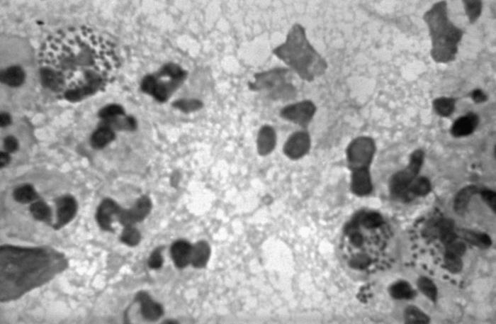
A neutrophil contains paired round bacteria. Would that observation mean the same thing in symptomatic male urethral discharge, a pharyngeal swab, and cerebrospinal fluid? Compare the site before transferring the interpretation. [17][19]
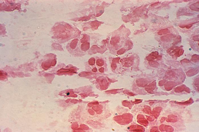
Find the intracellular pairs. This archival image is grayscale, so it teaches shape and location, not purple-versus-pink interpretation. The CDC identifies the organisms as N. gonorrhoeae. Image. Image: CDC / Bill Schwartz. Public domain. CDC original and reuse record. Original downloaded image.
Neisseria gonorrhoeae is an oxidase-positive Gram-negative diplococcus. Intracellular diplococci in urethral discharge from a symptomatic man can be diagnostic, but smear interpretation is less reliable at cervical, rectal, and pharyngeal sites. Use nucleic-acid amplification testing at the exposed anatomical sites. Suspected treatment failure also requires culture and antimicrobial susceptibility assessment. Pili and antigenic variation aid attachment and immune evasion; IgA protease and lipooligosaccharide contribute to pathogenesis. Gonococci lack the prominent polysaccharide capsule characteristic of meningococci. [17][4][2]
Uncomplicated gonorrhea is not automatically dual therapy. The CDC regimen is ceftriaxone 500 mg intramuscularly once for a person weighing less than 150 kg, or 1 g at 150 kg or above. Add chlamydia treatment when chlamydia has not been excluded. Doxycycline is used for most nonpregnant adults; pregnancy requires its own chlamydia regimen. Disseminated disease with dermatitis, tenosynovitis, or arthritis has different dosing and duration requirements. [17][22]
Pelvic inflammatory disease is an upper genital tract syndrome, not merely a positive gonorrhea test. Cervical motion, uterine, or adnexal tenderness in the appropriate setting can justify empiric treatment even when cervical gonorrhea and chlamydia tests are negative. A common outpatient regimen is ceftriaxone plus fourteen days of doxycycline and metronidazole. Anaerobic coverage matters; pregnancy, severe illness, or an abscess can require admission and parenteral care. [18]
N. meningitidis has a polysaccharide capsule and lipooligosaccharide whose lipid A promotes an intense inflammatory response. Meningitis with shock, purpura, disseminated coagulation abnormalities, and adrenal hemorrhage is an emergency. Ceftriaxone provides empiric treatment; penicillin is an option only after appropriate susceptibility evidence. Suspected invasive disease requires prompt droplet precautions and public-health coordination. [19][20][2]
Household contacts and people with relevant direct exposure to respiratory secretions may need prophylaxis. Start it promptly, ideally within twenty-four hours of identifying the index patient, rather than counting from the first symptom. Regimen choice considers pregnancy and local resistance; ceftriaxone is appropriate in pregnancy. Vaccination does not replace indicated exposure prophylaxis. MenACWY and MenB vaccines cover different meningococcal groups. [19][20]
Moraxella catarrhalis is another oxidase-positive Gram-negative diplococcus, associated with otitis, sinusitis, and respiratory exacerbations in susceptible hosts. Beta-lactamase production is common. An explicitly beta-lactamase-producing isolate should not be treated with amoxicillin alone; amoxicillin-clavulanate can be appropriate when the syndrome calls for it. The diplococcus shape does not make every respiratory isolate a meningococcus or gonococcus. [4][2]
Apply the site comparison. Paired intracellular cocci in symptomatic male urethral discharge support gonorrhea. In a pharyngeal specimen, other Neisseria species make morphology less specific, so site-appropriate molecular testing matters. In cerebrospinal fluid with meningitis and shock, urgent invasive-infection management takes priority over waiting to finish every identification test. [17][19]
Narrow Gram-negative rods without skipping the syndrome
Pink rods occur in cystitis, bowel abscesses, pneumonia, and bloodstream infection. First separate the exposure and organ system. Then ask whether the organism invades tissue, produces a toxin, or persists because treatment does not reach or inhibit it. [4][2]
Compare these elongated organisms with the paired cocci in the preceding image. The CDC description explicitly identifies intracellular Gram-negative rods, not gonococci. It does not establish a rod species. Image. Image: CDC / Joe Miller. Public domain. CDC original and reuse record. Original downloaded image.
Enteric organisms and urinary disease
Escherichia coli typically ferments lactose and is oxidase-negative. Adhesive fimbriae help urinary strains colonize the urinary tract; P fimbriae are associated with upper-tract disease. The K1 capsule is an important neonatal meningitis association. Different intestinal pathotypes do different jobs. ETEC heat-labile toxin activates the Gs and cyclic-AMP pathway, whereas its heat-stable toxin stimulates a guanylate-cyclase and cyclic-GMP pathway. Both can drive watery diarrhea. [4][2]
Shiga toxin-producing E. coli, including non-O157 strains, damages ribosomal RNA and can injure vascular endothelium. Bloody diarrhea followed by microangiopathic hemolytic anemia, thrombocytopenia, and acute kidney injury suggests hemolytic uremic syndrome. Request testing that detects Shiga toxin or its genes rather than relying on an O157 culture phenotype alone. Support hydration and renal care; avoid antibiotics and antimotility agents in suspected or confirmed STEC because they can increase harm. [30]
Klebsiella pneumoniae is commonly a nonmotile, encapsulated, lactose-fermenting rod. Mucoid colonies reflect capsule-related material. Pneumonia, urinary infection, bacteremia, and invasive liver abscess disease can occur, not only in people with alcohol-related disease. A positive colony string test describes hypermucoviscosity; it is neither a test on pus nor definitive proof of a particular capsule type or hypervirulent lineage. [4][2]
Extended-spectrum beta-lactamases can hydrolyze third-generation cephalosporins. Severe invasive ESBL-producing Enterobacterales infection generally favors an active carbapenem, but the infection site and susceptibility results still matter. ESBL production does not itself mean carbapenem resistance. Carbapenem resistance may involve carbapenemases or combinations of other enzymes with reduced permeability. Do not infer a specific resistance gene from a Gram stain or a single drug result. [9]
Proteus mirabilis is classically a motile, lactose-nonfermenting, urease-positive rod with swarming growth. Urease converts urea to ammonia, increasing urinary pH and favoring magnesium ammonium phosphate stones, whose crystals classically resemble coffin lids. A branching staghorn stone plus recurrent alkaline urinary infection makes an infection stone plausible, but not every staghorn calculus is struvite and Proteus is not the only urease producer. Obstruction or an infected stone can require source control in addition to active antibiotics. [4][2]
Shigella spreads efficiently with a low infectious inoculum, invades colonic mucosa, and can cause fever, tenesmus, and bloody diarrhea. Intracellular spread differs from ETEC secretion without the same invasive pattern. S. dysenteriae type 1 classically produces Shiga toxin. Treatment is not a universal empiric antibiotic rule: severity, transmission concerns, local resistance, and susceptibility testing matter. Avoid antimotility treatment in invasive bloody diarrhea. [4][2]
Salmonella is usually motile, lactose-nonfermenting, and hydrogen-sulfide-producing. Nontyphoidal gastroenteritis commonly needs supportive care, with antibiotics reserved for invasive illness or patients at elevated risk. Typhi causes a different systemic enteric-fever syndrome involving survival in host phagocytes; sustained fever, abdominal symptoms, and occasional rose spots can occur. Treatment must account for travel-related resistance. Salmonella is associated with osteomyelitis in sickle cell disease, but S. aureus remains an important competitor. [4][2][31]
Yersinia enterocolitica can follow pork exposure and cause terminal ileitis or mesenteric adenitis that resembles appendicitis. Cold-tolerant growth contributes to transfusion-related concern. Y. pestis has a different ecology: infected rodents and fleas can precede painful buboes, while pneumonic disease can spread through respiratory exposure. Bipolar staining is a classic special-stain observation, not a stand-alone species diagnosis. F1 capsule and Yop effector proteins support virulence. Suspected plague requires urgent treatment and public-health coordination. [4][2]
Respiratory, water-associated, and healthcare organisms
Pseudomonas aeruginosa is a nonfermenting, oxidase-positive rod. Pyocyanin and pyoverdine can generate characteristic pigments, but color or odor is not definitive identification. Exotoxin A inhibits elongation factor 2. Alginate-associated biofilm formation supports chronic airway persistence in cystic fibrosis and bronchiectasis. A history of chronic sinus disease, infertility, and reversed organ position instead suggests impaired ciliary clearance as the underlying host problem. [4][2]
Burn wounds, neutropenia, ventilator-associated pneumonia, hot-tub folliculitis, and puncture-related bone infection are useful contexts. Reduced porin entry, efflux, and beta-lactamases are distinct from biofilm-associated tolerance. Select an active antipseudomonal agent using site and susceptibility evidence; cefepime, piperacillin-tazobactam, or meropenem are possible choices in appropriate isolates. Ertapenem is not an antipseudomonal substitute. [9][2]
Haemophilus influenzae is a small pleomorphic coccobacillus classically requiring X factor, hemin, and V factor, NAD. Chocolate-medium growth and satellitism are historical identification concepts. The type b polysaccharide capsule explains the target of Hib vaccines; nontypeable strains can still cause otitis and respiratory disease. Invasive infection includes meningitis, epiglottitis, and bacteremia. A drooling child in a tripod posture needs controlled expert airway management, not a forced throat examination or a delay for confirmatory imaging. [4][43][2]
Acinetobacter baumannii can appear as a plump coccobacillus and sometimes stain variably. Healthcare-associated pneumonia or bloodstream infection requires clinical evidence of infection rather than colonization alone. Carbapenem-resistant isolates may carry OXA-type enzymes, among other mechanisms. Current IDSA guidance favors a sulbactam-durlobactam-based combination with imipenem-cilastatin or meropenem for appropriate invasive CRAB infection. Resistance testing and specialist input remain essential; this is not a treatment selected by shape alone. [9]
Curved rods and toxin-mediated secretion
Campylobacter jejuni is a curved or gull-wing-shaped organism associated with poultry exposure and inflammatory diarrhea. Microaerophilic growth and growth at higher incubation temperatures are classic identification properties, not reasons to attempt special cultures without laboratory protocols. Following infection, immune cross-reactivity between bacterial lipooligosaccharide and peripheral nerve gangliosides can cause Guillain-Barre syndrome. Both axonal and demyelinating variants occur. Most enteritis resolves supportively; selected severe or high-risk cases need antibiotics, often azithromycin, with resistance considered. [4][32][2]
Helicobacter pylori uses urease to generate local ammonia and buffer its gastric environment. Persistent inflammation contributes to peptic ulcer disease, gastric adenocarcinoma risk, and gastric MALT lymphoma; CagA and VacA are important virulence factors. Some early MALT lymphomas regress after eradication, but not every lymphoma does. Urea breath testing and stool antigen testing detect active infection; alarm features may require endoscopic evaluation rather than a test-and-treat shortcut. [4][2][21]
When antibiotic susceptibility is unknown, the 2024 ACG guidance favors fourteen-day optimized bismuth quadruple therapy with acid suppression, bismuth, tetracycline, and metronidazole. Do not routinely prescribe empiric clarithromycin triple therapy without evidence of susceptibility. Confirm eradication at least four weeks after antibiotics, with proton-pump inhibitors withheld for at least two weeks before the test. Symptom improvement alone does not establish eradication. [21]
Vibrio cholerae can cause profuse watery diarrhea when cholera toxin keeps the Gs pathway active, increasing cyclic AMP and intestinal chloride secretion. Severe dehydration is the immediate threat. Oral rehydration solution is central when feasible; severe dehydration requires intravenous fluids. Selected antibiotics are adjuncts, not substitutes for fluid replacement. In contrast, V. vulnificus can cause severe wound infection or sepsis after coastal water or raw seafood exposure, especially with liver disease. V. parahaemolyticus more commonly produces seafood-associated gastroenteritis, although species associations are not absolute. Hemorrhagic bullae and rapidly progressive illness require immediate antibiotics and surgical assessment. [4][42][28]
Bacteroides fragilis is an anaerobic gut organism with a capsule that contributes to abscess formation. It need not be the most abundant member of the microbiome to cause substantial disease after bowel leakage. Beta-lactamase production can undermine simple penicillin treatment. A postoperative intra-abdominal abscess may contain both anaerobic and aerobic bacteria; drainage and a regimen covering the full expected flora are needed. Metronidazole alone does not supply broad aerobic coverage. [4][2][39]
Use a changed specimen to test the method. Ascitic fluid with 500 leukocytes per microliter and 50 percent neutrophils contains 250 neutrophils per microliter. In a patient with cirrhosis and no surgically treatable abdominal source, that meets the threshold for spontaneous bacterial peritonitis even if the culture is negative. Treat promptly using community or healthcare exposure and resistance risk. Consider indicated albumin separately; do not confuse treating the current episode with prophylaxis against another episode. [41]
Now contrast that patient with one who has free air, focal peritonitis, or a polymicrobial abdominal specimen after bowel surgery. Those findings raise concern for a secondary source requiring investigation and source control. Counting neutrophils is useful, but it must not erase the anatomical explanation. [41][39]
A poor Gram stain does not imply a missing cell wall
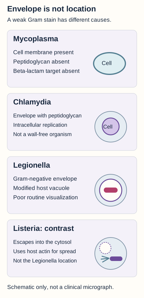
A patient has pneumonia and many inflammatory cells, but no convincing organisms on the routine stain. Compare three explanations before choosing one: the organism has no peptidoglycan, it has an envelope but is poorly visualized, or the specimen simply failed to show the pathogen. Those explanations do not predict the same treatment. [1][2][23]
Separate cell-envelope biology from intracellular location. A weak routine stain is not evidence that every organism shown lacks a cell wall.
Wall-free and intracellular organisms
Mycoplasma pneumoniae lacks peptidoglycan and has a sterol-containing cell membrane. Beta-lactams therefore lack their usual cell-wall target. A persistent cough and interstitial respiratory findings can suggest it, but clinical appearance is not specific. Cold-reactive antibodies against red-cell I antigen may occur and are not a definitive diagnostic test. Molecular testing can help. Macrolides inhibit the 50S ribosomal subunit, whereas doxycycline inhibits the 30S subunit; do not assign both to the same target. [2]
Chlamydia trachomatis has a Gram-negative-type envelope and does synthesize peptidoglycan. It is not simply a wall-free counterpart of Mycoplasma. Infectious elementary bodies establish infection, while reticulate bodies support intracellular replication; these developmental forms should not be described as organisms incapable of all independent metabolic activity. Serovars A through C cause trachoma, D through K cause common genital and neonatal infections, and L1 through L3 cause lymphogranuloma venereum. C. pneumoniae causes a separate respiratory infection. [23][2]
Use site-appropriate molecular testing for genital chlamydia. Doxycycline is recommended for most nonpregnant adults; azithromycin is the recommended pregnancy regimen, with alternatives under the guideline. The existence of a pregnancy-specific amoxicillin alternative is another reason to avoid the false claim that all chlamydiae lack a beta-lactam target. [22][23]
Rickettsia rickettsii targets vascular endothelium, producing vasculitis and capillary leak. Fever, severe headache, thrombocytopenia, low sodium, and a compatible tick exposure can precede the classic rash spreading from wrists or ankles toward the trunk, palms, and soles. The rash may be absent or late. Start doxycycline for suspected Rocky Mountain spotted fever in children as well as adults without waiting for serologic confirmation. Early negative serology and the historical Weil-Felix test cannot safely exclude disease. [24][2]
Legionella can be poorly seen on routine Gram stain despite a Gram-negative envelope. It survives in a modified intracellular vacuole; this differs from Listeria escaping into the cytosol. Contaminated water aerosols can transmit infection, including from cooling towers and building water systems. Ordinary room air conditioners are not the usual water-system source. Pneumonia with diarrhea, confusion, or hyponatremia may fit, but no single associated laboratory value establishes the diagnosis or proves SIADH.
A urinary antigen test mainly detects L. pneumophila serogroup 1. Pair it with lower respiratory molecular testing or appropriate culture; a negative urinary result does not exclude other Legionella infections. Buffered charcoal yeast extract, cysteine, and iron are classic laboratory associations. An active macrolide or fluoroquinolone provides intracellular treatment. [27][2]
Spirochetes and exposure-directed testing
Treponema pallidum is too thin for dependable routine Gram-stain identification. Primary syphilis may produce a painless chancre; secondary disease can cause a diffuse rash involving palms or soles and condylomata lata. Latency can precede late gummatous, cardiovascular, or neurologic complications. Neurosyphilis and ocular disease can occur at any stage, not only after decades. Suspected neurologic or ocular involvement requires a CNS-appropriate penicillin regimen rather than assuming intramuscular benzathine penicillin is sufficient. [38][25]
Interpret syphilis serology as a pair of questions. Nontreponemal tests such as RPR or VDRL provide quantitative titers useful for follow-up, but false positives occur. Treponemal tests support specificity and often stay reactive after successful treatment. A rise or fall in a comparable nontreponemal titer has a different meaning from a persistently positive treponemal result. Fever and myalgia shortly after treatment can be a Jarisch-Herxheimer reaction; urticaria, airway symptoms, or circulatory collapse require assessment for an immediate allergy or another emergency. [38]
Congenital syphilis can lead to abnormalities including Hutchinson teeth, saddle-nose deformity, and saber shins. Those late findings do not substitute for appropriate maternal screening and infant evaluation. Diagnosis and treatment depend on the maternal record, infant findings, and recommended testing rather than the presence of a complete historical triad. [2][38]
Borrelia burgdorferi causes Lyme disease after an infected Ixodes tick bite. Erythema migrans is an expanding lesion, not necessarily a target with central clearing, and a typical lesion in the right setting is diagnosed clinically. Attachment duration contributes to transmission risk and prophylaxis decisions; a thirty-six-hour high-risk prophylaxis criterion is not a guarantee of zero transmission before that time.
Later disease can cause facial palsy, meningitis, atrioventricular block, or large-joint arthritis. Use recommended two-tier serology when indicated, including validated modified two-enzyme-immunoassay algorithms; a routine stained blood film is not the standard Lyme test. Many presentations can be treated orally, whereas severe carditis or selected neurologic disease can require intravenous treatment and monitoring. [33]
Leptospira enters through mucosa or injured skin after contact with water contaminated by animal urine. Fever with calf pain, conjunctival suffusion, kidney injury, and jaundice suggests leptospirosis, with severe multiorgan disease often termed Weil disease. Early blood PCR and later antibody or urine testing answer different timing questions. Treat a strongly suspected infection without waiting for confirmation; severe disease can require intravenous penicillin or ceftriaxone and organ support. [34]
Brucella is a small Gram-negative coccobacillus and a facultative intracellular pathogen, not an obligate intracellular organism. Unpasteurized dairy or occupational animal exposure can precede prolonged fever, sweats, arthralgia, and hepatosplenic findings. Alert the laboratory to the suspected organism. A common uncomplicated regimen combines doxycycline and rifampin for at least six weeks, but RB51 is rifampin-resistant and complicated infection needs a different specialist plan. Francisella tularensis can follow tick or rabbit exposure with an inoculation ulcer and regional adenopathy. It also warrants laboratory notification and prompt syndrome-appropriate treatment. [35][44][2]
Acid-fast organisms and nonbacterial alternatives
Mycobacterium tuberculosis has a lipid-rich envelope containing mycolic acids, so acid-fast staining is more useful than ordinary Gram staining. Chronic cough, weight loss, and upper-lung cavitation support investigation, but an acid-fast smear does not by itself distinguish tuberculosis from nontuberculous mycobacteria. Molecular testing, culture, and susceptibility evidence guide therapy. The conventional initial isoniazid, rifampin, pyrazinamide, and ethambutol regimen remains important, but eligible patients may receive newer shorter regimens. Isoniazid inhibits mycolic-acid synthesis after KatG-dependent activation, with InhA as an important target; people at risk of neuropathy need pyridoxine. [2][36]
M. leprae disease varies with cell-mediated immunity. Stronger cellular containment produces more localized, often anesthetic lesions with fewer bacilli and granulomas; weak containment permits widespread lepromatous disease with many bacilli and foamy macrophages. Peripheral nerve injury explains sensory loss. The historical lepromin response is not a stand-alone diagnostic test. WHO program guidance uses rifampicin, dapsone, and clofazimine for both paucibacillary and multibacillary disease, for six and twelve months respectively. United States specialist regimens differ, so the WHO schedule is not a universal prescription. [2][29][46]
Bacterial vaginosis is polymicrobial dysbiosis, not proof of a single Gardnerella infection. A higher vaginal pH, thin discharge, an amine reaction, and epithelial cells coated with adherent bacteria support the diagnosis. At least three of these four Amsel criteria support BV. Gram-stain Nugent scoring is useful here; the stain is not useless simply because some organisms are variable. Compare this with candidiasis, which commonly has a normal vaginal pH and yeast forms, and trichomoniasis, which is a protozoal infection. Metronidazole is one recommended treatment for symptomatic BV. [26]
Cryptococcus is an encapsulated yeast, not a Gram-negative bacterium. Subacute meningitis in advanced HIV warrants cryptococcal antigen testing and assessment of cerebrospinal fluid pressure. An India ink halo is a classic observation, but antigen testing is more sensitive. Induction commonly uses liposomal amphotericin B with flucytosine in resource-available settings, followed by fluconazole-based therapy; raised pressure needs its own management. A negative bacterial stain should not close the fungal differential. [37]
A viral mimic also matters. Epstein-Barr virus can cause pharyngitis with posterior cervical nodes and splenic enlargement. An aminopenicillin-associated rash in that setting is not automatically evidence of immediate IgE-mediated penicillin allergy, but a future allergy assessment may still be appropriate. Likewise, negative blood cultures in suspected endocarditis do not automatically identify HACEK organisms. Prior antibiotics are common contributors; exposure and clinical context may direct testing for Coxiella, Bartonella, Brucella, Tropheryma whipplei, or other fastidious pathogens. Modern routine blood-culture systems often recover HACEK organisms. [2][40]
Apply the initial pneumonia comparison to a new result. A negative Legionella urinary antigen does not establish Mycoplasma or justify stopping all relevant testing. Ask what the assay detects, what it misses, and whether the proposed drug reaches the organism and has a target. That final check keeps a useful chart from becoming an unsafe shortcut. [27][23][2]
Try it here · Checkpoint 3 of 3
Make your prediction before reading the choices. A first attempt is just a starting point.
Case 32
Show answer and explanations for case 32
A. Exclude Legionella and use routine sputum microscopy alone (Why this does not fit)
Routine microscopy is useful for many bacterial pneumonias. Legionella can stain poorly, and the negative urinary result does not exclude all Legionella infections.
Reasoning steps for option A
When does routine sputum microscopy help identify bacterial pneumonia?
Routine microscopy is useful for many bacterial pneumonias.
Why do a bacteria-negative Gram stain and negative urinary antigen not eliminate Legionella here?
Legionella can stain poorly, and the negative urinary result does not exclude all Legionella infections.
B. Repeat the same urinary test as the only investigation (Why this does not fit)
Repeating a test can help with selected collection or timing problems. It does not correct the restricted organism and serogroup coverage of this assay.
Reasoning steps for option B
When might repeating a urine antigen help with a collection or timing issue?
Repeating a test can help with selected collection or timing problems.
Why does repeating the same assay alone fail to assess Legionella outside its main serogroup target?
It does not correct the restricted organism and serogroup coverage of this assay.
C. Lower respiratory molecular testing and Legionella culture (Best answer)
Respiratory molecular testing or culture can detect Legionella infections missed by a serogroup-limited urine assay. The urinary test mainly detects L. pneumophila serogroup 1 and does not exclude the full differential.
Reasoning steps for option C
Which Legionella species/serogroup does the urinary antigen principally detect?
It mainly detects L. pneumophila serogroup 1.
What infections remain possible despite its negative result in this patient?
Other Legionella infections and an imperfectly detected infection remain possible.
What lower respiratory tests broaden the investigation while active therapy continues?
Pair appropriate lower respiratory molecular testing or culture with the existing urinary test rather than treating it as a genus-wide exclusion.
D. Use cold agglutinins to establish the respiratory pathogen (Why this does not fit)
Cold agglutinins can occur in Mycoplasma-associated illness. They are nonspecific and do not resolve the serogroup limitation or establish an alternative pathogen.
Reasoning steps for option D
What other atypical respiratory illness may produce cold agglutinins?
Cold agglutinins can occur in Mycoplasma-associated illness.
Why cannot cold agglutinins resolve the Legionella urinary test’s serogroup limitation or establish another pathogen?
They are nonspecific and do not resolve the serogroup limitation or establish an alternative pathogen.
Takeaway: Pair appropriate lower respiratory molecular testing or culture with the existing urinary test rather than treating it as a genus-wide exclusion.
Use the supplied history and laboratory findings to choose the best answer. Identify the finding that separates your choice from its strongest competitor, then inspect the option-specific explanations. These original educational cases are not recalled examination questions.
Case 2
Show answer and explanations for case 2
A. Stop treatment until a repeat stain shows an organism (Why this does not fit)
Additional sampling sometimes helps resolve uncertain diagnoses. Waiting for a positive stain would leave a strongly supported bacterial infection untreated.
Reasoning steps for option A
Would another positive smear be required before treating this febrile patient with neck stiffness and neutrophilic CSF?
Repeat sampling can clarify uncertain meningitis diagnoses, but treatment need not await visible organisms in CSF after antibiotics.
What risk follows from withholding antibiotics while repeating a stain after ceftriaxone and vancomycin?
Waiting for a positive stain would leave a strongly supported bacterial infection untreated.
B. Stop antibacterial therapy and provide supportive care (Why this does not fit)
Supportive care alone can be appropriate for selected uncomplicated viral meningitis. The CSF pattern and preceding antibiotics make that conclusion unsafe here.
Reasoning steps for option B
When might supportive care alone suffice for meningitis?
Supportive care alone may fit selected uncomplicated viral meningitis, unlike this marked neutrophilic pleocytosis and CSF-to-serum glucose ratio of 0.2.
Why do 1,450 CSF leukocytes with 88% neutrophils and glucose 18 versus serum 90 oppose a viral-only plan?
The CSF pattern and preceding antibiotics make that conclusion unsafe here.
C. Continue antibiotics and pursue further microbiologic testing (Best answer)
Marked neutrophilic inflammation and low paired CSF glucose support bacterial meningitis. Prior antibiotics and an imperfectly sensitive stain prevent the negative smear from excluding it.
Reasoning steps for option C
What does CSF glucose 18 mg/dL against serum glucose 90 mg/dL indicate?
CSF glucose is only one fifth of the serum value.
How do 1,450 leukocytes/µL with 88% neutrophils shift concern?
The marked neutrophilic inflammation reinforces concern for bacterial meningitis.
Does a negative CSF stain two hours after antibiotics justify stopping therapy?
It does not justify stopping antibacterial therapy while appropriate microbiologic testing continues.
D. Replace antibacterial treatment with antifungal monotherapy alone (Why this does not fit)
Fungal disease can cause meningitis and low CSF glucose. The acute syndrome and strongly neutrophilic pattern do not justify abandoning bacterial coverage solely because the smear is negative.
Reasoning steps for option D
Can fungal meningitis also depress CSF glucose?
Fungal meningitis can lower CSF glucose, but this one-day syndrome with 88% neutrophils still demands bacterial coverage.
Why does a negative stain two hours after antibiotics not support switching this acute neutrophilic illness to antifungal monotherapy?
The acute syndrome and strongly neutrophilic pattern do not justify abandoning bacterial coverage solely because the smear is negative.
Takeaway: It does not justify stopping antibacterial therapy while appropriate microbiologic testing continues.
A. Conversion into dormant endospores on the valve (Why this does not fit)
Endospores permit persistence by certain Gram-positive rods. The cultured organism is a staphylococcus, not an endospore-forming bacillus.
Reasoning steps for option A
Which bacteria could persist as endospores, and does this coagulase-negative staphylococcus qualify?
Certain Gram-positive rods form dormant endospores; the repeatedly isolated coagulase-negative staphylococcus does not.
Why are recurrent cultures from a prosthetic valve not explained by staphylococcal sporulation?
The cultured organism is a staphylococcus, not an endospore-forming bacillus.
B. Production of an intestinal preformed enterotoxin (Why this does not fit)
Preformed staphylococcal enterotoxin can produce abrupt vomiting. It does not explain sustained bacteremia localized to prosthetic material.
Reasoning steps for option B
What clinical pattern would a preformed staphylococcal enterotoxin cause?
Preformed staphylococcal enterotoxin causes abrupt vomiting after contaminated food, not a three-week valve-associated bacteremia.
Why do three concordant blood-culture sets and a new perivalvular lesion require another mechanism?
It does not explain sustained bacteremia localized to prosthetic material.
C. Loss of all peptidoglycan during bloodstream growth (Why this does not fit)
A wall-free organism can evade drugs that require a cell-wall target. This identified staphylococcus instead has a device-associated persistence setting, not evidence of complete wall loss.
Reasoning steps for option C
How could genuine loss of peptidoglycan affect a cell-wall-active drug?
A genuinely wall-free organism can evade a cell-wall-targeting drug, but a cultured staphylococcus attached to prosthetic material suggests biofilm.
What feature of a cultured valve-associated staphylococcus points away from a wall-free organism?
This identified staphylococcus instead has a device-associated persistence setting, not evidence of complete wall loss.
D. Surface attachment and persistence in a biofilm (Best answer)
Coagulase-negative staphylococci can persist on implanted material. Concordant cultures and a prosthetic-valve lesion support device infection rather than incidental skin contamination.
Reasoning steps for option D
What do three separately positive sets for the same coagulase-negative staphylococcus suggest?
They support a reproducible bloodstream infection.
Why is the new perivalvular abnormality important four months after implantation?
It identifies a surface where the organism can persist.
How can recurrence despite in-vitro inhibition reflect a persistent valve-surface community?
Biofilm-associated persistence requires attention to the infected prosthetic source as well as antimicrobial activity.
Takeaway: Biofilm-associated persistence requires attention to the infected prosthetic source as well as antimicrobial activity.
Linezolid has activity against many resistant Gram-positive organisms. An active antistaphylococcal beta-lactam is preferred for this MSSA bloodstream infection.
Reasoning steps for option A
When does linezolid activity against resistant Gram-positive bacteria become relevant?
Linezolid covers many resistant Gram-positive organisms, but the blood and wound isolate is oxacillin-susceptible S. aureus.
Why favor a targeted beta-lactam over linezolid for oxacillin-susceptible S. aureus bacteremia?
An active antistaphylococcal beta-lactam is preferred for this MSSA bloodstream infection.
B. Vancomycin (Why this does not fit)
Vancomycin is useful for serious MRSA infection or certain beta-lactam contraindications. This isolate is methicillin-susceptible and the patient has no supplied beta-lactam contraindication.
Reasoning steps for option B
When is vancomycin a useful choice for invasive staphylococcal infection?
Vancomycin may treat serious MRSA or be used with certain beta-lactam contraindications; neither is supplied here.
What do negative mecA testing and absence of beta-lactam allergy change here?
This isolate is methicillin-susceptible and the patient has no supplied beta-lactam contraindication.
C. Daptomycin (Why this does not fit)
Daptomycin is useful for selected bloodstream infections, including certain resistant Gram-positive infections. With methicillin susceptibility and no beta-lactam contraindication, cefazolin is the preferred targeted option here.
Reasoning steps for option C
When might daptomycin serve a bloodstream Gram-positive infection?
Daptomycin can treat selected Gram-positive bloodstream infections, but this patient has MSSA and no beta-lactam allergy.
Why choose cefazolin instead after this coagulase-positive isolate proves oxacillin-susceptible?
With methicillin susceptibility and no beta-lactam contraindication, cefazolin is the preferred targeted option here.
D. Cefazolin (Best answer)
An antistaphylococcal beta-lactam is preferred for suitable invasive MSSA infection. The species identification and oxacillin susceptibility allow narrowing from empiric MRSA coverage.
Reasoning steps for option D
Which pathogen do coagulase-positive clustered cocci in blood and wound cultures identify?
S. aureus is the bloodstream pathogen.
How do oxacillin susceptibility and absent mecA refine empiric MRSA coverage?
A methicillin-susceptible treatment pathway is appropriate.
What definitive drug is suitable given no beta-lactam allergy?
Cefazolin is an appropriate targeted beta-lactam for this MSSA bloodstream infection.
Takeaway: Cefazolin is an appropriate targeted beta-lactam for this MSSA bloodstream infection.
A. Pauci-immune injury from antineutrophil cytoplasmic antibodies (Why this does not fit)
ANCA-associated vasculitis can cause nephritic urine and renal dysfunction. The supplied postinfectious timing and low complement are better explained by immune-complex glomerulonephritis.
Reasoning steps for option A
Can ANCA vasculitis produce hematuria and red-cell casts in a child?
ANCA vasculitis can cause nephritic urine, but it does not better explain this post-skin-infection delay with low complement.
Why do low C3 and nephritis three weeks after crusted sores favor immune complexes instead?
The supplied postinfectious timing and low complement are better explained by immune-complex glomerulonephritis.
B. Antibody binding directly to glomerular basement membrane (Why this does not fit)
Anti-basement-membrane disease can cause rapidly progressive nephritis. The preceding streptococcal skin infection and complement pattern favor immune-complex disease rather than direct basement-membrane targeting.
Reasoning steps for option B
Can anti-GBM antibodies cause rapidly progressive nephritis with casts?
Anti-GBM disease can rapidly injure glomeruli; increased anti-DNase B after skin sores and low C3 instead point toward immune-complex nephritis.
Why do increased anti-DNase B and low C3 favor post-streptococcal injury over direct GBM binding?
The preceding streptococcal skin infection and complement pattern favor immune-complex disease rather than direct basement-membrane targeting.
C. Mesangial IgA deposition during mucosal infection (Why this does not fit)
IgA nephropathy can produce gross hematuria around a respiratory illness. The three-week delay after skin infection and low C3 instead support a postinfectious process.
Reasoning steps for option C
When does IgA nephropathy typically produce visible hematuria relative to an infection?
IgA nephropathy can cause gross hematuria around a respiratory infection, unlike this delayed nephritis following a skin infection.
How does a three-week gap after skin sores with low C3 distinguish this child from synpharyngitic IgA disease?
The three-week delay after skin infection and low C3 instead support a postinfectious process.
D. Immune-complex deposition with complement activation (Best answer)
A delayed nephritic illness after streptococcal infection can reflect immune-complex injury. The skin-infection interval, red-cell casts, and low C3 support post-streptococcal glomerulonephritis.
Reasoning steps for option D
What do red-cell casts explain about this boy’s dark urine?
The bleeding arises from an inflammatory glomerular process.
How do the three-week skin-sore interval and elevated anti-DNase B fit together?
A recent streptococcal skin infection preceded the nephritis.
Why does C3 fall in post-streptococcal glomerulonephritis?
A. Give oral amoxicillin after delivery (Why this does not fit)
Oral antibiotics may be appropriate for selected maternal urinary infections. Postpartum treatment does not replace intrapartum prophylaxis for neonatal exposure during delivery.
Reasoning steps for option A
Could oral amoxicillin treat a maternal urinary infection detected at 13 weeks?
Oral antibiotics can treat selected maternal urinary infections, as her earlier infection was treated, but postpartum dosing cannot prevent exposure in labor.
Why would giving it only after delivery miss the newborn exposure during labor?
Postpartum treatment does not replace intrapartum prophylaxis for neonatal exposure during delivery.
B. Give intravenous penicillin during labor (Best answer)
GBS bacteriuria during the current pregnancy is an indication for intrapartum prophylaxis. A later negative rectovaginal screen does not erase that indication.
Reasoning steps for option B
What does GBS in urine at 13 weeks establish for this pregnancy?
GBS was documented in urine during this pregnancy.
Does a negative rectovaginal culture at 36 weeks 5 days override that earlier bacteriuria?
No. Intrapartum prophylaxis is still indicated.
When should penicillin be administered to limit early-onset newborn disease?
Administer indicated maternal intravenous penicillin during labor rather than replacing it with postpartum treatment.
C. Withhold prophylaxis because the late culture was negative (Why this does not fit)
A negative appropriately timed screen can avoid prophylaxis when no overriding indication exists. Documented GBS bacteriuria in this pregnancy is an overriding indication.
Reasoning steps for option C
When can a negative 36-week rectovaginal GBS screen permit no intrapartum antibiotics?
A negative late rectovaginal screen can avert prophylaxis if no overriding indication exists; GBS in urine at 13 weeks is such an indication.
What does the earlier GBS-positive urine culture establish despite the negative late screen?
Documented GBS bacteriuria in this pregnancy is an overriding indication.
D. Give the newborn prophylactic antibiotics after birth (Why this does not fit)
A symptomatic or otherwise high-risk newborn may need neonatal evaluation and treatment. Routine neonatal antibiotics are not a substitute for the indicated maternal intrapartum prophylaxis.
Reasoning steps for option D
When would neonatal antibiotics be needed after birth?
A symptomatic or high-risk neonate may need evaluation and antibiotics, but this is not a substitute for the mother’s intrapartum GBS prophylaxis.
Why do they not replace maternal penicillin during this GBS-bacteriuric pregnancy?
Routine neonatal antibiotics are not a substitute for the indicated maternal intrapartum prophylaxis.
Takeaway: Administer indicated maternal intravenous penicillin during labor rather than replacing it with postpartum treatment.
Ceftriaxone covers several causes of bacterial meningitis outside the neonatal limitations. It does not correct the Listeria gap and has additional neonatal safety concerns.
Reasoning steps for option A
Does ceftriaxone cover common nonneonatal meningitis pathogens?
Ceftriaxone covers several nonneonatal bacterial meningitis pathogens, but does not reliably cover the Listeria suggested by these rods.
Why would adding it to cefotaxime fail to cover small beta-hemolytic Gram-positive rods in this 12-day-old?
It does not correct the Listeria gap and has additional neonatal safety concerns.
B. Ampicillin (Best answer)
Ampicillin provides important coverage for invasive Listeria infection. A neonate with meningitis and small Gram-positive rods needs coverage that cefotaxime does not reliably supply.
Reasoning steps for option B
Which neonatal meningitis pathogen is suggested by small beta-hemolytic Gram-positive rods?
Listeria is an important concern.
Why does the existing cefotaxime leave this pathogen uncovered?
It does not provide dependable Listeria coverage.
Which addition provides reliable Listeria-directed coverage?
Ampicillin supplies the needed Listeria-active component while the full neonatal meningitis regimen is managed.
C. Gentamicin alone (Why this does not fit)
Gentamicin can provide synergistic activity in selected combination regimens. It is not an adequate stand-alone correction for the central nervous system coverage gap described.
Reasoning steps for option C
What role can gentamicin play in a Listeria-directed combination?
Gentamicin can contribute synergy in selected combinations, not stand alone as dependable CNS coverage for suspected neonatal Listeria.
Why is gentamicin alone inadequate for this infant’s CSF infection while cefotaxime lacks Listeria coverage?
It is not an adequate stand-alone correction for the central nervous system coverage gap described.
D. Metronidazole (Why this does not fit)
Metronidazole treats selected anaerobic infections. Listeria is not the obligate anaerobic target that would make this drug an appropriate correction.
Reasoning steps for option D
Which type of infection would make metronidazole useful?
Metronidazole treats selected anaerobes, whereas the small beta-hemolytic rods in neonatal CSF suggest Listeria rather than an obligate anaerobe.
Why is a neonatal CSF isolate suspicious for Listeria not that anaerobic target?
Listeria is not the obligate anaerobic target that would make this drug an appropriate correction.
Takeaway: Ampicillin supplies the needed Listeria-active component while the full neonatal meningitis regimen is managed.
A. Splenic clearance of opsonized encapsulated bacteria (Best answer)
The spleen helps clear antibody-coated encapsulated organisms from blood. The culture phenotype supports pneumococcus, whose capsule makes this clearance function particularly important.
Reasoning steps for option A
Which organism matches alpha hemolysis, lancet-shaped diplococci, and bile solubility?
Streptococcus pneumoniae is strongly supported.
Which pneumococcal structure impedes phagocytosis?
Its polysaccharide capsule impedes effective phagocytosis.
Why does her traumatic splenectomy elevate the risk of invasive pneumococcal infection?
Absent splenic clearance increases susceptibility to invasive encapsulated bacterial infection.
B. Formation of the terminal complement membrane attack complex (Why this does not fit)
Terminal complement defects predispose strongly to invasive Neisseria infection. The supplied Gram-positive pneumococcal phenotype and splenectomy point to a different defense problem.
Reasoning steps for option B
Which invasive bacterial infections are classically associated with loss of terminal complement function?
Terminal complement defects strongly predispose to invasive Neisseria, not the bile-soluble Gram-positive diplococci recovered here.
Why do bile-soluble lancet-shaped Gram-positive diplococci after splenectomy favor impaired splenic clearance instead?
The supplied Gram-positive pneumococcal phenotype and splenectomy point to a different defense problem.
C. Generation of the neutrophil oxidative burst (Why this does not fit)
Oxidative-burst defects increase susceptibility to selected catalase-positive infections. Splenectomy does not establish that defect, and the isolate fits pneumococcus rather than that pattern.
Reasoning steps for option C
Which infections become more likely when neutrophils cannot generate an oxidative burst?
Oxidative-burst defects predispose to certain catalase-positive infections; prior splenectomy does not imply such a defect.
Why does splenectomy with pneumococcal bacteremia not establish that neutrophil defect?
Splenectomy does not establish that defect, and the isolate fits pneumococcus rather than that pattern.
D. Activation of macrophages by antigen-specific T cells (Why this does not fit)
Cell-mediated immunity is important against many intracellular pathogens. The described encapsulated extracellular bloodstream infection is more directly linked to splenic clearance.
Reasoning steps for option D
What sort of pathogen relies especially on antigen-specific T-cell macrophage activation for control?
Antigen-specific T-cell activation helps control intracellular pathogens, whereas the cultured encapsulated pneumococcus is extracellular.
Why does an encapsulated extracellular pneumococcus in an asplenic host implicate a different defense?
The described encapsulated extracellular bloodstream infection is more directly linked to splenic clearance.
Vancomycin has activity against susceptible enterococci and can be considered in specific intolerance settings. There is no stated beta-lactam contraindication, and the susceptible E. faecalis combination strategy is better supported here.
Reasoning steps for option A
When might vancomycin be considered for enterococcal infection?
Vancomycin has activity against susceptible enterococci and may be considered in intolerance settings, but this isolate is ampicillin-susceptible.
Why does ampicillin susceptibility without a beta-lactam contraindication favor a synergistic regimen for this vegetation?
There is no stated beta-lactam contraindication, and the susceptible E. faecalis combination strategy is better supported here.
B. Ceftriaxone monotherapy (Why this does not fit)
Ceftriaxone treats several other susceptible endocarditis pathogens. Enterococcus is intrinsically unreliable as a target for cephalosporin monotherapy; the combination effect cannot be assumed for ceftriaxone alone.
Reasoning steps for option B
Can ceftriaxone alone treat some other susceptible causes of endocarditis?
Ceftriaxone treats several other susceptible endocarditis pathogens, but E. faecalis cannot be assumed susceptible to cephalosporin alone.
Why does E. faecalis require ampicillin as the partner rather than relying on ceftriaxone alone?
Enterococcus is intrinsically unreliable as a target for cephalosporin monotherapy; the combination effect cannot be assumed for ceftriaxone alone.
C. Ampicillin plus gentamicin (Why this does not fit)
Aminoglycoside synergy can be useful in selected susceptible enterococcal endocarditis. The high-level resistance removes that expected benefit while impaired renal function increases toxicity concern.
Reasoning steps for option C
Under what susceptibility conditions can gentamicin add synergy in enterococcal endocarditis?
Gentamicin can provide synergy for susceptible enterococci, but this isolate has high-level resistance and the patient has impaired renal function.
How do high-level gentamicin resistance and eGFR 28 change the benefit-risk balance?
The high-level resistance removes that expected benefit while impaired renal function increases toxicity concern.
D. Ampicillin plus ceftriaxone (Best answer)
This combination can provide complementary beta-lactam synergy for suitable E. faecalis endocarditis. It avoids relying on gentamicin synergy that high-level resistance has defeated.
Reasoning steps for option D
What does high-level gentamicin resistance do to the proposed ampicillin-gentamicin synergy?
It defeats the expected aminoglycoside synergy.
How can ceftriaxone help despite unreliable cephalosporin monotherapy against E. faecalis?
It can act as a synergistic partner with ampicillin in appropriate E. faecalis infection, even though ceftriaxone alone is unreliable.
Which regimen avoids aminoglycoside toxicity with eGFR 28 while treating valve infection?
Ampicillin plus ceftriaxone is the suitable synergistic choice among the supplied options.
Takeaway: Ampicillin plus ceftriaxone is the suitable synergistic choice among the supplied options.
Coxiella can cause endocarditis that is not detected by routine blood culture. Exposure to birthing farm animals and a culture-negative valve infection strongly supports directed testing.
Reasoning steps for option A
What do a new regurgitant murmur and echocardiographic vegetation establish despite negative cultures?
The clinical and echocardiographic findings support endocarditis.
Which aspect of assisting goat deliveries identifies a fastidious pathogen exposure?
Contact with birthing farm animals supports Coxiella exposure.
Which directed assay should accompany the culture-negative endocarditis evaluation?
Obtain directed Coxiella serology in the blood-culture-negative endocarditis evaluation.
B. Heterophile antibody testing (Why this does not fit)
Heterophile antibodies can support Epstein-Barr virus mononucleosis in an appropriate patient. A valve vegetation with farm-birth exposure is not explained by uncomplicated mononucleosis.
Reasoning steps for option B
What syndrome might heterophile antibodies help identify?
Heterophile antibodies can support EBV mononucleosis, not explain this aortic vegetation with repeated negative cultures.
Why do aortic vegetation and goat-delivery exposure not fit uncomplicated EBV mononucleosis?
A valve vegetation with farm-birth exposure is not explained by uncomplicated mononucleosis.
C. Urinary Legionella serogroup 1 antigen (Why this does not fit)
This antigen test supports diagnosis of a subset of Legionella respiratory infections. The exposure and isolated valve syndrome do not indicate that respiratory test.
Reasoning steps for option C
What illness does a Legionella serogroup 1 urinary antigen assay investigate?
The urinary antigen supports a subset of Legionella respiratory infections, not this isolated culture-negative valve syndrome.
Why does a culture-negative aortic vegetation without a respiratory syndrome call for exposure-directed testing instead?
The exposure and isolated valve syndrome do not indicate that respiratory test.
D. Repeat routine cultures solely to identify HACEK (Why this does not fit)
Repeat cultures can be useful, and HACEK organisms can cause endocarditis. Modern systems often recover HACEK; the specific animal exposure warrants directed Coxiella testing rather than that sole assumption.
Reasoning steps for option D
Can HACEK bacteria cause endocarditis with negative initial cultures?
HACEK bacteria can cause endocarditis and further cultures sometimes help, but modern routine systems often recover them; goat deliveries suggest Coxiella.
Why do properly collected negative cultures without prior antibiotics plus goat births favor directed Coxiella serology rather than only more routine HACEK cultures?
Modern systems often recover HACEK; the specific animal exposure warrants directed Coxiella testing rather than that sole assumption.
Takeaway: Obtain directed Coxiella serology in the blood-culture-negative endocarditis evaluation.
A. Oral single-agent therapy with outpatient reassessment (Why this does not fit)
Selected localized infections can be treated with an oral regimen under appropriate guidance. Shock and altered mentation make this a systemic emergency rather than an outpatient localized infection.
Reasoning steps for option A
When could a localized anthrax skin lesion be managed with an oral regimen?
Selected localized anthrax infections may be treated orally under guidance, but this worker already has shock and bacteremia.
Why do broad rods in blood, hypotension, and altered mentation rule out outpatient monotherapy?
Shock and altered mentation make this a systemic emergency rather than an outpatient localized infection.
B. Intravenous multidrug therapy; assess antitoxin (Best answer)
Systemic anthrax requires a multidrug and toxin-directed assessment rather than a routine skin-infection plan. Bacteremia, shock, and altered mentation also require evaluation for central nervous system involvement.
Reasoning steps for option B
What do blood-culture rods and hypotension indicate beyond a cutaneous eschar?
Bacteremia and shock establish systemic involvement.
Why does altered mentation in suspected systemic anthrax prompt CNS assessment?
Central nervous system involvement must be assessed.
Which treatment intensity addresses bacteremia and toxin effects while resuscitation continues?
Use systemic anthrax combination treatment with antitoxin assessment rather than a prophylaxis or routine outpatient plan.
C. A prophylaxis schedule without active-disease evaluation (Why this does not fit)
Postexposure prophylaxis addresses risk before symptomatic disease develops. This patient already has bacteremia and systemic illness requiring active-disease treatment.
Reasoning steps for option C
When is a postexposure prophylaxis schedule appropriate after contact with animal hides?
Postexposure prophylaxis addresses asymptomatic risk after exposure, not this patient’s bacteremia and hemodynamic instability.
Why are fever, bacteremia, and shock already active systemic anthrax rather than exposure alone?
This patient already has bacteremia and systemic illness requiring active-disease treatment.
D. Antitoxin alone with observation of the skin lesion (Why this does not fit)
Antitoxin can address circulating toxin in severe anthrax. It does not replace antimicrobial treatment and organ support for established systemic infection.
Reasoning steps for option D
What can anthrax antitoxin address in severe disease?
Antitoxin addresses circulating anthrax toxin but cannot replace antibiotics and organ support for established bacteremic disease.
Why can antitoxin alone not treat bacteremia and shock from this hide-associated infection?
It does not replace antimicrobial treatment and organ support for established systemic infection.
Takeaway: Use systemic anthrax combination treatment with antitoxin assessment rather than a prophylaxis or routine outpatient plan.
A. ADP-ribosylation of Gs by a secretory enterotoxin (Why this does not fit)
Gs activation explains cholera and ETEC heat-labile toxin effects. The organism features and emetic time course instead fit preformed B. cereus toxin.
Reasoning steps for option A
Does the rice-associated rod explain a Gs-mediated secretory process?
Gs activation explains cholera and ETEC heat-labile toxin effects, not the described aerobic spore-forming rod.
Why does vomiting two hours after the meal argue against that choice?
The short emetic interval fits preformed B. cereus toxin instead of this secretory mechanism.
B. Ingestion of a preformed heat-stable emetic toxin (Best answer)
Preformed cereulide can cause rapid vomiting in B. cereus food poisoning. The short incubation and aerobic spore-forming rod fit the emetic rather than the later enterotoxin syndrome.
Reasoning steps for option B
What does vomiting two hours after shared rice imply?
The short incubation implies toxin was present in the food before ingestion.
Which syndrome does an aerobic spore-forming rod cause with prominent early emesis?
The B. cereus emetic syndrome fits vomiting in several diners without fever or bloody stool.
Why not invoke intestinal pore-forming toxin production?
Preformed heat-stable cereulide fits the rapid emetic illness; intestinally produced toxins fit later diarrhea.
C. Invasion of colonic mucosa with intracellular spread (Why this does not fit)
An invasive enteric pathogen can cause inflammatory diarrhea. The rapid afebrile vomiting without bloody stool does not support colonic invasion.
Reasoning steps for option C
What stool pattern would colonic invasion predict in this shared-meal illness?
Colonic invasion can produce inflammatory diarrhea, often with bloody stool, rather than isolated rapid vomiting.
What in this shared-meal episode argues against invasion?
The friends have rapid afebrile vomiting without bloody stool, not an invasive colitis pattern.
D. Production of a pore-forming enterotoxin after intestinal growth (Why this does not fit)
B. cereus can cause diarrhea through enterotoxins produced after ingestion. That mechanism better fits a later diarrheal syndrome than prominent vomiting within two hours.
Reasoning steps for option D
Which B. cereus illness involves toxin production after intestinal growth?
Its diarrheal syndrome can result from enterotoxins produced after ingestion.
Why does the two-hour vomiting onset favor another B. cereus mechanism?
Preformed emetic toxin fits this early vomiting better than later pore-forming diarrheal toxins.
Takeaway: Preformed heat-stable cereulide explains this presentation rather than the later pore-forming diarrheal toxins.
A. ADP-ribosylation of elongation factor 2 (Why this does not fit)
Diphtheria toxin and Pseudomonas exotoxin A inhibit protein synthesis through EF-2. The large Gram-positive rods, muscle gas, and hemolytic pattern favor clostridial membrane injury.
Reasoning steps for option A
Which toxins act through EF-2 rather than the membrane injury suggested by hemolysis?
Diphtheria toxin and Pseudomonas exotoxin A inhibit protein synthesis through EF-2; that mechanism does not best explain the gas-filled thigh with intravascular hemolysis.
Which paired operative and blood findings instead point to membrane injury?
Large Gram-positive rods with muscle gas and intravascular hemolysis favor clostridial membrane destruction.
B. Cleavage of vesicle-release proteins at cholinergic terminals (Why this does not fit)
Botulinum toxins can cause flaccid paralysis by disrupting acetylcholine release. A release defect does not explain destructive myonecrosis and acute intravascular hemolysis.
Reasoning steps for option B
What deficit would botulinum cleavage of release proteins produce?
Disrupted acetylcholine release at cholinergic terminals causes flaccid paralysis.
Why is that deficit mismatched to this crush-injury syndrome?
It cannot account for destructive myonecrosis and acute intravascular hemolysis.
C. Hydrolysis of cell-membrane phospholipids (Best answer)
C. perfringens alpha toxin acts as phospholipase C and injures membranes. The combination of myonecrosis and intravascular hemolysis supports that membrane-destructive activity.
Reasoning steps for option C
How do hemoglobinemia and high LDH qualify the hemoglobin drop?
A fall from 13.8 to 8.1 g/dL without matching bleeding indicates intravascular hemolysis.
Which organism fits gas in injured muscle with large Gram-positive rods?
Clostridial myonecrosis, particularly C. perfringens, fits the operative findings.
What toxin activity links erythrocyte lysis and muscle necrosis?
C. perfringens alpha-toxin phospholipase damages both cell membranes.
D. Persistent activation of adenylate cyclase through Gs (Why this does not fit)
Certain enterotoxins increase intestinal cyclic AMP and fluid secretion. A secretory intestinal mechanism does not account for this muscle and red-cell membrane injury.
Reasoning steps for option D
What organ effect would persistent Gs activation predict instead of muscle and red-cell injury?
Certain enterotoxins activate Gs, raising intestinal cyclic AMP and causing fluid secretion rather than destructive myonecrosis and hemolysis.
What makes that mechanism inadequate for the falling hemoglobin?
An intestinal secretory mechanism cannot explain simultaneous muscle and red-cell membrane injury.
A. Reduced acetylcholine release at peripheral motor terminals (Why this does not fit)
Peripheral acetylcholine-release failure produces flaccid weakness. That pattern is opposite to the stimulus-triggered spasms and jaw stiffness described.
Reasoning steps for option A
What motor phenotype follows reduced peripheral acetylcholine release?
Does that phenotype match touch-triggered jaw and generalized spasms?
No. His contractions are excessive, not weakened by peripheral transmission failure.
B. Reduced inhibitory transmitter release in spinal circuits (Best answer)
Loss of GABA and glycine release removes restraint from motor activity. The wound interval, trismus, and stimulus-triggered spasms support tetanus rather than peripheral transmission failure.
Reasoning steps for option B
Do the triggered spasms indicate too little or excessive muscle contraction?
Where would loss of restraint produce that motor pattern?
Reduced inhibitory input in spinal circuits permits excessive motor activity.
How does tetanospasmin differ from a peripheral acetylcholine-release block?
It reduces inhibitory neurotransmitter release, rather than blocking peripheral acetylcholine release and causing flaccidity.
C. Blocked postsynaptic acetylcholine receptors at skeletal muscle (Why this does not fit)
Postsynaptic receptor blockade can weaken skeletal muscle contraction. It does not explain generalized disinhibited contractions after a contaminated wound.
Reasoning steps for option C
What would blockade of muscle acetylcholine receptors do?
A. Provide respiratory support and urgently obtain antitoxin (Best answer)
Botulism can progress to respiratory failure before confirmation is available. The compatible cranial, autonomic, and descending motor pattern supports prompt antitoxin consultation without waiting for laboratory results.
Reasoning steps for option A
Why do two diners with descending weakness and dry mouth suggest botulism?
The shared preserved meal, cranial symptoms, autonomic dryness, and descending weakness strongly support food-associated botulism.
Will antitoxin immediately restore her weak cough?
No. Established weakness still needs respiratory support as bedside measurements deteriorate.
What should proceed before toxin test results return?
Arrange urgent antitoxin consultation and respiratory care without waiting for laboratory confirmation.
B. Give antitoxin and postpone respiratory intervention (Why this does not fit)
Antitoxin can prevent further toxin action. It does not immediately reverse established weakness, so deteriorating ventilation still requires respiratory care.
Reasoning steps for option B
What can botulism antitoxin still prevent?
Antitoxin can prevent further toxin action.
Why can her deteriorating respiratory measurements not wait after antitoxin?
It cannot immediately reverse established weakness; weak cough and worsening ventilation require respiratory care.
C. Treat as myasthenia and await the toxin test (Why this does not fit)
Myasthenia can cause bulbar weakness and respiratory impairment. The shared food exposure, dry mouth, and descending pattern favor botulism and require toxin-directed action now.
Reasoning steps for option C
Which feature could resemble myasthenia?
Myasthenia can cause bulbar weakness and respiratory impairment.
What shared exposure and autonomic findings change the priority?
Another affected diner, dry mouth, and descending weakness favor botulism requiring immediate toxin-directed action.
D. Provide respiratory support but defer antitoxin until confirmation (Why this does not fit)
Respiratory support addresses the immediate threat. Deferring antitoxin despite strong clinical suspicion permits additional toxin binding.
Reasoning steps for option D
What immediate danger does respiratory support address?
It addresses her weak cough and deteriorating ventilation.
Why not wait for toxin confirmation before antitoxin consultation?
Strong clinical suspicion warrants antitoxin before additional toxin binds.
Takeaway: Arrange urgent antitoxin consultation and respiratory care without waiting for laboratory confirmation.
A. The result requires immediate fidaxomicin treatment (Why this does not fit)
Fidaxomicin is a recommended treatment for appropriate adult C. difficile infection. A treatment recommendation for symptomatic disease cannot be applied to an isolated positive test in an asymptomatic patient.
Reasoning steps for option A
When is fidaxomicin appropriate for C. difficile?
It is recommended for appropriate symptomatic adult C. difficile infection.
Does a positive assay on this man's formed stool establish that illness?
No. One formed movement daily without pain, fever, or leukocytosis does not justify treatment from the assay alone.
B. The result should be followed by daily tests until negative (Why this does not fit)
Repeat testing can occasionally serve a defined diagnostic purpose. Testing for clearance is not recommended, and persistent assay positivity does not necessarily mean ongoing disease.
Reasoning steps for option B
Can another C. difficile test ever have a diagnostic purpose?
Repeat testing can occasionally serve a defined diagnostic purpose.
Would daily assays document clearance after his positive formed-stool test?
No. Testing for clearance is not recommended; persistent positivity need not indicate ongoing disease.
C. The result establishes a fulminant toxin-mediated infection (Why this does not fit)
Fulminant infection involves severe clinical findings such as shock or ileus. Neither compatible diarrhea nor fulminant clinical findings are present.
Reasoning steps for option C
What clinical signs would substantiate fulminant C. difficile illness?
Shock or ileus are severe findings in fulminant disease.
Are those or compatible diarrhea present during this elective admission?
No. He has daily formed stool and no compatible diarrheal or fulminant syndrome.
D. The result may reflect colonization without infection (Best answer)
A nucleic-acid test detects the genetic potential for toxin production. Without compatible symptoms, this result does not establish C. difficile infection or justify treatment by itself.
Reasoning steps for option D
What exactly does a positive C. difficile nucleic-acid assay detect?
It detects genetic material associated with a toxigenic organism, not proof of active toxin-mediated illness.
Which symptoms are missing despite recent antibiotic use?
He has formed stool once daily, without abdominal pain, fever, leukocytosis, diarrhea, or fulminant findings.
How should the result from formed stool be interpreted?
It can reflect colonization and alone does not justify treatment.
Takeaway: The positive assay can represent colonization and does not justify treatment by itself.
A. Start antitoxin and antibiotics while toxigenicity testing proceeds (Best answer)
Respiratory diphtheria requires both toxin-directed treatment and bacterial eradication. The membrane, immunization history, and cardiac findings make waiting for toxin confirmation unsafe.
Reasoning steps for option A
What do an adherent gray membrane and new conduction abnormality suggest?
In this incompletely immunized patient they raise concern for respiratory diphtheria with systemic toxin effects.
Can antibiotics neutralize toxin already released?
No. Antitoxin provides the toxin-directed treatment, while antibiotics eradicate bacteria.
What should happen while toxigenicity testing continues?
Start antitoxin and antibiotics without delaying airway or cardiac care.
B. Defer antitoxin and antibiotics until toxigenicity is confirmed (Why this does not fit)
Toxigenicity testing distinguishes a toxigenic strain from species identification alone. That diagnostic distinction does not justify delaying treatment in a clinically supported respiratory emergency.
Reasoning steps for option B
What does the pending toxigenicity test establish beyond coryneform isolation?
It distinguishes a toxigenic strain from identification alone.
Why not wait for that result with a gray membrane and new ECG change?
The clinically supported respiratory emergency requires treatment before confirmation.
C. Start antibiotics but defer antitoxin until toxigenicity is confirmed (Why this does not fit)
Antibiotics help eradicate the organism and limit transmission. They do not neutralize circulating toxin, so antitoxin should not be delayed in this strongly suspected respiratory disease.
Reasoning steps for option C
What does antibiotic therapy accomplish in suspected diphtheria?
It eradicates the organism and limits transmission.
What danger persists if antitoxin waits despite the conduction abnormality?
Antibiotics cannot neutralize circulating toxin; antitoxin should not be delayed.
D. Start antitoxin but defer antibiotics until toxigenicity is confirmed (Why this does not fit)
Antitoxin addresses unbound toxin. It does not eradicate the organism, so the antimicrobial component is also needed.
Reasoning steps for option D
What is the role of antitoxin in this membrane-associated illness?
It addresses unbound toxin.
Why still give antibiotics before toxigenicity returns?
Antitoxin cannot eradicate the organism; antimicrobial treatment is also needed.
Takeaway: Start appropriate antitoxin and antibiotics without delaying airway and cardiac care.
A. Ceftriaxone 500 mg intramuscularly once, without doxycycline (Why this does not fit)
Omitting chlamydia treatment is appropriate after a negative test. The 500 mg ceftriaxone dose is for patients below 150 kg, whereas this patient weighs 165 kg.
Reasoning steps for option A
Does the negative chlamydia assay permit omitting doxycycline?
Yes. Excluded chlamydia does not require routine doxycycline.
Why is the 500 mg ceftriaxone dose inadequate at 165 kg?
That dose applies below 150 kg; his weight requires 1 g intramuscularly.
B. Ceftriaxone 500 mg intramuscularly once, plus doxycycline for seven days (Why this does not fit)
This combination can fit an uncomplicated infection below 150 kg when chlamydia has not been excluded. Neither condition applies: his weight requires 1 g ceftriaxone, and his negative chlamydia test makes routine doxycycline unnecessary.
Reasoning steps for option B
When could 500 mg ceftriaxone plus doxycycline fit?
It can fit uncomplicated infection below 150 kg if chlamydia remains unexcluded.
Which two measured results invalidate that regimen here?
He weighs 165 kg, requiring 1 g, and his chlamydia assay is negative, making doxycycline unnecessary.
C. Ceftriaxone 1 g intramuscularly once, without doxycycline (Best answer)
The ceftriaxone dose is 1 g for patients weighing at least 150 kg. Chlamydia has been excluded, so a routine doxycycline course is unnecessary.
Reasoning steps for option C
Which weight-based ceftriaxone category includes 165 kg?
At least 150 kg requires ceftriaxone 1 g intramuscularly once.
Does his negative chlamydia nucleic-acid result call for doxycycline?
No. Chlamydia has been excluded, so routine doxycycline is unnecessary.
What regimen meets both the weight and assay findings?
Give ceftriaxone 1 g intramuscularly once without routine doxycycline.
D. Ceftriaxone 1 g intramuscularly once, plus doxycycline for seven days (Why this does not fit)
The ceftriaxone dose correctly accounts for his weight. Doxycycline is added when chlamydia has not been excluded; the negative nucleic-acid result removes that indication here.
Reasoning steps for option D
Does 1 g ceftriaxone account for this patient's weight?
Yes. His 165 kg weight exceeds the 150 kg threshold.
What result removes the added doxycycline indication?
Negative chlamydia nucleic-acid testing excludes the infection for which doxycycline would otherwise be added.
Takeaway: Give ceftriaxone 1 g intramuscularly once without routine doxycycline.
A. Ceftriaxone, doxycycline, and metronidazole (Best answer)
This regimen treats a common outpatient PID presentation and includes anaerobic coverage. Negative cervical testing does not exclude upper genital tract infection when the clinical criteria support empiric treatment.
Reasoning steps for option A
What do cervical motion and uterine tenderness establish despite negative cervical assays?
Together with lower abdominal pain they support a clinical PID syndrome; cervical tests cannot exclude upper-tract infection.
Why is narrow cervicitis treatment inadequate here?
The suspected upper-tract infection needs broader empiric coverage including an anaerobic component.
Which outpatient course fits this stable nonpregnant patient?
Ceftriaxone plus fourteen days of doxycycline and metronidazole is appropriate in this setting.
B. Metronidazole alone with urine culture follow-up (Why this does not fit)
Metronidazole treats BV and supplies useful anaerobic coverage. It is not sufficient alone for this clinically supported pelvic inflammatory syndrome.
Reasoning steps for option B
Which component of the pelvic presentation might metronidazole address?
It treats BV and supplies useful anaerobic coverage.
Why cannot it alone cover cervical motion and uterine tenderness?
Those findings support PID, for which metronidazole monotherapy is insufficient.
C. Ceftriaxone and one dose of azithromycin (Why this does not fit)
A limited regimen can address selected uncomplicated cervical infections. It does not provide the recommended duration and anaerobic component for the upper genital tract syndrome.
Reasoning steps for option C
What narrower syndrome might a brief ceftriaxone-azithromycin regimen address?
A limited regimen can address selected uncomplicated cervical infections.
What is missing for her clinically supported upper-tract infection?
It lacks the recommended duration and anaerobic component for PID.
D. Doxycycline alone with repeated cervical testing (Why this does not fit)
Doxycycline treats uncomplicated chlamydial infection in many nonpregnant adults. The clinical PID syndrome requires broader coverage rather than waiting on another cervical test.
Reasoning steps for option D
For which narrower genital infection could doxycycline alone suffice in a nonpregnant adult?
Doxycycline alone treats uncomplicated chlamydial infection in many nonpregnant adults, not the broader clinical PID syndrome described here.
Why should negative cervical testing not prompt doxycycline alone and retesting?
Her cervical motion and uterine tenderness support PID requiring broader empiric coverage.
Takeaway: Ceftriaxone plus fourteen days of doxycycline and metronidazole is an appropriate outpatient PID regimen in this setting.
Ceftriaxone is an appropriate meningococcal postexposure prophylaxis choice in pregnancy. Her household exposure meets the relevant contact category despite the absence of symptoms.
Reasoning steps for option A
Does absence of symptoms remove the indication after household meningococcal exposure?
No. Prophylaxis prevents invasive disease in close contacts exposed before the partner was admitted.
How does pregnancy at 18 weeks guide the agent choice?
Pregnancy favors ceftriaxone for indicated prophylaxis.
What should the treating or public-health team arrange now?
Prompt ceftriaxone prophylaxis, without waiting for symptoms.
B. Ciprofloxacin (Why this does not fit)
Ciprofloxacin can be used for selected contacts when local resistance guidance allows. Pregnancy and the availability of ceftriaxone make it a less suitable choice here.
Reasoning steps for option B
When can ciprofloxacin be used after meningococcal exposure?
It can be used for selected contacts if local resistance guidance allows.
Why choose another agent for this household contact?
At 18 weeks pregnant, she is better served by available ceftriaxone prophylaxis.
C. Azithromycin (Why this does not fit)
Azithromycin may be used in particular resistance-related prophylaxis situations. It is not the routine preferred choice for this pregnant household contact when ceftriaxone can be given.
Reasoning steps for option C
When might azithromycin enter meningococcal contact prophylaxis?
It may be used in particular resistance-related situations.
Does this pregnant, asymptomatic household contact need it instead of ceftriaxone?
No. Ceftriaxone is the routine preferred choice when it can be given.
D. Rifampin (Why this does not fit)
Rifampin is a prophylactic option for many nonpregnant close contacts. Pregnancy makes ceftriaxone the preferred choice in this patient.
Reasoning steps for option D
Which meningococcal household contacts could receive rifampin rather than the pregnancy-preferred agent?
Rifampin is a prophylactic option for many nonpregnant close contacts; this contact is pregnant, favoring ceftriaxone.
What changes the choice for this partner's household exposure?
Her pregnancy favors ceftriaxone rather than rifampin.
Takeaway: Give indicated ceftriaxone prophylaxis promptly through the treating or public-health team.
Penicillin V is appropriate for selected susceptible Gram-positive respiratory infections. It does not adequately address this beta-lactamase-producing Moraxella infection.
Reasoning steps for option A
What infections might penicillin V reasonably treat?
Why does this Moraxella sample argue against penicillin V?
The reported beta-lactamase-producing Gram-negative diplococcus is not adequately addressed by it.
B. Amoxicillin-clavulanate (Best answer)
Clavulanate can inhibit the beta-lactamase that would otherwise undermine amoxicillin. The reported mechanism supports using the inhibitor-containing option among these choices.
Reasoning steps for option B
What resistance mechanism was documented in the respiratory Moraxella isolate?
The Gram-negative diplococcus produces beta-lactamase.
What role does clavulanate play alongside amoxicillin?
It inhibits susceptible beta-lactamase activity that could undermine amoxicillin alone.
Which listed oral agent therefore targets the reported mechanism?
Amoxicillin-clavulanate is the appropriate inhibitor-containing option.
C. Amoxicillin (Why this does not fit)
Amoxicillin can treat susceptible respiratory bacteria. The explicit beta-lactamase result makes amoxicillin alone unreliable for this isolate.
Reasoning steps for option C
When could amoxicillin treat a respiratory isolate?
It can treat susceptible respiratory bacteria.
What does the positive beta-lactamase assay predict here?
Amoxicillin alone is unreliable against this Moraxella isolate.
D. Cephalexin (Why this does not fit)
Cephalexin is useful for selected skin infections and other susceptible organisms. It is not the appropriate targeted oral choice for the reported beta-lactamase-producing respiratory isolate.
Reasoning steps for option D
How does the usual role of cephalexin differ from this beta-lactamase-positive Moraxella infection?
Cephalexin treats selected skin infections and other susceptible organisms, but is not the appropriate targeted choice for this respiratory isolate.
Why not target this COPD exacerbation's isolate with cephalexin?
It is not the appropriate targeted oral choice for beta-lactamase-producing respiratory Moraxella.
Takeaway: Amoxicillin-clavulanate is the appropriate inhibitor-containing option for this clinical comparison.
A. Loperamide to reduce continuing intestinal fluid losses (Why this does not fit)
Antimotility drugs can relieve selected noninflammatory diarrheal symptoms. They should not be used with STEC or bloody inflammatory diarrhea.
Reasoning steps for option A
When can slowing intestinal transit help diarrhea without blood or invasive features?
Antimotility drugs can relieve selected noninflammatory diarrheal symptoms.
Why is loperamide unsafe after this child’s bloody diarrhea and positive Shiga toxin test?
They should not be used with STEC or bloody inflammatory diarrhea.
B. Ciprofloxacin with repeat stool testing for clearance (Why this does not fit)
Antibiotics can be indicated for selected invasive bacterial diarrheal illnesses. The Shiga toxin result and HUS pattern make routine antibiotic treatment potentially harmful here.
Reasoning steps for option B
When might ciprofloxacin be considered for bacterial diarrhea?
Antibiotics can be indicated for selected invasive bacterial diarrheal illnesses.
Why do schistocytes, thrombocytopenia, renal injury, and Shiga toxin argue against routine ciprofloxacin here?
The Shiga toxin result and HUS pattern make routine antibiotic treatment potentially harmful here.
C. Supportive care with renal and hematologic monitoring (Best answer)
The diarrheal prodrome and toxin result support STEC-associated hemolytic uremic syndrome. Fluid, renal, and hematologic care are central; antibiotics and antimotility agents should be avoided for STEC.
Reasoning steps for option C
What do hemoglobin 8.2, platelets 48,000, schistocytes, and creatinine 2.1 indicate together?
They support a thrombotic microangiopathy with acute kidney injury.
How do bloody diarrhea five days earlier and the stool Shiga toxin gene identify the cause?
The diarrheal prodrome and Shiga toxin result support STEC-associated HUS.
What management avoids worsening STEC-associated HUS while monitoring kidney and blood complications?
Provide supportive renal and hematologic care rather than routine antibiotics or antimotility drugs for STEC.
D. Plasma exchange for presumed immune-mediated TTP (Why this does not fit)
Plasma exchange is urgent in an appropriate immune TTP presentation. The postdiarrheal Shiga toxin-associated renal syndrome specifically supports HUS rather than assuming immune TTP.
Reasoning steps for option D
In what different thrombotic microangiopathy would urgent plasma exchange be appropriate?
Plasma exchange is urgent in an appropriate immune TTP presentation.
Why does this child’s Shiga toxin-positive diarrheal prodrome with prominent renal injury favor HUS over presumed immune TTP?
The postdiarrheal Shiga toxin-associated renal syndrome specifically supports HUS rather than assuming immune TTP.
Takeaway: Provide supportive renal and hematologic care rather than routine antibiotics or antimotility drugs for STEC.
An active carbapenem is preferred for serious invasive ESBL-producing Enterobacterales infection. Ongoing shock and bacteremia make this an initial intravenous treatment decision, not an oral step-down situation.
Reasoning steps for option A
How do an ESBL gene and ceftriaxone resistance characterize this E. coli isolate?
The isolate follows an ESBL-producing Enterobacterales pathway.
Why does ongoing vasopressor use after resuscitation require systemic treatment now?
Bacteremia with ongoing shock requires appropriate initial systemic therapy.
Which reported active drug is preferred for this ESBL bloodstream infection with shock?
Use active meropenem rather than relying on a nonpreferred susceptible alternative or premature oral step-down.
B. Oral trimethoprim-sulfamethoxazole (Why this does not fit)
This agent can be useful for selected susceptible urinary infections or appropriate oral step-down. Persistent shock makes an oral step-down approach inappropriate at this point.
Reasoning steps for option B
When could susceptible oral trimethoprim-sulfamethoxazole be used for a urinary isolate?
This agent can be useful for selected susceptible urinary infections or appropriate oral step-down.
Why is oral step-down premature while this woman still requires a vasopressor?
Persistent shock makes an oral step-down approach inappropriate at this point.
C. Piperacillin-tazobactam (Why this does not fit)
This drug can cover many urinary and intra-abdominal pathogens. It is not preferred over an active carbapenem for the severe ESBL bacteremia described.
Reasoning steps for option C
What usual coverage makes piperacillin-tazobactam plausible in urinary infection?
This drug can cover many urinary and intra-abdominal pathogens.
Why does reported susceptibility not make it preferred over meropenem for ESBL bacteremia with shock?
It is not preferred over an active carbapenem for the severe ESBL bacteremia described.
D. Cefepime (Why this does not fit)
Cefepime can treat selected susceptible Gram-negative infections. A susceptible report does not make it the preferred agent for this severe ESBL bloodstream infection.
Reasoning steps for option D
When might a cefepime-susceptible Gram-negative isolate be treated with cefepime?
Cefepime can treat selected susceptible Gram-negative infections.
Why is cefepime susceptibility insufficient to prefer it in this severe ESBL bloodstream infection?
A susceptible report does not make it the preferred agent for this severe ESBL bloodstream infection.
Takeaway: Use active meropenem rather than relying on a nonpreferred susceptible alternative or premature oral step-down.
A. Excess intestinal oxalate absorption after fat malabsorption (Why this does not fit)
Enteric hyperoxaluria can promote calcium oxalate stones. The urease-positive isolate and alkaline urine supply a more direct explanation here.
Reasoning steps for option A
What noninfectious stone mechanism would fat malabsorption suggest instead of urease activity?
Excess intestinal oxalate absorption can produce enteric hyperoxaluria and calcium oxalate stones, rather than explaining the urease-associated alkaline urine.
Why do Proteus-like urease activity and urine pH 8.8 better explain this branching calculus?
The urease-positive isolate and alkaline urine supply a more direct explanation here.
B. Uric acid precipitation in persistently acidic urine (Why this does not fit)
Uric acid stones are favored by low urinary pH. The markedly alkaline infected urine points in the opposite direction.
Reasoning steps for option B
What urine chemistry favors uric acid precipitation?
Uric acid stones are favored by low urinary pH.
How does measured pH 8.8 contradict an acidic-urine uric acid mechanism?
The markedly alkaline infected urine points in the opposite direction.
C. Inherited failure of proximal tubular cystine transport (Why this does not fit)
Cystinuria can cause recurrent stones. It does not explain the linked urease-positive infection and markedly alkaline urine.
Reasoning steps for option C
What inherited tubular defect can produce recurrent cystine calculi?
Cystinuria involves defective proximal tubular cystine transport and can produce recurrent cystine stones.
Why does cystinuria fail to account for febrile Proteus-like infection and alkaline urine?
It does not explain the linked urease-positive infection and markedly alkaline urine.
D. Urea breakdown with ammonia generation (Best answer)
Urease raises urinary pH through ammonia production. That environment favors magnesium ammonium phosphate precipitation in this recurrent Proteus-associated infection.
Reasoning steps for option D
What does urease from the cultured Proteus-phenotype rod release from urinary urea?
Urease generates ammonia from urea.
How does generated ammonia alter this patient’s urine pH?
It raises the pH.
Which calculus precipitates in infected alkaline urine and can form a branching renal-pelvis stone?
Alkaline infected urine favors struvite, or magnesium ammonium phosphate, precipitation.
A. Impaired CFTR function with alginate-associated biofilm persistence (Why this does not fit)
Cystic fibrosis can cause bronchiectasis with mucoid Pseudomonas. The lifelong laterality abnormality and accompanying history more specifically support ciliary dysfunction.
Reasoning steps for option A
Why might bronchiectasis and mucoid Pseudomonas initially suggest CFTR disease?
Cystic fibrosis can cause bronchiectasis with mucoid Pseudomonas.
Which lifelong features instead favor ciliary dysfunction over CFTR dysfunction?
The lifelong laterality abnormality and accompanying history more specifically support ciliary dysfunction.
B. Impaired CFTR function with K1-capsule-dependent persistence (Why this does not fit)
CFTR disease can predispose to chronic airway infection, and K1 is a bacterial capsule association. Situs inversus favors a ciliary disorder, while K1 is not the relevant mucoid Pseudomonas explanation.
Reasoning steps for option B
Which parts of the CFTR/K1 pairing resemble recognized infection mechanisms?
CFTR disease can predispose to chronic airway infection, and K1 is a bacterial capsule association; neither explains the combination of situs inversus and mucoid Pseudomonas here.
Why do situs inversus and mucoid pigment-producing Pseudomonas undermine the CFTR/K1 pairing?
Situs inversus favors a ciliary disorder, while K1 is not the relevant mucoid Pseudomonas explanation.
C. Impaired ciliary function with IgA-protease-dependent persistence (Why this does not fit)
Ciliary dysfunction fits the lifelong airway and laterality findings. The mucoid nonfermenter instead points to Pseudomonas alginate-associated persistence, not an IgA-protease explanation.
Reasoning steps for option C
Which lifelong airway and laterality findings support the ciliary half of this option?
Lifelong sinus disease, bronchiectasis, infertility, and situs inversus support ciliary dysfunction, but do not make IgA protease the relevant bacterial persistence mechanism.
Why is IgA protease the wrong persistence mechanism for this mucoid oxidase-positive nonfermenter?
The mucoid nonfermenter instead points to Pseudomonas alginate-associated persistence, not an IgA-protease explanation.
D. Impaired ciliary function with alginate-associated biofilm persistence (Best answer)
The host findings fit primary ciliary dysfunction, and mucoid Pseudomonas can persist in biofilms. Situs inversus distinguishes this host pattern from an ordinary CFTR-associated presentation.
Reasoning steps for option D
What host defect links sinusitis, bronchiectasis, infertility, and situs inversus?
Primary ciliary dysfunction explains their combination.
Which organism fits an oxidase-positive nonfermenter with blue-green pigment?
Pseudomonas aeruginosa is strongly supported.
How do defective ciliary clearance and alginate-associated biofilm jointly explain recurrent mucoid isolates?
A. Arrange controlled airway management by an experienced team (Best answer)
The position, drooling, and progressive work of breathing suggest a threatened upper airway. A normal oxygen saturation at this moment does not make forced examination or delay safe.
Reasoning steps for option A
What do drooling, forward positioning, refusal to lie down, and worsening work of breathing suggest?
They suggest a threatened upper airway, including possible epiglottitis.
Why is room-air oxygen saturation of 97 percent not reassuring enough to defer action?
Preserved saturation does not exclude impending airway obstruction when drooling, forward positioning, and increasing work of breathing indicate a threatened upper airway.
Who should assess and secure this child’s threatened airway before confirmatory testing?
Arrange controlled assessment and airway management by an experienced team.
B. Obtain a bedside throat swab before calling the airway team (Why this does not fit)
Microbiologic sampling can guide later antimicrobial treatment. Manipulating a threatened upper airway before expert planning can precipitate deterioration.
Reasoning steps for option B
When might a throat sample help direct antimicrobial therapy?
Microbiologic sampling can guide later antimicrobial treatment.
Why could a bedside throat swab before expert airway planning destabilize this drooling child?
Manipulating a threatened upper airway before expert planning can precipitate deterioration.
C. Observe until the oxygen saturation falls below normal (Why this does not fit)
Oxygen saturation helps monitor respiratory status. Waiting for desaturation ignores the clinical evidence of an evolving upper-airway emergency.
Reasoning steps for option C
What useful information does pulse oximetry provide during respiratory assessment?
A. Direct invasion of anterior horn cells by the cultured organism (Why this does not fit)
Anterior horn injury can cause flaccid weakness. The supplied Campylobacter-associated timing and CSF findings instead support immune-mediated peripheral neuropathy.
Reasoning steps for option A
What pattern of motor impairment can anterior horn cell damage cause?
Anterior horn injury can cause flaccid weakness.
Why do two-week post-Campylobacter timing, areflexia, and high-CSF-protein/low-cell findings favor peripheral immune injury instead?
The supplied Campylobacter-associated timing and CSF findings instead support immune-mediated peripheral neuropathy.
B. Loss of inhibitory transmitter release in spinal interneurons (Why this does not fit)
Tetanus toxin causes disinhibited muscle activity. It produces spasms and rigidity rather than this flaccid areflexic postinfectious syndrome.
Reasoning steps for option B
What effect does impaired inhibitory transmitter release have in tetanus?
Why do flaccid ascending weakness and absent ankle reflexes differ from tetanic spasms and rigidity?
It produces spasms and rigidity rather than this flaccid areflexic postinfectious syndrome.
C. Immune cross-reactivity with peripheral nerve gangliosides (Best answer)
Campylobacter infection can precede an immune-mediated Guillain-Barre syndrome. The delayed ascending weakness, areflexia, and CSF pattern support postinfectious peripheral nerve injury.
Reasoning steps for option C
What diagnosis unifies ascending leg weakness, areflexia, and elevated CSF protein with few cells?
Guillain-Barre syndrome is supported by the weakness, reflex loss, and CSF pattern.
Why is the earlier poultry-associated curved rod relevant two weeks later?
Campylobacter can trigger a delayed immune response affecting peripheral nerves.
What molecular mimicry target connects Campylobacter surface structures to peripheral nerve damage?
Cross-reactivity between bacterial surface structures and nerve gangliosides can produce peripheral nerve injury.
D. Direct toxin blockade of peripheral acetylcholine release (Why this does not fit)
Botulinum toxin can cause weakness through presynaptic release failure. The delayed postenteritis ascending pattern and areflexia fit an immune neuropathy rather than acute descending botulism.
Reasoning steps for option D
How does botulinum toxin interfere with neuromuscular transmission?
Botulinum toxin can cause weakness through presynaptic release failure.
Why does delayed ascending areflexic weakness after bloody enteritis differ from acute descending botulism?
The delayed postenteritis ascending pattern and areflexia fit an immune neuropathy rather than acute descending botulism.
Takeaway: Cross-reactivity between bacterial surface structures and nerve gangliosides can produce peripheral nerve injury.
A. Proton-pump inhibitor, bismuth, tetracycline, and metronidazole (Best answer)
Optimized bismuth quadruple therapy is a preferred empiric approach when susceptibility is unknown. The prior macrolide exposure adds a reason not to rely on empiric clarithromycin therapy.
Reasoning steps for option A
What finding identifies an infection needing eradication in this patient with a duodenal ulcer?
A positive test for active H. pylori identifies the infection accompanying her ulcer.
Why do prior macrolide courses and unavailable susceptibility testing argue against empiric clarithromycin?
Clarithromycin susceptibility is not established, and prior macrolide exposure adds a reason to avoid relying on empiric clarithromycin therapy.
Which complete fourteen-day regimen is preferred when H. pylori susceptibility is unknown?
Use optimized bismuth quadruple therapy with a proton-pump inhibitor, bismuth, tetracycline, and metronidazole for fourteen days, then confirm eradication with appropriately timed testing.
B. Proton-pump inhibitor, amoxicillin, and levofloxacin (Why this does not fit)
Levofloxacin-containing regimens can have a role with appropriate susceptibility information. No susceptibility evidence supports choosing this empiric alternative here.
Reasoning steps for option B
When can a levofloxacin-containing H. pylori regimen be considered?
Levofloxacin-containing regimens can have a role with appropriate susceptibility information.
Why is empiric levofloxacin triple therapy not supported without susceptibility information in this patient?
No susceptibility evidence supports choosing this empiric alternative here.
C. Proton-pump inhibitor, amoxicillin, and clarithromycin (Why this does not fit)
Clarithromycin triple therapy can be appropriate when susceptibility is established. Susceptibility is unknown, and prior macrolide exposure makes empiric reliance on clarithromycin unsuitable.
Reasoning steps for option C
When can amoxicillin/clarithromycin triple therapy appropriately target H. pylori?
Clarithromycin triple therapy can be appropriate when susceptibility is established.
Why do this woman’s previous macrolide courses and unknown clarithromycin susceptibility disfavor it?
Susceptibility is unknown, and prior macrolide exposure makes empiric reliance on clarithromycin unsuitable.
D. Proton-pump inhibitor, tetracycline, and metronidazole (Why this does not fit)
Tetracycline and metronidazole are components of bismuth quadruple treatment. Omitting bismuth does not reproduce the recommended optimized regimen.
Reasoning steps for option D
Which two antibiotic components of optimized bismuth quadruple therapy are retained in this three-drug option?
Tetracycline and metronidazole are retained, but the proton-pump inhibitor plus these antibiotics does not replace the complete bismuth-containing regimen.
Which omitted ingredient prevents PPI/tetracycline/metronidazole from being the complete recommended regimen?
Omitting bismuth does not reproduce the recommended optimized regimen.
Takeaway: Use fourteen-day optimized bismuth quadruple therapy, then confirm eradication with appropriately timed testing.
A. Drain the collection and cover anaerobic plus aerobic enteric flora (Best answer)
A drainable bowel-source abscess needs source control and coverage of its expected mixed flora. B. fragilis identification does not establish that the collection is monomicrobial.
Reasoning steps for option A
Why does a 6-cm postoperative collection remain a source of infection despite resuscitation?
A sizeable collection remains available as an infectious source.
Why should identification of B. fragilis not narrow treatment to one anaerobe?
The bowel-source aspirate shows mixed flora, so identifying B. fragilis does not remove the need to cover accompanying aerobic enteric organisms.
Which combination addresses both this drainable source and mixed bowel flora?
Provide drainage plus antimicrobial coverage for the expected anaerobic and aerobic enteric organisms.
B. Drain the collection and use metronidazole alone (Why this does not fit)
Drainage addresses the anatomical source and metronidazole covers many anaerobes. It does not adequately cover the accompanying aerobic enteric organisms.
Reasoning steps for option B
What does drainage accomplish, and what does metronidazole cover?
Drainage addresses the anatomical source and metronidazole covers many anaerobes.
What organisms in this mixed perforated-colon aspirate would metronidazole alone leave inadequately covered?
It does not adequately cover the accompanying aerobic enteric organisms.
C. Drain the collection and use cefazolin alone (Why this does not fit)
Cefazolin can treat selected susceptible organisms. It does not provide the needed anaerobic and broad enteric coverage for this mixed bowel-source abscess.
Reasoning steps for option C
Why might cefazolin seem plausible as an antibiotic after abscess drainage?
Cefazolin treats selected susceptible organisms, but does not supply the anaerobic and broad enteric coverage required for this perforated-colon abscess.
Why is cefazolin alone inadequate for anaerobic and broader enteric organisms in this bowel-source abscess?
It does not provide the needed anaerobic and broad enteric coverage for this mixed bowel-source abscess.
D. Leave the collection undrained and use broad antibiotics (Why this does not fit)
Broad antibiotics can cover expected pathogens. They do not replace indicated source control for the sizeable drainable collection.
Reasoning steps for option D
What part of this postoperative infection could broad antibiotics address?
Broad antibiotics can cover expected pathogens.
Why must the 6-cm drainable collection also undergo source control?
They do not replace indicated source control for the sizeable drainable collection.
Takeaway: Provide drainage plus antimicrobial coverage for the expected anaerobic and aerobic enteric organisms.
A. Urinary Legionella serogroup 1 antigen (Why this does not fit)
This test can help diagnose a subset of Legionella pneumonia. Floodwater exposure with suffusion, jaundice, and renal injury supports leptospirosis rather than a serogroup-specific respiratory assay.
Reasoning steps for option A
For what respiratory infection is a serogroup 1 urinary antigen useful?
This test can help diagnose a subset of Legionella pneumonia.
Why do floodwater exposure, calf pain, conjunctival suffusion, jaundice, and renal injury point elsewhere?
Floodwater exposure with suffusion, jaundice, and renal injury supports leptospirosis rather than a serogroup-specific respiratory assay.
B. Stool Shiga toxin testing as the primary investigation (Why this does not fit)
Shiga toxin testing is relevant to a postdiarrheal HUS presentation. The exposure and febrile suffusion syndrome point to leptospirosis rather than the supplied HUS pathway.
Reasoning steps for option B
What postdiarrheal syndrome makes stool Shiga toxin testing relevant?
Shiga toxin testing is relevant to a postdiarrheal HUS presentation.
Why does this day-four febrile floodwater syndrome not primarily follow a diarrheal HUS pathway?
The exposure and febrile suffusion syndrome point to leptospirosis rather than the supplied HUS pathway.
C. Leptospira PCR on an acute blood specimen (Best answer)
Blood PCR can detect leptospiral DNA during the early septicemic phase. The short symptom duration makes direct blood detection useful while early antibody testing may still be insensitive.
Reasoning steps for option C
Which infection unifies floodwater cleanup with suffusion, calf pain, jaundice, and kidney injury?
Severe leptospirosis is strongly supported.
Why might antibody testing be negative on day four of fever?
An antibody response may not yet be reliably detectable.
Which specimen and assay directly test the early septicemic phase while treatment starts?
Use acute blood PCR while treatment proceeds, with additional stage-appropriate testing as indicated.
D. Microscopic agglutination serology as the sole exclusion test (Why this does not fit)
Agglutination serology is important in leptospirosis diagnosis. An early negative antibody result cannot serve as the sole exclusion test at this stage.
Reasoning steps for option D
What role can microscopic agglutination serology play later in leptospirosis?
Agglutination serology is important in leptospirosis diagnosis.
Why cannot a negative antibody test alone exclude leptospirosis on this fourth day of fever?
An early negative antibody result cannot serve as the sole exclusion test at this stage.
Takeaway: Use acute blood PCR while treatment proceeds, with additional stage-appropriate testing as indicated.
A. Production of a penicillin-hydrolyzing beta-lactamase (Why this does not fit)
Beta-lactamases explain resistance in many other bacteria. The relevant Mycoplasma limitation is absence of the target rather than the supplied enzyme mechanism.
Reasoning steps for option A
How does beta-lactamase-mediated penicillin resistance differ from Mycoplasma target absence?
Penicillin-hydrolyzing beta-lactamase can inactivate the drug in other bacteria; Mycoplasma instead lacks the peptidoglycan target needed for amoxicillin activity.
Why does molecular identification of Mycoplasma make missing cell-wall target more relevant than beta-lactamase?
The relevant Mycoplasma limitation is absence of the target rather than the supplied enzyme mechanism.
B. Absence of the peptidoglycan target (Best answer)
Mycoplasma lacks peptidoglycan. A beta-lactam cannot inhibit a cell-wall synthesis target that this organism does not possess.
Reasoning steps for option B
Which cell-wall synthesis process does amoxicillin normally inhibit?
Amoxicillin inhibits peptidoglycan synthesis through bacterial penicillin-binding proteins.
Does PCR-identified Mycoplasma pneumoniae have peptidoglycan for that process?
No. It lacks peptidoglycan.
Why did amoxicillin fail despite this patient’s persistent cough and interstitial opacities?
Amoxicillin lacks the necessary target in Mycoplasma; choose an appropriate active class rather than adding a beta-lactamase inhibitor.
C. Formation of a thick polysaccharide capsule (Why this does not fit)
Capsules can interfere with host phagocytosis. They do not explain the missing beta-lactam target in the identified organism.
Reasoning steps for option C
How can a thick capsule benefit a bacterial pathogen?
Capsules can interfere with host phagocytosis.
Why would a capsule not explain amoxicillin failure against wall-free Mycoplasma?
They do not explain the missing beta-lactam target in the identified organism.
D. Replacement of D-Ala-D-Ala with D-Ala-D-Lac (Why this does not fit)
This change can mediate vancomycin resistance in enterococci. It does not explain the wall-free Mycoplasma response to amoxicillin.
Reasoning steps for option D
In which resistance setting does D-Ala-D-Ala replacement with D-Ala-D-Lac matter?
This change can mediate vancomycin resistance in enterococci.
Why does that vancomycin-resistance mechanism not explain Mycoplasma’s amoxicillin insensitivity?
It does not explain the wall-free Mycoplasma response to amoxicillin.
Takeaway: Amoxicillin lacks the necessary target in Mycoplasma; choose an appropriate active class rather than adding a beta-lactamase inhibitor.
A. Peptidoglycan is absent; use the routine doxycycline regimen (Why this does not fit)
Doxycycline is an important chlamydia treatment outside the pregnancy-specific pathway. Both the wall-free claim and the unqualified regimen transfer are inappropriate here.
Reasoning steps for option A
Why might doxycycline seem reasonable for confirmed chlamydial cervicitis in a nonpregnant adult?
Doxycycline is an important chlamydia treatment outside the pregnancy-specific pathway.
Why do both the absent-peptidoglycan claim and doxycycline fail for this pregnant patient?
Both the wall-free claim and the unqualified regimen transfer are inappropriate here.
B. Peptidoglycan is present; use the pregnancy azithromycin regimen (Best answer)
Chlamydia does synthesize peptidoglycan, and azithromycin is the recommended pregnancy treatment. It should not be grouped with wall-free Mycoplasma or treated by ignoring pregnancy.
Reasoning steps for option B
Does the organism causing this patient's cervicitis lack peptidoglycan as Mycoplasma does?
No. It has a specialized envelope and synthesizes peptidoglycan.
How does her pregnancy alter the usual adult chlamydia prescription?
Pregnancy calls for the recommended azithromycin regimen rather than simply applying the routine nonpregnant doxycycline regimen.
Which envelope statement and antibiotic fit her confirmed infection without allergy?
Acknowledge chlamydial peptidoglycan and use the recommended pregnancy azithromycin regimen.
C. Peptidoglycan is absent; use the pregnancy azithromycin regimen (Why this does not fit)
Azithromycin is appropriate in pregnancy for the confirmed infection. The claim that Chlamydia lacks peptidoglycan is incorrect.
Reasoning steps for option C
Why is azithromycin the appealing half of this pairing for the pregnant patient?
Azithromycin is appropriate in pregnancy for the confirmed infection.
What is wrong with saying her Chlamydia has no peptidoglycan?
The claim that Chlamydia lacks peptidoglycan is incorrect.
D. Peptidoglycan is present; use the routine doxycycline regimen (Why this does not fit)
Chlamydial peptidoglycan is present, and doxycycline is used in many nonpregnant adults. The pregnancy-specific recommendation in this case is azithromycin rather than simply transferring the nonpregnant regimen.
Reasoning steps for option D
Which parts of the peptidoglycan-plus-doxycycline pairing hold outside pregnancy?
Chlamydial peptidoglycan is present, and doxycycline is used in many nonpregnant adults.
Why does pregnancy make doxycycline the wrong regimen despite correct envelope biology?
The pregnancy-specific recommendation in this case is azithromycin rather than simply transferring the nonpregnant regimen.
Takeaway: Acknowledge chlamydial peptidoglycan and use the recommended pregnancy azithromycin regimen.
A. Exclude rickettsial disease and provide antipyretics alone (Why this does not fit)
Supportive treatment is suitable for some self-limited febrile illnesses. A negative early test does not exclude the serious syndrome supported by the exposure, rash, and laboratory findings.
Reasoning steps for option A
When would antipyretics alone suffice for a febrile child after an outdoor exposure?
Supportive treatment is suitable for some self-limited febrile illnesses.
Why do the recent woodland trip, distal rash, low platelets, and hyponatremia outweigh this child's early negative serology?
A negative early test does not exclude the serious syndrome supported by the exposure, rash, and laboratory findings.
B. Wait for paired serology before starting antimicrobial treatment (Why this does not fit)
Paired serology can confirm a recent infection retrospectively. Waiting risks progression of a clinically supported rickettsial infection.
Reasoning steps for option B
What can paired rickettsial serology eventually establish after this child's febrile rash?
Paired serology can confirm a recent infection retrospectively.
Why must therapy not await paired titers in this child with suspected RMSF?
Waiting risks progression of a clinically supported rickettsial infection.
C. Start amoxicillin to avoid tetracycline exposure in a young child (Why this does not fit)
Amoxicillin treats selected pediatric bacterial infections. Avoiding indicated short-course doxycycline because of age would leave suspected RMSF inadequately treated.
Reasoning steps for option C
Why might amoxicillin be considered in a child with a bacterial fever?
A. One intramuscular benzathine penicillin dose (Why this does not fit)
That regimen treats certain early syphilis presentations without neurologic or ocular involvement. Confirmed ocular disease requires the CNS-appropriate regimen rather than that simpler pathway.
Reasoning steps for option A
In what syphilis presentation would a single benzathine penicillin injection be appropriate?
That regimen treats certain early syphilis presentations without neurologic or ocular involvement.
Why does documented uveitis with reactive RPR and treponemal testing defeat the single-dose plan?
Confirmed ocular disease requires the CNS-appropriate regimen rather than that simpler pathway.
B. Intravenous aqueous crystalline penicillin G (Best answer)
Ocular syphilis is treated with a neurosyphilis regimen. Compatible ocular findings are not negated by a nonreactive CSF VDRL.
Reasoning steps for option B
What do the uveitis and two reactive syphilis tests establish beyond uncomplicated latent disease?
The confirmed ocular inflammatory disease accompanies supportive syphilis serology.
Does this patient's negative CSF VDRL permit treatment as uncomplicated syphilis?
No. Ocular syphilis requires appropriate treatment even with normal CSF findings.
Which penicillin regimen treats his ocular involvement without a reported allergy?
Treat with intravenous aqueous crystalline penicillin G using the neurosyphilis regimen.
C. Three weekly intramuscular benzathine penicillin doses (Why this does not fit)
Weekly benzathine treatment is used for appropriate late latent or unknown-duration infection. Increasing the number of benzathine doses does not substitute for the ocular syphilis regimen.
Reasoning steps for option C
When would three weekly benzathine injections be considered in syphilis?
Weekly benzathine treatment is used for appropriate late latent or unknown-duration infection.
Why cannot three intramuscular doses replace CNS-appropriate therapy for his uveitis?
Increasing the number of benzathine doses does not substitute for the ocular syphilis regimen.
D. Oral doxycycline without parenteral therapy (Why this does not fit)
Doxycycline is an alternative in selected non-neurologic syphilis situations. It is not the preferred treatment for the confirmed ocular disease in a patient without penicillin allergy.
Reasoning steps for option D
Where can oral doxycycline serve as a syphilis alternative?
Doxycycline is an alternative in selected non-neurologic syphilis situations.
Why is oral doxycycline alone inappropriate for confirmed ocular syphilis without penicillin allergy?
It is not the preferred treatment for the confirmed ocular disease in a patient without penicillin allergy.
Takeaway: Treat with intravenous aqueous crystalline penicillin G using the neurosyphilis regimen.
A. Treat as isolated sinus-node disease without antimicrobial therapy (Why this does not fit)
Primary conduction disease can produce presyncope. The compatible recent infection and atrioventricular pattern require treatment for the infectious cause rather than dismissing it.
Reasoning steps for option A
What noninfectious conduction disorder might initially explain presyncope?
Primary conduction disease can produce presyncope.
Why do the recent expanding lesion and AV block argue against isolated sinus-node disease?
The compatible recent infection and atrioventricular pattern require treatment for the infectious cause rather than dismissing it.
B. Hospitalize with monitoring and begin intravenous ceftriaxone (Best answer)
Severe Lyme carditis can progress through changing degrees of block. The markedly prolonged PR interval and intermittent higher-grade block require monitoring and initial intravenous treatment.
Reasoning steps for option B
What complication links the prior expanding lesion, compatible serology, and new AV conduction defect?
The findings support Lyme carditis affecting atrioventricular conduction.
How do PR 340 ms and intermittent higher-grade block change the site of care?
Marked PR prolongation and intermittent higher-grade block risk worsening conduction, requiring hospital monitoring rather than routine outpatient follow-up.
What initial antibiotic and monitoring plan fits his presyncope and severe conduction disease?
Hospitalize for monitoring and begin intravenous ceftriaxone, with subsequent treatment adjusted to clinical recovery.
C. Give oral doxycycline with routine outpatient follow-up (Why this does not fit)
Oral doxycycline treats many uncomplicated Lyme presentations. The significant conduction abnormality makes this a monitored severe-carditis situation rather than an ordinary outpatient course.
Reasoning steps for option C
Which less severe Lyme presentations can receive outpatient oral doxycycline?
Oral doxycycline treats many uncomplicated Lyme presentations.
Why does this patient's PR of 340 ms with intermittent higher-grade block preclude routine outpatient care?
The significant conduction abnormality makes this a monitored severe-carditis situation rather than an ordinary outpatient course.
D. Observe without antibiotics until the skin lesion returns (Why this does not fit)
A prior compatible lesion can support the exposure history. Its disappearance does not negate active cardiac involvement requiring treatment.
Reasoning steps for option D
What diagnostic value does the earlier expanding skin lesion retain after fading?
A prior compatible lesion can support the exposure history.
Why not wait for the lesion to recur when presyncope and AV block are present?
Its disappearance does not negate active cardiac involvement requiring treatment.
Takeaway: Hospitalize for monitoring and begin intravenous ceftriaxone, with subsequent treatment adjusted to clinical recovery.
Isoniazid can create a functional vitamin B6 problem that contributes to neuropathy. Malnutrition increases the relevance of recommended pyridoxine supplementation.
Reasoning steps for option A
Which component of his tuberculosis regimen is linked to symmetric distal neuropathy?
Isoniazid is the relevant component.
Which vitamin cofactor does isoniazid interfere with, especially in malnutrition?
Vitamin B6, supplied as pyridoxine, is involved.
What supplement and symptom assessment should accompany this new neuropathy?
Provide indicated pyridoxine and assess the neuropathy rather than assuming the symptom is an unavoidable treatment effect.
B. Folate (Why this does not fit)
Folate replacement is appropriate for certain deficiencies and drug effects. It does not address the characteristic isoniazid-associated neuropathy risk.
Reasoning steps for option B
Why might folate replacement enter consideration in a malnourished patient?
Folate replacement can address certain deficiencies and drug effects, but it does not target the vitamin B6 problem associated with isoniazid neuropathy.
Why does folate not directly address neuropathy emerging during isoniazid therapy?
It does not address the characteristic isoniazid-associated neuropathy risk.
C. Cobalamin (Why this does not fit)
Vitamin B12 deficiency can cause peripheral neurologic findings. The supplied drug exposure highlights the B6-associated risk rather than establishing B12 deficiency.
Reasoning steps for option C
Why might cobalamin deficiency be considered for symmetric distal sensory symptoms?
Vitamin B12 deficiency can cause peripheral neurologic findings.
What in this treatment history favors pyridoxine rather than assuming B12 deficiency?
The supplied drug exposure highlights the B6-associated risk rather than establishing B12 deficiency.
D. Thiamine (Why this does not fit)
Thiamine deficiency can contribute to neurologic illness in malnutrition. The specific tuberculosis-drug association in this question points to isoniazid and pyridoxine.
Reasoning steps for option D
How could thiamine deficiency enter the differential in a malnourished patient?
Thiamine deficiency can contribute to neurologic illness in malnutrition.
Which specific tuberculosis drug points instead to a pyridoxine-related neuropathy?
The specific tuberculosis-drug association in this question points to isoniazid and pyridoxine.
Takeaway: Provide indicated pyridoxine and assess the neuropathy rather than assuming the symptom is an unavoidable treatment effect.
A. Gonococcal cervicitis treated with ceftriaxone (Why this does not fit)
Gonorrhea can produce mucopurulent cervical discharge. The supplied vaginal microscopy and pH instead support BV, not a gonococcal diagnosis from this specimen.
Reasoning steps for option A
What cervical finding might make gonococcal cervicitis and ceftriaxone plausible?
Gonorrhea can produce mucopurulent cervical discharge.
Why do pH 5.5 and bacteria-coated vaginal epithelial cells not diagnose gonorrhea?
The supplied vaginal microscopy and pH instead support BV, not a gonococcal diagnosis from this specimen.
B. Candidal overgrowth treated with fluconazole (Why this does not fit)
Candidiasis can produce vaginal symptoms and responds to antifungal therapy. The elevated pH, thin discharge, and bacterial epithelial coating are not the typical candidal pattern.
Reasoning steps for option B
Why might an antifungal be considered for a patient with vaginal symptoms?
Candidiasis can produce vaginal symptoms and responds to antifungal therapy.
How do thin gray discharge, elevated pH, and absent yeast argue against candidiasis?
The elevated pH, thin discharge, and bacterial epithelial coating are not the typical candidal pattern.
C. Polymicrobial dysbiosis treated with metronidazole (Best answer)
The discharge, elevated pH, and bacteria-coated epithelial cells support bacterial vaginosis. This is a community-composition disorder rather than proof that one isolated organism is the sole cause.
Reasoning steps for option C
What diagnosis combines thin gray discharge, pH 5.5, and clue-like bacteria-coated cells?
Bacterial vaginosis is supported by the combined findings.
Do bacteria coating epithelial cells establish a single bacterial cause?
No. BV is polymicrobial dysbiosis.
Which treatment addresses this symptomatic polymicrobial vaginal syndrome?
Treat symptomatic BV with a recommended regimen such as metronidazole rather than relabeling it as candidiasis, gonorrhea, or a protozoal infection.
D. Protozoal vaginitis treated with metronidazole (Why this does not fit)
Trichomoniasis can cause an elevated pH and may respond to metronidazole. The characteristic bacteria-coated epithelial cells favor BV; choosing the same drug does not make the protozoal diagnosis correct.
Reasoning steps for option D
What vaginal chemistry and treatment overlap could make trichomoniasis seem plausible?
Trichomoniasis can also cause elevated vaginal pH and respond to metronidazole, but the bacteria-coated epithelial cells here favor BV.
Why do bacteria-coated cells and no observed protozoa favor BV despite metronidazole appearing in both options?
The characteristic bacteria-coated epithelial cells favor BV; choosing the same drug does not make the protozoal diagnosis correct.
Takeaway: Treat symptomatic BV with a recommended regimen such as metronidazole rather than relabeling it as candidiasis, gonorrhea, or a protozoal infection.
A. Use fluconazole alone and substitute mannitol for CSF drainage (Why this does not fit)
Fluconazole is used in later treatment phases and selected other cryptococcal settings. Neither that monotherapy choice nor routine mannitol substitution addresses this initial meningitis and pressure-management situation appropriately.
Reasoning steps for option A
At what stage of cryptococcal treatment might fluconazole be used?
Fluconazole is used in later treatment phases and selected other cryptococcal settings.
Why do positive CSF antigen and opening pressure 38 cm H2O call for more than fluconazole and mannitol?
Neither that monotherapy choice nor routine mannitol substitution addresses this initial meningitis and pressure-management situation appropriately.
B. Use fluconazole alone and treat raised CSF pressure (Why this does not fit)
Pressure management addresses an important complication. Fluconazole alone is not the preferred induction when the recommended combination is available for this severe meningitis.
Reasoning steps for option B
What problem would active CSF-pressure treatment address in this patient?
It would address symptomatic raised CSF pressure, documented by an opening pressure of 38 cm H2O, without by itself providing adequate antifungal induction.
Why is fluconazole monotherapy insufficient for induction when combination drugs are available?
Fluconazole alone is not the preferred induction when the recommended combination is available for this severe meningitis.
C. Use amphotericin plus flucytosine; treat high CSF pressure (Best answer)
Combination induction treats the fungal infection, while therapeutic CSF drainage can address symptomatic raised pressure. Antifungal treatment does not eliminate the need to manage the documented pressure problem.
Reasoning steps for option C
What does a positive CSF cryptococcal antigen establish in advanced HIV with subacute meningitis?
Cryptococcal meningitis is established.
What does an opening pressure of 38 cm H2O add to the diagnosis?
The opening pressure is markedly elevated and accompanies pressure-related symptoms.
Which initial plan treats both cryptococcal burden and symptomatic elevated pressure?
Provide appropriate combination antifungal induction and active management of raised CSF pressure.
D. Use amphotericin plus flucytosine but defer pressure treatment (Why this does not fit)
The antifungal combination is appropriate induction therapy. The symptomatic high opening pressure requires its own management rather than waiting for fungal treatment alone.
Reasoning steps for option D
What part of this cryptococcal meningitis plan is correctly addressed by amphotericin plus flucytosine?
The combination provides appropriate antifungal induction, but does not replace separate management of the elevated CSF pressure.
Why must symptomatic pressure of 38 cm H2O be addressed separately rather than deferred?
The symptomatic high opening pressure requires its own management rather than waiting for fungal treatment alone.
Takeaway: Provide appropriate combination antifungal induction and active management of raised CSF pressure.
A. Rifampin plus azithromycin (Why this does not fit)
Combination treatment is important for brucellosis. Including rifampin does not address the characteristic RB51 resistance, and this is not the appropriate alternative regimen.
Reasoning steps for option A
Why does a two-drug approach initially sound reasonable for Brucella infection?
Combination treatment is important for brucellosis.
Why does adding rifampin to azithromycin fail after the reference laboratory identifies RB51?
Including rifampin does not address the characteristic RB51 resistance, and this is not the appropriate alternative regimen.
B. Amoxicillin plus clavulanate (Why this does not fit)
An inhibitor-containing beta-lactam treats some susceptible extracellular bacterial infections. It is not the recommended targeted combination for this identified intracellular Brucella infection.
Reasoning steps for option B
What type of infection might an amoxicillin-clavulanate regimen otherwise treat?
An inhibitor-containing beta-lactam treats some susceptible extracellular bacterial infections.
Why is this beta-lactam combination not targeted therapy for the identified intracellular RB51 strain?
It is not the recommended targeted combination for this identified intracellular Brucella infection.
C. Doxycycline plus rifampin (Why this does not fit)
This is a common combination for suitable uncomplicated brucellosis. The identified RB51 strain is a specific rifampin-resistant exception.
Reasoning steps for option C
For which uncomplicated Brucella infections is doxycycline plus rifampin familiar?
This is a common combination for suitable uncomplicated brucellosis.
Which RB51-specific resistance invalidates rifampin in this vaccine-associated exposure?
The identified RB51 strain is a specific rifampin-resistant exception.
D. Doxycycline plus trimethoprim-sulfamethoxazole (Best answer)
An active alternative to rifampin is needed for RB51 infection. RB51 is rifampin-resistant, so the usual doxycycline-rifampin pairing cannot be transferred unchanged.
Reasoning steps for option D
What treatment-relevant property follows from reference identification of Brucella RB51?
RB51 has characteristic rifampin resistance.
Why cannot the usual rifampin component be carried into this patient's regimen?
The rifampin component cannot be assumed active.
Which listed rifampin-free combination suits RB51 when active alternatives are tolerated?
Doxycycline with trimethoprim-sulfamethoxazole is the suitable listed alternative, managed with the appropriate prolonged specialist-guided course.
Takeaway: Doxycycline with trimethoprim-sulfamethoxazole is the suitable listed alternative, managed with the appropriate prolonged specialist-guided course.
A. Colistin plus meropenem (Why this does not fit)
Polymyxin-containing regimens have been used for resistant Acinetobacter infections. They are not preferred over an available active sulbactam-durlobactam-based regimen in this comparison.
Reasoning steps for option A
What historical treatment role makes colistin-based therapy plausible for resistant Acinetobacter?
Polymyxin-containing combinations have been used for resistant Acinetobacter, but are not preferred over the available active sulbactam-durlobactam regimen here.
Why is colistin plus meropenem not preferred when this bloodstream isolate is susceptible to available sulbactam-durlobactam?
They are not preferred over an available active sulbactam-durlobactam-based regimen in this comparison.
B. Ampicillin-sulbactam plus minocycline (Why this does not fit)
High-dose sulbactam-based alternatives can be considered in constrained treatment situations. The preferred sulbactam-durlobactam option is available and active here; this alternative is not the preferred comparison choice.
Reasoning steps for option B
When might high-dose ampicillin-sulbactam combinations be considered for resistant Acinetobacter?
High-dose sulbactam-based alternatives can be considered in constrained treatment situations.
What available active drug makes that fallback less appropriate for this patient's bacteremic pneumonia?
The preferred sulbactam-durlobactam option is available and active here; this alternative is not the preferred comparison choice.
C. Tigecycline plus meropenem (Why this does not fit)
Tigecycline can have a role in selected resistant-infection combinations. Bloodstream infection and the available preferred active regimen make this a less suitable choice.
Reasoning steps for option C
Why might tigecycline enter consideration for selected resistant infections?
Tigecycline can have a role in selected resistant-infection combinations.
Why do matching bacteremia and active sulbactam-durlobactam weigh against tigecycline plus meropenem here?
Bloodstream infection and the available preferred active regimen make this a less suitable choice.
D. Sulbactam-durlobactam plus meropenem (Best answer)
A sulbactam-durlobactam combination with meropenem or imipenem-cilastatin is preferred for suitable invasive CRAB infection. Matching bloodstream and respiratory isolates with a septic syndrome establish infection rather than an incidental respiratory culture.
Reasoning steps for option D
Do sepsis, a new ventilator-associated infiltrate, and matching blood culture suggest mere airway colonization?
No. The septic syndrome, new infiltrate, and matching bacteremia support invasive infection.
How does carbapenem resistance change treatment selection for this Acinetobacter isolate?
The isolate requires a carbapenem-resistant Acinetobacter treatment strategy.
Which preferred combination uses the available active sulbactam-durlobactam for invasive infection?
Use an active sulbactam-durlobactam-based combination with meropenem or imipenem-cilastatin under current guidance.
Takeaway: Use an active sulbactam-durlobactam-based combination with meropenem or imipenem-cilastatin under current guidance.
A. Repeat the tap because the count must exceed 250 (Why this does not fit)
Repeat paracentesis can be useful for defined follow-up or diagnostic questions. The usual threshold is at least 250 neutrophils per microliter, not strictly greater than 250.
Reasoning steps for option A
When might repeat paracentesis be useful in a patient with ascites?
Repeat paracentesis can be useful for defined follow-up or diagnostic questions.
Why is repeating the tap to wait for a count above 250 mistaken when 500 cells times 50% equals 250 neutrophils per microliter?
The usual threshold is at least 250 neutrophils per microliter, not strictly greater than 250.
B. Give a prophylactic regimen without treating this episode (Why this does not fit)
Prophylaxis can be indicated to reduce future SBP risk. A symptomatic episode meeting the neutrophil threshold requires treatment, not substitution of a prevention regimen.
Reasoning steps for option B
What role might SBP prophylaxis play after or apart from an acute episode?
Prophylaxis can be indicated to reduce future SBP risk.
Why do symptoms and exactly 250 ascitic neutrophils call for treatment rather than prophylaxis alone?
A symptomatic episode meeting the neutrophil threshold requires treatment, not substitution of a prevention regimen.
C. Wait for culture growth before giving antibiotics (Why this does not fit)
Culture results help identify the organism and refine therapy. A qualifying neutrophil count supports prompt treatment without waiting for culture positivity.
Reasoning steps for option C
How could pending ascitic culture eventually guide antibiotic selection?
Culture results help identify the organism and refine therapy.
Why should this cirrhotic patient not wait for culture growth with 250 neutrophils per microliter?
A qualifying neutrophil count supports prompt treatment without waiting for culture positivity.
D. Begin empiric treatment for spontaneous bacterial peritonitis (Best answer)
The absolute ascitic neutrophil count is 250 per microliter. That meets the inclusive diagnostic treatment threshold even before culture results are available.
Reasoning steps for option D
How many ascitic neutrophils result from 500 leukocytes per microliter with 50% neutrophils?
500 multiplied by 0.50 equals 250 neutrophils per microliter.
Does the inclusive SBP cutoff include this calculated value of exactly 250?
Yes. The threshold includes 250.
What should begin now for symptomatic cirrhosis with no secondary source while culture is pending?
Begin appropriate empiric treatment for SBP and refine management using exposure, resistance risk, and subsequent results.
Takeaway: Begin appropriate empiric treatment for SBP and refine management using exposure, resistance risk, and subsequent results.
A. Rifampicin, dapsone, and clofazimine for twelve months (Why this does not fit)
This three-drug duration is used for multibacillary disease in WHO program guidance. The supplied program classification is paucibacillary.
Reasoning steps for option A
For which WHO leprosy classification is the three-drug twelve-month course intended?
This three-drug duration is used for multibacillary disease in WHO program guidance.
Why is twelve months too long for this patient's confirmed paucibacillary program classification?
The supplied program classification is paucibacillary.
B. Rifampicin, dapsone, and clofazimine for six months (Best answer)
WHO guidance uses all three drugs for paucibacillary disease. The shorter paucibacillary duration does not mean that clofazimine is omitted.
Reasoning steps for option B
Which program's leprosy schedule governs this patient?
The patient is treated in a WHO-guided national program.
Does current WHO paucibacillary treatment omit clofazimine?
No. It includes rifampicin, dapsone, and clofazimine.
How long should the WHO three-drug course run for this classification?
Use the WHO three-drug paucibacillary regimen for six months; other jurisdictions may use different specialist protocols.
C. Rifampicin and dapsone for six months (Why this does not fit)
A two-drug paucibacillary regimen appears in older teaching material. Current WHO guidance includes clofazimine for paucibacillary as well as multibacillary disease.
Reasoning steps for option C
Why might older paucibacillary teaching suggest six months of rifampicin and dapsone?
A two-drug paucibacillary regimen appears in older teaching material.
Which third agent does current WHO guidance add even for paucibacillary disease?
Current WHO guidance includes clofazimine for paucibacillary as well as multibacillary disease.
D. Rifampicin and dapsone for twelve months (Why this does not fit)
Rifampicin and dapsone are active components of multidrug treatment. This choice omits the third recommended drug and does not match the supplied WHO paucibacillary schedule.
Reasoning steps for option D
Which two active drugs does the proposed twelve-month two-drug course contain?
Rifampicin and dapsone are active components of multidrug treatment.
Why do both its missing clofazimine and twelve-month duration conflict with the WHO paucibacillary schedule?
This choice omits the third recommended drug and does not match the supplied WHO paucibacillary schedule.
Takeaway: Use the WHO three-drug paucibacillary regimen for six months; other jurisdictions may use different specialist protocols.