Where the big arteries break. Which floor of the brain goes dark, and what falls with it.
The Core Clue
Big artery, big territory. Each cortical vessel feeds a specific district of brain real estate. Kill the artery, and the function of that district falls out in one beat. The map is the diagnosis: face plus arm, leg alone, vision with macular sparing, or quadriplegic but awake.
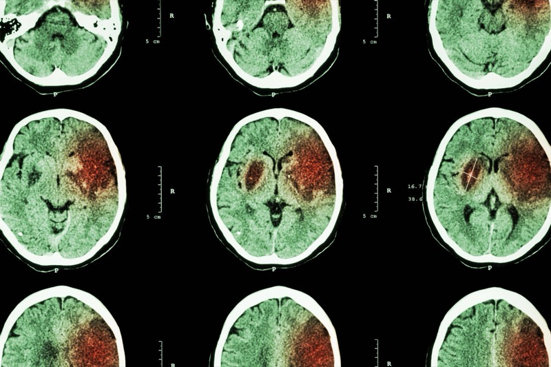
CT brain · ischemic infarct shows as a hypodense (dark) wedge · tap to expand
The Opening Case
One vignette. One vessel. Read the deficit pattern, name the artery.
Chart · ED · 72-year-old
A 72-year-old woman with atrial fibrillation is brought in after suddenly collapsing in her kitchen. On exam she has weakness of the right leg far worse than the right arm. The right side of her face is unaffected. She is incontinent of urine for the first time. Family notes she is flat, slow to respond, and seems “not herself.” Speech is preserved. Visual fields are full.
Which artery is occluded, and which side?
Left anterior cerebral artery (ACA). The leg sensorimotor strip lives on the medial wall of the hemisphere, in ACA territory, while the arm and face strips live laterally in MCA territory. Leg-dominant contralateral weakness with a spared arm and face IS the ACA fingerprint. Add the medial frontal lobe damage (urinary continence center, prefrontal cortex) and you get new urinary incontinence plus abulia, exactly what the family is describing. Atrial fibrillation tells you the mechanism: a clot launched from the left atrium, lodged in the left ACA.
The Willis
Inferior view. Frontal lobes up. Tap any artery to light up its territory and read the syndrome.
Inferior view · Circle of Willis · Public-domain trace
The Willis
Six vessels. Six districts. Tap a vessel to walk the territory it feeds and the syndrome it leaves behind.
Tap any artery
ARTERY
Name
Territory
Syndrome
MC Cause
Board Pearl
Cortical strokes are contralateral. Cortex sits above the medullary decussations, so a cortical injury hits the opposite side of the body, every time. If you see ipsilateral findings paired with contralateral findings (crossed deficits), think brainstem instead. That distinction sorts every cortical stem from every basilar stem in one breath.
The Four Syndromes
Territory, clue, cause, trap. One card each.
Artery 1 · ACA
Anterior Cerebral Artery (ACA)
Leg dominant. Medial frontal-parietal cortex.
Region
Anteromedial brain. Medial frontal lobe and medial parietal lobe. Sits on the inside wall of the hemisphere, against the falx.
Feeds
The leg portion of the motor and sensory strips (medial sensorimotor cortex). Supplementary motor area. The medial frontal continence center. Prefrontal cortex.
See It
Contralateral motor + sensory loss of the LOWER extremity, face and arm spared. New urinary incontinence. Flat affect, abulia, “not themselves.” Foot drop on the affected side.
MC Cause
Embolic. Cardioembolic from atrial fibrillation is the textbook source (clot leaves the left atrium, sails up the ICA, lodges in the ACA). Artery-to-artery embolism from an ICA atherosclerotic plaque is the runner-up.
Trap
Calling leg-dominant weakness “just a small stroke.” ACA hits the medial frontal lobe, so personality change and incontinence ride along with the motor pattern.
Artery 2 · MCA
Middle Cerebral Artery (MCA)
Most common cortical stroke. Face plus arm.
Region
Anterolateral brain. Lateral frontal lobe + lateral parietal lobe + lateral temporal lobe. The big slice that wraps the side of the hemisphere.
Feeds
Face and UPPER extremity portions of the sensorimotor strip. Broca area (superior division) and Wernicke area (inferior division) on the dominant side. Optic radiations through temporal and parietal lobes.
See It
Contralateral motor + sensory loss of FACE and UPPER extremity, leg spared. Gaze deviated toward the lesion. Contralateral homonymous hemianopia.
MC Cause
Atherosclerotic thromboembolism. Cardioembolic from atrial fibrillation. ICA plaque shooting a clot down into the MCA.
Parietal lobe involvement gives hemineglect (hemispatial neglect): the patient ignores the entire left side of the world. Will shave only the right cheek, eat from only the right side of the plate, draw a clock with all the numbers crammed on the right.
Non-dominant · hemineglect
Trap
Calling fluent jargon speech “intact language.” Wernicke aphasia is fluent but meaningless. The words flow; the meaning is gone. And do not miss Gerstmann: ask the patient to write, calculate, and name their fingers, not just to talk.
Artery 3 · PCA
Posterior Cerebral Artery (PCA)
Vision and memory. Macular sparing.
Feeds
Occipital cortex (vision). Medial inferior temporal lobe (memory). Thalamus. Splenium of corpus callosum.
See It
Contralateral homonymous hemianopia with macular sparing (the macula gets MCA collateral, so the central tip is preserved). Alexia without agraphia (dominant). Prosopagnosia (bilateral inferior temporal). Thalamic pain weeks later (Dejerine-Roussy).
MC Cause
Atherosclerosis of the vertebrobasilar system. Embolism from the basilar tip or vertebral artery plaque.
Trap
Forgetting the macula is spared. A homonymous hemianopia that takes out central vision too is more likely a parietal or occipital tumor than a clean PCA stroke.
Artery 4 · Basilar
Basilar Artery
Top of basilar · Locked-in.
Feeds
Brainstem (especially ventral pons), midbrain, thalamus, both occipital lobes through the PCAs that branch off its top.
See It
Top of basilar: bilateral visual loss, altered consciousness, oculomotor problems, memory loss. Locked-in (ventral pons): quadriplegic, no speech, no swallow, but FULLY awake. Only vertical eye movement and blinking preserved.
MC Cause
Vertebrobasilar atherosclerosis. Basilar tip embolism. Cardiac source throwing a clot through the vertebral system.
Trap
Calling a locked-in patient unresponsive and writing them off. They are aware of the room. Use vertical gaze and blinking to communicate.
The Homunculus Anchor
ACA owns the medial strip, so it owns the leg. MCA owns the lateral wedge, so it owns the face and arm. Tap to flip the body.
Why this works
The motor strip wraps the brain like a headband. The leg portion sits on the medial wall (ACA real estate). The face and arm portions sit on the lateral surface (MCA real estate). Same strip, different artery.
The M-Person
One stick figure. Two arteries. The torso is an M for MCA. The legs are a red column for ACA. Tap a stroke and watch what falls.
Mnemonic · Tap to strike the artery
The M-Person
Upper body shaped like the letter M (rose) is MCA territory: face plus upper extremity. The red column underneath is ACA territory: legs.
Why this sticks. The motor strip wraps the brain like a headband: medial side = leg, lateral side = face and arm. The M sits on the side of the head (lateral, MCA real estate). The legs hang in the middle (medial, ACA real estate). One figure, two arteries, the entire homunculus geography in a single picture.
MCA = M = anterolateral brain = face + upper extremity + (left) language or (right) neglect. ACA = red column = anteromedial brain = lower extremity + bladder + abulia.
Anton and Locked-in
Two board favorites. Both look bizarre. Both are cortical-vascular signatures.
Bilateral PCA · Anton syndrome
Cortical blindness with denial
Bilateral occipital infarcts blow out both visual cortices. The patient is completely blind, yet they insist their vision is fine and confabulate the room around them, bumping into furniture as they describe it. The visual cortex is gone, but the brain has not gotten the memo.
Pearl: a patient who walks into the wall and tells you what the wall looks like has Anton syndrome until proven otherwise. Bilateral PCA territory infarct.
Basilar pons · Locked-in
Aware. Trapped.
A ventral pontine infarct from basilar occlusion knocks out every motor pathway running through the pons (corticospinal and corticobulbar). Quadriplegic, no speech, no swallow. The dorsal pontine reticular formation is spared, so consciousness is fully intact. CN III in the midbrain is spared, so vertical gaze and blinking still work.
Pearl: a quadriplegic patient who blinks once for yes is locked-in, not clinical subjectose. Speak to them like a person who hears every word.
MCA Aphasia Map
Dominant (left) MCA territory. Tap a card to reveal the bedside pattern.
Superior division · Broca
Broca (expressive) aphasia
Frontal lobe operculum. Motor speech blueprint gone.
Non-fluent, effortful, telegraphic speech. The patient knows what they want to say and gets frustrated trying to push it out. Comprehension is preserved. Right face plus arm weakness rides along because the precentral gyrus motor strip sits right next to Broca. Repetition is impaired.
Tap to reveal
Inferior division · Wernicke
Wernicke (receptive) aphasia
Superior temporal gyrus. Language meaning gone.
Fluent jargon. Words pour out, but they do not connect to meaning. The patient does not know they are not making sense. Comprehension is shot, so they cannot follow commands. Often a right superior visual quadrant cut rides along (optic radiations through the temporal lobe). Repetition is impaired.
Tap to reveal
Full MCA · Global
Global aphasia
Both divisions. Almost no language at all.
Combined Broca plus Wernicke. The patient produces little or no meaningful speech and cannot understand questions. Right face plus arm plus often leg weakness rides along because the whole MCA territory is down. Repetition is impaired. This is the classic large left MCA infarct or proximal M1 occlusion.
Tap to reveal
Cross-reference
Deeper map of cortical localization, including how the arcuate fasciculus turns this into conduction aphasia, lives on the cerebral cortex page.
The Discriminator Card
Read the stem. Match the pattern. Name the vessel.
Stem cue
Vessel
Face plus arm weakness, leg spared. Aphasia or neglect.
MCA
Leg weakness greater than arm. Face spared. New incontinence, flat affect.
ACA
Contralateral homonymous hemianopia with macular sparing. Alexia without agraphia.
PCA
Quadriplegia. Awake. Vertical gaze and blinking only.
Basilar (locked-in)
Cortical blindness, patient denies it.
Bilateral PCA (Anton)
Bilateral visual loss + altered consciousness + oculomotor signs.
Top of the basilar
One-line rule
Face plus arm goes MCA. Leg alone goes ACA. Hemianopia with macular sparing goes PCA. Quadriplegic but awake goes basilar.
Watershed Areas
The end-zones of arterial supply. The last pixels of brain to get blood. First to fall when pressure drops.
The Core Clue
A watershed area is the border between two arterial territories. It is the last neighborhood at the end of the water main. When systemic pressure crashes, the rest of the brain still gets perfusion, but the watershed is left dry. ACA / MCA border and MCA / PCA border are the two zones to know.
Lateral view · two watershed zones
ACA / MCA border
Man-in-a-barrel syndrome
The ACA / MCA watershed sits right where the motor strip transitions from leg (medial) to face and arm (lateral). When this strip falls out, the patient loses proximal motor function on both arms (and sometimes proximal legs) while the distal hands and feet are spared. The result looks like a person stuck in a barrel: shoulders pinned at their sides, hips locked, but the hands and feet still wiggle.
Proximal > distal weakness
MCA / PCA border
Visual + cognitive deficits
The MCA / PCA watershed sits over the posterior parietal and lateral occipital cortex. When this strip falls out, the patient develops higher-order visual and cognitive problems: visual disorientation, simultanagnosia (cannot see the whole picture), trouble naming objects by sight, and reading deficits. Vision is technically “intact” but the brain cannot use it.
Visual association + spatial
MC Scenario
Severe hypotension, cardiac arrest with delayed return of spontaneous circulation, hemorrhagic shock, prolonged surgery with low MAP, or bilateral carotid stenosis. The watershed zones are vulnerable because they sit at the end of the arterial line. When the pressure head drops, the rest of the brain still gets enough flow, but the end-zones run dry first.
Cross-reference
Watershed strokes are bilateral and proximal. Cardiac-arrest survivor with new shoulder-girdle weakness and preserved hand grip is man-in-a-barrel until proven otherwise.
Localize the Stroke
Read the deficit pattern. Pick the vessel. The answer unfolds under your choice.
The Rule
Each cortical artery owns a specific piece of brain real estate. Kill the artery: that district goes dark. The deficit pattern IS the localization.
Contralateral LEG greater than arm and face weakness. Cortical sensory loss in the leg. No aphasia. No neglect. Which vessel?
Anterior Cerebral Artery (callosomarginal branch)
Correct. The ACA feeds the medial hemisphere surface, where the leg motor and sensory strips live on the inside wall. Leg-dominant deficit with cortical sensory loss and a spared face is the ACA fingerprint. The arm and face live laterally in MCA territory and escape.
Discriminator: leg greater than face and arm, cortical sensory loss in the leg = ACA.
Not MCA
MCA feeds the LATERAL cortex: face and arm dominate. An MCA stroke produces face plus arm greater than leg. When the leg is worse than the face and arm, the lesion is on the MEDIAL surface, which is ACA territory. Rethink the hemisphere side.
Not PCA
PCA feeds the occipital lobe and medial temporal lobe. Its signature is a visual field cut with macular sparing, not motor weakness. There is no motor strip in PCA territory.
Not Lacunar
A lacunar infarct from the lenticulostriate arteries hits the internal capsule and produces EQUAL weakness of face, arm, and leg with NO cortical sensory findings. Leg-only deficit with cortical sensory loss is a cortical stroke, not a lacunar one.
Contralateral FACE and ARM greater than leg weakness. Dominant hemisphere: aphasia. Nondominant hemisphere: hemineglect. Which vessel?
Middle Cerebral Artery
Correct. The MCA feeds the lateral cortex: face area, hand and arm area, Broca and Wernicke language cortex (dominant left hemisphere), and spatial attention cortex (nondominant right). Face plus arm greater than leg, with language or attention deficit, is the MCA pattern.
Discriminator: face and arm greater than leg, with aphasia (dominant) or neglect (nondominant) = MCA.
Not ACA
ACA feeds the MEDIAL surface. Leg greater than arm and face is the ACA pattern. Face plus arm greater than leg points to the LATERAL cortex, which is MCA territory.
Not PCA
PCA territory does not include lateral motor or sensory cortex. PCA stroke produces a visual field cut, not face-arm motor deficits with aphasia. If there is arm weakness plus language breakdown, this is MCA.
Not Lacunar
Lacunar infarcts produce pure motor or pure sensory deficits with equal distribution across face, arm, and leg. Aphasia and neglect require cortex. If cortical signs are present, this is cortical MCA, not a lacunar internal capsule event.
Homonymous hemianopia with macular sparing. Alexia without agraphia. No arm or leg weakness. Which vessel?
Posterior Cerebral Artery
Correct. The PCA feeds primary visual cortex (calcarine fissure, occipital lobe). Its stroke signature is contralateral homonymous hemianopia with macular sparing, because the macula has dual supply from both PCA and MCA at the occipital pole. Alexia without agraphia is a PCA variant where the infarct plus splenium of the corpus callosum disconnects visual cortex from language cortex: the patient cannot decode written symbols, but motor writing is intact.
Discriminator: visual field cut with macular sparing, alexia without agraphia, no motor deficit = PCA.
Not MCA
MCA produces lateral cortex signs: face plus arm weakness and language deficits. A pure visual field cut without motor deficit does not fit the MCA pattern. Visual cortex is in the occipital lobe, which is PCA territory.
Not ACA
ACA territory does not include visual cortex. ACA strokes produce leg-dominant weakness and medial frontal signs. A pure visual field cut with no motor deficit points to occipital cortex, which is PCA.
Not Basilar
Basilar top occlusion can cause BILATERAL visual loss and Anton syndrome (denial of blindness). A unilateral hemianopia with preserved motor function and intact consciousness is a unilateral PCA stroke, not a basilar event.
Pure motor hemiplegia: face, arm, and leg ALL equally weak. No sensory loss. No cortical signs. Hypertensive patient. Which vessel?
Lacunar Infarct (lenticulostriate arteries)
Correct. The lenticulostriate arteries are tiny perforating vessels off the MCA mainstem that drill into the internal capsule. Hypertension causes lipohyalinosis, narrowing these vessels. When one occludes, the corticospinal and corticobulbar tracts passing through the capsule fall together: face, arm, and leg all drop equally. No sensory loss (sensory tracts travel separately through the posterior limb). No aphasia or neglect (cortex is intact).
Discriminator: equal face plus arm plus leg pure motor, no cortical signs, hypertensive = lacunar infarct.
Not the classic MCA cortical pattern
MCA cortex infarcts produce face plus arm greater than leg with cortical signs (aphasia or neglect). Equal deficit across all three without any cortical signs points to the internal capsule (white matter), not MCA cortex. The lenticulostriate arteries branch from the MCA but supply deep structures, not cortex.
Not ACA
ACA produces leg-dominant weakness from the medial motor strip. Equal involvement of face, arm, and leg cannot come from ACA territory because the arm and face live laterally in MCA territory, far from the ACA distribution.
Not PCA
PCA territory is the occipital lobe and medial temporal lobe, neither of which carries the corticospinal tract. Pure motor hemiplegia without visual loss is a subcortical internal capsule event, not a PCA stroke.
Quiz
Eight vignettes. Eight reads. Tap to commit, read the explanation.
Medically reviewed by Kaitlyn Cocuzzo, MD and Fatima Ali, DO · Last reviewed June 2026
Bone Wizardry is an independent educational resource for visual learning in the medical sciences. It is not affiliated with, endorsed by, or sponsored by any licensing or examination board, contains no real or recalled examination questions, and does not guarantee any educational or examination outcome.