T1-T12. Where spinal curves group. Where visceral reflexes are loudest. Where OMM has the most clinical medicine traps.
Opening Challenge
A patient has a thoracic vertebra that is neutral, sidebent LEFT, and rotated RIGHT. There is no flexion or extension restriction. Which of the following best describes this somatic dysfunction?
scroll to begin
Section 1
Fryette's Laws
The two rules that govern all thoracic somatic dysfunction diagnosis. Get these wrong, every notation falls apart.
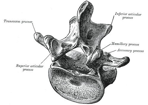
Thoracic vertebra · superior view
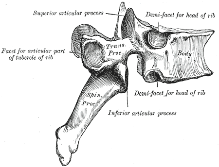
Thoracic vertebra · lateral view
First thoracic vertebra · T1
Twelfth thoracic vertebra · T12
Palpatory Diagnosis
TART: the four findings of somatic dysfunction
T = Tissue texture change (bogginess, ropiness, warmth acutely; cool, fibrotic, stringy chronically). A = Asymmetry (one transverse process sits more posterior than its pair). R = Restriction of motion (the segment will not move into one direction). T = Tenderness (the patient's report of pain on palpation; the most subjective).
Any ONE of the four can be present, but the diagnosis of somatic dysfunction needs at least one objective finding. Tenderness alone is not enough.
Landmark Reading
Posterior rib angle palpation
The rib angle is the most posterior, most lateral curve of the rib, roughly two finger-widths lateral to the transverse process tips. It is the easiest place to feel a rib lag or lead during respiration.
Inhalation dysfunction: the rib stays elevated, will not drop on exhalation. The most caudad rib of a group holds the key. Exhalation dysfunction: the rib stays depressed, will not rise on inhalation. The most cephalad rib holds the key.
Rib angles also sit directly over the paravertebral sympathetic chain, which is why rib-angle pressure is used for sympathetic modulation.
Rule of Threes: spinous process vs transverse process level
T1-T3Spinous process is at the level of its own transverse process (same level).
T4-T6Spinous process is half a segment below its own transverse process.
T7-T9Spinous process is a full segment below its own transverse process.
T10Behaves like T7-T9: a full segment below.
T11Behaves like T4-T6: half a segment below.
T12Behaves like T1-T3: at its own level again.
◆
Why the Rule of Threes matters: to find the transverse processes of a given segment you palpate from the spinous process and shift up by the offset. If you mis-locate the transverse processes, you read rotation at the wrong level and your whole diagnosis collapses. Count down in threes: 1, half, full, then mirror back up: full, half, level.
Law I · Type I · Group
NEUTRAL spine OPPOSITE directions
When vertebrae are in neutral position (no F or E restriction), sidebending and rotation move in opposite directions.
Multiple vertebrae are involved as a group curve.
Sidebent Right → Rotated Left
Written: N SR RL
Law II · Type II · Non-Group
FLEXED or EXTENDED SAME side
When a vertebra is flexed or extended (non-neutral), sidebending and rotation move to the same side.
Single vertebra involved.
ERSR = Extended, Rotated Right, Sidebent Right
FRSL = Flexed, Rotated Left, Sidebent Left
★
Memory anchor: Law I = Neutral = Opposite (N and O are both round-ish, no sharp edges). Law II = F or E = same side. Three words: Neutral Opposites, Non-neutral Sames.
Type I · Group
Group Curve Mechanics
Position: Neutral (no F/E restriction) Rotation vs sidebending: Opposite directions Number of vertebrae: Multiple (group) Cause: Postural compensation Onset: Chronic, gradual
Example: T4-T8 as a group, neutral, sidebent right, rotated left. Written: T4-T8 N SR RL
Type II · Non-Group
Single Vertebra Mechanics
Position: Flexed OR Extended (non-neutral) Rotation vs sidebending: Same side Number of vertebrae: Single Cause: Traumatic or acute injury Onset: Often acute
Fryette's Law III: Initiating motion in one plane reduces available motion in all other planes. This is the mechanical basis for why restrictions compound, and why you treat the most restricted segment first.
⚠
clinical medicine trap: A vertebra described as "restricted in flexion" is STUCK IN EXTENSION (can't flex). A vertebra "restricted in extension" is STUCK IN FLEXION (can't extend). The restriction names the movement it cannot perform, not the position it's locked in. Always flip it.
1 / 6
Section 2
Notation System
How to read and write thoracic SD diagnoses. Every clinical medicine OMM question is either asking you to decode a notation or pick the right one.
Notation Key
F / E / NF = Flexed (stuck in flexion) · E = Extended (stuck in extension) · N = Neutral
R / LR = rotated right · L = rotated left
SR / SLSR = sidebent right · SL = sidebent left
Reading Examples
T5 ERSRType IIT5 is extended (stuck in extension), rotated right, sidebent right. Same-side = Type II. To treat: engage in flexion, left rotation, left sidebending.
T5 FRSLType IIT5 is flexed (stuck in flexion), rotated left, sidebent left. Same-side = Type II. To treat: engage in extension, right rotation, right sidebending.
T4-T8 NSRRLType IGroup curve T4-T8, neutral, sidebent right, rotated left. Opposite-side = Type I. Treat by addressing the apex of the group curve.
T6 ERSLType IIT6 extended, rotated left, sidebent left. Same-side = Type II. Visceral concern: T6 associates with gallbladder and stomach.
Segment Diagnostic Builder · set the motion, read the diagnosis
Sagittal position (flexion / extension)
Rotation
Sidebending
Type I · Fryette's Law I
NSRRL
★
Read it back yourself: set the model to Extended, Right rotation, Right sidebending. Same-side coupling lights up Type II and the name reads ERRSR. Now flip sidebending to Left while staying Extended: the tool refuses to call it a clean Fryette lesion, because non-neutral segments must couple to the same side. The model only produces lesions the spine can actually make.
◆
How to find the notation from the exam: The vertebra moves freely into one direction and is restricted in the other. Restricted in extension = stuck in flexion = starts with F. Rotated and sidebent = check same vs opposite to determine Law I or Law II.
Mobility Test
Finding the Diagnosis
Patient seated. Physician places fingers on transverse processes bilaterally at the level being tested.
Step 1: Flex the spine. Does the TP become symmetric or asymmetric? Step 2: Extend the spine. Same question. Step 3: Where the asymmetry is worse = position of dysfunction (F or E). Result: Rotation is always toward the side where the TP is more posterior (more prominent).
Treatment Logic
Barrier Concept
HVLA moves through the restrictive barrier (the direction it won't go).
ERRSR: Stuck in extension, rotated right, sidebent right.
Barrier is in: flexion, left rotation, left sidebending.
Thrust: into flexion + left rotation + left sidebend.
FRLSL: Stuck in flexion, rotated left, sidebent left.
Thrust: into extension + right rotation + right sidebend.
1 / 6
Section 3
Thoracic HVLA Techniques
Three core techniques. Each targets a different region of T1-T12. Know which technique goes where and why.
T4-T10 · Supine
Dog Technique (Crossed Arm)
Patient position: Supine, arms crossed over chest. Physician: Passes arm under the patient's axilla. Places the opposite hand at the target transverse process, palm up under the patient's back. Action: Rolls patient toward physician to load the contact hand against the TP. Leans body weight through the patient's crossed arms to deliver the thrust on exhalation.
Best for: Mid-thoracic T4-T10. Good for most Type II ERS/FRS dysfunctions in this range.
T1-T4 · Seated
Kirksville Crunch
Patient position: Seated, interlocks fingers behind the neck (prisoner position). Physician: Stands behind patient. Reaches through the patient's arms from behind. Thenar eminences contact the transverse processes at the dysfunctional level. Action: Patient exhales. Physician delivers an anterior thrust through the thenar contacts on exhalation.
Best for: Upper thoracic T1-T4. The neck-interlaced position locks out the cervical spine and focuses force at the target level.
T6-T12 · Prone
Prone Thoracic HVLA
Patient position: Prone on the treatment table. Physician: Applies thumb or pisiform contact to the transverse process of the dysfunctional vertebra. Body positioned lateral to the patient. Direction: Thrust directed anteriorly and slightly cephalad at approximately 45 degrees.
Best for: Lower thoracic T6-T12. Especially useful when supine positioning is contraindicated.
Cavitation
The Pop Explained
The audible pop during HVLA is caused by nitrogen gas cavitation within the synovial fluid of the zygapophyseal joint. Rapid intra-articular pressure drop during joint gapping allows dissolved nitrogen gas to flash out of solution.
This is NOT: ligament tearing, bone cracking, or a sign of damage. It is the same mechanism as knuckle cracking.
Post-procedure soreness: Normal. Similar to post-exercise soreness. Resolves within 24-48 hours.
⚠
Thrust direction rule: For any ERSR lesion (rotated right, extended), the physician engages the barrier in flexion, left rotation, left sidebending, then thrusts through that barrier. The technique matches the notation. Identify the position of dysfunction, then do the opposite.
⚠
HVLA contraindications: Osteoporosis, fracture, osteomyelitis, malignancy involving the spine, active disc herniation with neurological deficit, anticoagulation, vertebrobasilar insufficiency (cervical), severe spondylosis with instability. Active visceral infection at the corresponding spinal level is a relative contraindication.
1 / 6
Section 4
Viscerosomatic Reflexes
Thoracic SD is not always musculoskeletal. The board wants you to link spinal levels to organs and reason backward from spine to viscera.
Spinal Level
Associated Organs
Clinical Clue
T1-T4
Heart (T1-T5), Upper extremity (C8-T1 for hand/forearm), Lung apex
Chest pain, palpitations, arm numbness
T2-T7
Lungs, Upper GI, Esophagus
Dyspnea, dysphagia, regurgitation
T5-T9
Heart (T1-T5), Stomach, Esophagus
Epigastric pain, heartburn, cardiac referred pain
T6-T9
Gallbladder (right-sided), Liver, Stomach
Right upper quadrant pain, jaundice, nausea after fatty meals
Pelvic pain, dysmenorrhea, testicular torsion referred pain
★
Viscerosomatic rule: When a patient has thoracic SD at levels linked to an organ AND has visceral symptoms, the spine is likely a secondary finding of visceral disease. Treat the organ first. Spinal SD may resolve when the underlying cause is addressed. Never manipulate without ruling out visceral pathology first.
Classic Pattern
Right T4-T6 SD + Chest Pain
Right-sided T4-T6 somatic dysfunction in a patient with chest pain should prompt cardiac workup. T1-T5 is the cardiac sympathetic level. The SD is the secondary finding of myocardial ischemia or pericarditis, not the primary problem.
Rule: Treat the spine AND work up the cardiac cause in parallel. Do not dismiss chest pain as musculoskeletal because there is a spinal finding.
Classic Pattern
T10 SD + Periumbilical Pain
T10 is the classic viscerosomatic level for early appendicitis. The periumbilical pain of early appendicitis (before peritoneal involvement) is visceral pain referred via T10 sympathetic afferents.
Somatic findings at T10 in a patient with fever and periumbilical pain: think appendicitis. The SD at T10 is the somatic expression of the visceral injury. Treat the appendix.
ⓘ
Somatovisceral reflex (reverse direction): Spinal SD can also influence organ function via the same reflex arc in reverse. Sustained thoracic SD at T5-T9 may contribute to gastric hypermotility or cardiac arrhythmia. Treating the spine can improve visceral function. Both directions are tested in clinical practice.
Walk a segment to its diagnosis
Answer before you reveal. Each step locks once you commit.
Step 1. On flexion the transverse process asymmetry at T8 worsens; on extension it improves. What is the sagittal position of the dysfunction?
Step 2. The right transverse process of T8 is more posterior. Rotation and sidebending therefore go where, and how is the lesion written?
Memory hooks
Tap each card to bring it into focus.
Neutral Opposites, Non-neutral Sames
Law I is Neutral and Opposite (both round letters, no sharp edges, like a smooth group curve). Law II is non-neutral and SAME side. Three words carry both laws.
Tap to reveal
Restriction names what it cannot do
Restricted in flexion = stuck in EXTENSION. Restricted in extension = stuck in FLEXION. The label is the door that is locked, so the position is the opposite room. Always flip it before you write F or E.
Tap to reveal
Posterior process points at the rotation
The transverse process that sits more posterior is on the side the body rotated toward. More posterior on the right = rotated right. Then couple sidebending: same side if non-neutral, opposite side if neutral.
Tap to reveal
Green light to fatty meals: T6 to T9 on the right
Right-sided T6 to T9 somatic dysfunction plus right upper quadrant pain after fatty food points at the gallbladder before the spine. Treat the organ, then reassess the segment.
Tap to reveal
1 / 6
Section 5
Case Challenge
One case, three reveals. Tap each question to uncover the reasoning. Cover the answer and think first.
Case Vignette
A 52-year-old man presents with mid-back pain for 3 weeks. He also reports indigestion and mild epigastric discomfort after meals. Structural examination reveals T6 rotated right and sidebent right in an extended position. Range of motion testing shows that T6 becomes more restricted when moved into extension. There is no history of trauma.
Diagnosis
Type II (Non-group) somatic dysfunction: T6 ERSR
The single vertebra is stuck in an extended position (restricted when moved into more extension = it is already AS FAR into extension as it will go, and motion testing confirms this). Rotated right and sidebent right = same side = Type II mechanics (Law II: F or E yields same-side coupling).
T6 is within the T5-T9 (stomach, esophagus) and T6-T9 (gallbladder, liver) viscerosomatic zones.
Combined with his epigastric discomfort and indigestion, this warrants evaluation for peptic ulcer disease, GERD, or gallbladder pathology (biliary colic, cholecystitis) before attributing his symptoms to musculoskeletal causes.
The spinal SD at T6 is likely a secondary somatic manifestation of an upper GI or hepatobiliary process.
Treatment
T6 ERSR: stuck in extension, rotated right, sidebent right.
Engage the barrier: the opposite of the position of dysfunction.
Flexion (opposite of extension) + left rotation (opposite of right) + left sidebending (opposite of right).
Technique choice: T6 is mid-thoracic, so the Dog Technique (crossed arm, supine) is most appropriate. Position the physician's hand under the T6 transverse process. Flex the patient's thoracic spine slightly, introduce left rotation and left sidebending, then thrust on exhalation through the flexion-left rotation-left sidebending barrier.
Clinical note: Given the potential visceral involvement, OMM is adjunctive. Address the underlying GI or hepatobiliary cause first.
1 / 6
Section 6
Board Drill
Four clinical questions. No multiple-select. No copied content. Work through each before revealing the explanation.
Question 1 of 4
During a structural examination, a physician identifies that T4 through T7 form a group curve. The vertebrae are in a neutral position with no flexion or extension restriction. The entire group is sidebent to the LEFT.
According to Fryette's Law I, toward which direction should the vertebral bodies be rotated?
Fryette's Law I: Neutral vertebrae in a group couple sidebending and rotation in opposite directions. Group sidebent LEFT means bodies rotate RIGHT. Written as: T4-T7 N SL RR.
Good instinct if you chose A: same-side coupling does happen in thoracic vertebrae, but only under Law II when there is a flexion or extension restriction. C is wrong because the neutral position is exactly what defines Law I mechanics. D is wrong because all thoracic group curves include a rotational component by definition. Think of a parking lot where all the cars angled the same way without any brakes applied: that is the compensating group (Law I). One car with its wheel turned and brake stuck is the single-segment problem (Law II). Break it down: Law I = neutral group + opposite coupling (sidebend and rotation in opposite directions); same-side coupling belongs to Law II only.
Question 2 of 4
A physician identifies a somatic dysfunction at T8. Structural exam shows: T8 is restricted in extension (moves freely into flexion), rotated left, and sidebent left. The dysfunction becomes more pronounced when T8 is tested in extension.
How should this dysfunction be documented?
T8 FRSL. "Restricted in extension" means T8 cannot extend freely. It is stuck in flexion. Restricted in extension = position is flexed. Rotation and sidebending are both left = same side = Type II (non-neutral).
Good instinct if you chose ERSL (option C): left rotation and left sidebending do fit ERSL notation, but ERS means stuck in EXTENSION and unable to flex. This patient cannot extend freely, which means they are stuck in flexion. The critical decode: "restricted in X" = cannot do X = stuck in the OPPOSITE of X. Option A (NSRR) contradicts the F/E restriction. Option D is neutral notation, also wrong. Think of a gate stuck closed on one side: restricted from opening to the right means it is jammed on the left side. Restricted in extension = jammed in flexion. Break it down: "restricted in extension" = T8 is stuck in FLEXION (FRS notation); rotation and sidebend to the same side = Type II; FRSL = flexed, left rotation, left sidebend.
Question 3 of 4
A physician uses the seated Kirksville Crunch technique to treat a T2 somatic dysfunction. After the technique, the patient reports immediate relief of her neck stiffness. Reassessment shows the dysfunction has resolved. During the procedure, the patient heard a loud pop and now reports mild soreness specifically at T2.
The audible pop heard during HVLA is caused by which mechanism?
Nitrogen gas cavitation within the zygapophyseal joint synovial fluid. Rapid intra-articular pressure drop during joint gapping allows dissolved nitrogen to flash out of solution as gas bubbles. The same physics as knuckle cracking. The sound does NOT indicate tearing or fracture.
Good instinct if you chose ligamentous tearing: the loud pop and subsequent soreness make it sound like something tore. Ligamentous tearing would cause severe pain, instability, and worsening, not relief with mild soreness that clears in 24-48 hours. Endplate fracture requires grossly excessive force and presents with acute worsening. Tendons do not snap over thoracic transverse processes. Mild post-procedural soreness at the treated level is normal and expected. Think of popping a soda can: the pop is gas being released under pressure, not the can cracking. The synovial fluid releases dissolved nitrogen gas when joint pressure drops rapidly, exactly the physics of knuckle cracking. Break it down: HVLA pop = nitrogen gas cavitation in synovial fluid from rapid joint-gapping pressure drop; this is the same mechanism as cracking knuckles and does NOT indicate tearing, fracture, or injury.
Question 4 of 4
A 38-year-old woman presents with right-sided lower thoracic pain that began after a coughing episode. Structural examination reveals T9 ERSR. She also reports right flank pain, fever of 38.4°C, and nausea. Urine dipstick shows 3+ leukocyte esterase and 2+ nitrites.
What is the MOST appropriate next step in management?
Pyelonephritis with viscerosomatic reflex. The renal viscerosomatic zone is T10-L1; T9 sits at its upper margin. The patient has pyelonephritis confirmed by UA (+ LE + nitrites), fever, and CVA-distribution flank pain. The thoracic SD at T9 is a viscerosomatic reflex from the kidney infection, not a primary musculoskeletal problem. Antibiotics are the definitive treatment.
Good instinct if you chose HVLA: T9 ERSR is a real finding and treating it seems efficient. The problem is that the spinal dysfunction is a viscerosomatic reflex from the kidney infection, not a primary structural problem. Treating the reflex without treating the infection is like silencing a smoke alarm instead of finding the fire. Disc herniation does not cause fever or a positive UA. Spinal X-ray does not address the infection and delays appropriate care. Think of the spine as a warning light on the dashboard: you do not fix the car by unplugging the light while the engine is still overheating. Find the source, treat the source. The spinal SD typically resolves on its own once the infection clears. Break it down: T9-L1 is the renal viscerosomatic zone; spinal SD in this region + fever + positive UA = pyelonephritis with viscerosomatic reflex; treat the infection first; HVLA over an active infection site is relatively contraindicated.
1 / 6
Section 7
Board Walkthrough
Original clinical vignettes, one at a time, shuffled and never repeated until the bank exhausts. Right-click or long-press an option to cross it out. Double-click or double-tap to highlight.
1 / 6
Medically reviewed by Fatima Ali, DO and Kaitlyn Cocuzzo, MD · Last reviewed June 2026
Bone Wizardry is an independent educational resource for visual learning in the medical sciences. It is not affiliated with, endorsed by, or sponsored by any licensing or examination board, contains no real or recalled examination questions, and does not guarantee any educational or examination outcome.