Memory Hooks
Lock it in
Tap each card to reveal the hook.
Where does Charcot-Bouchard bleed? Deep or surface?
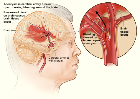
HTN is a wrecking ball that only reaches the basement. Lenticulostriate arteries are the deep-basement pipes. HTN pressure tunnels down into the brain's core: putamen, thalamus, pons, cerebellum. The cortex is upstairs, safe. Amyloid angiopathy is the upstairs tenant. If the bleed is in the cortex, HTN is not your answer.
Tap to reveal
Why is CT bright white (hyperdense) for hemorrhage but dark for ischemic stroke?
Blood clot = dense protein (hemoglobin) = stops X-rays = white on CT. Think of it like a lead apron on the beam. Ischemic stroke = water-swollen tissue = low density = dark. Hyperdense is your red flag for bleeding. You never need contrast to spot fresh blood. Hyperdense = it just happened.
Tap to reveal
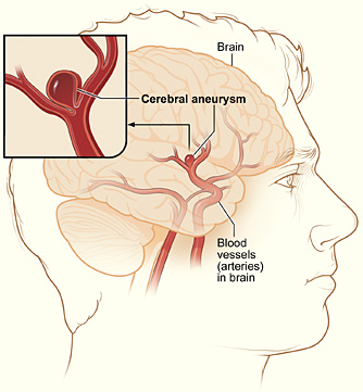
Why is angiography always negative for Charcot-Bouchard?
These microaneurysms are 50-200 microns wide. A human hair is 70 microns. Angiography resolves down to about 1-2 millimeters. You are trying to see a single hair strand with a camera designed for ropes. A negative angiogram in an elderly hypertensive with a deep bleed is NOT reassuring. It confirms the diagnosis. The absence of a visible aneurysm IS the finding.
Tap to reveal
What is lipohyalinosis and why does it matter?
Lipohyalinosis is what chronic HTN does to small arteries: smooth muscle dies and is replaced by hyaline fibrous material. The wall goes from elastic rubber to brittle glass. A rubber pipe stretches under pressure. A glass pipe cracks. Same pathology causes both lacunar infarcts (vessel occludes) and Charcot-Bouchard hemorrhage (vessel ruptures). Same cause, two endings.
Tap to reveal
Berry aneurysm vs Charcot-Bouchard: one sentence each
Berry = saccular, circle of Willis, subarachnoid blood, worst headache of life, angiography positive. Charcot-Bouchard = microaneurysm, deep penetrating arteries, parenchymal blood, chronic HTN, angiography negative. Big junction vessels = big, visible aneurysm = subarachnoid. End arteries = small, deep, invisible microaneurysm = parenchymal. Opposite ends of the vascular tree.
Tap to reveal
Clinical Images
Radiology & Anatomy
Tap any image for the full view.
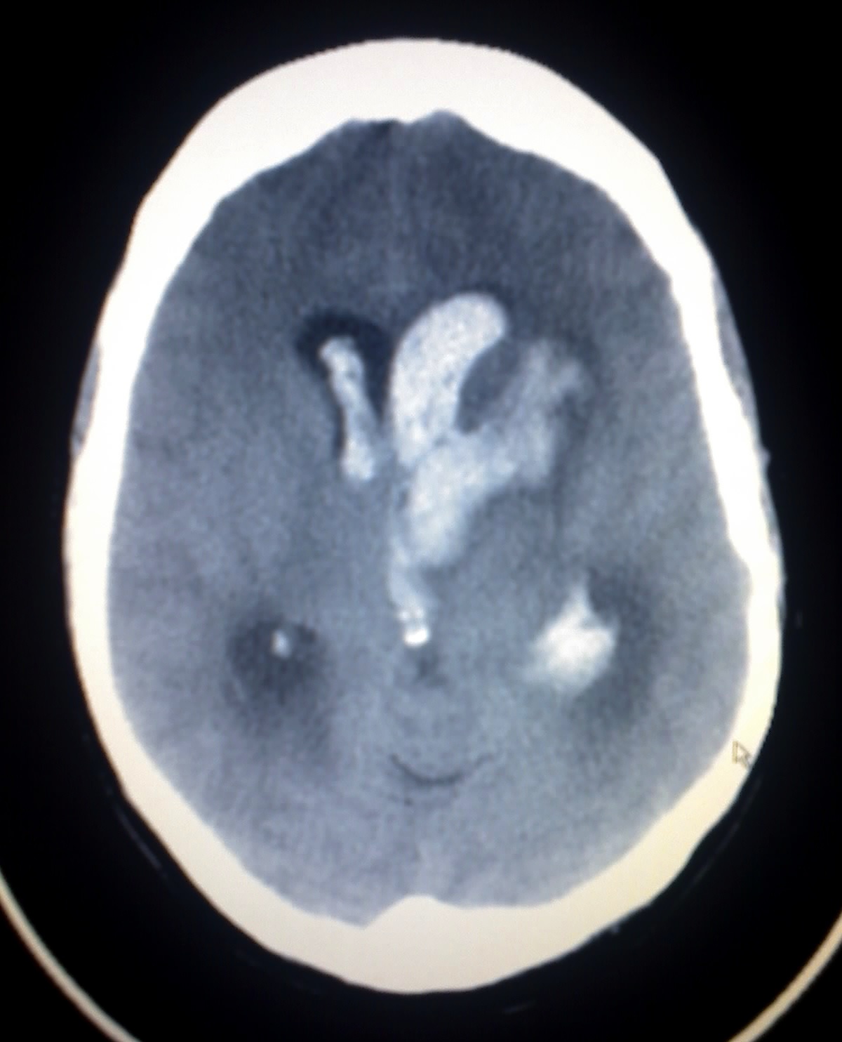
CT: Hyperdense Intracerebral Bleed
Hemorrhagic Stroke Anatomy
Cerebral Aneurysm Anatomy (NIH)
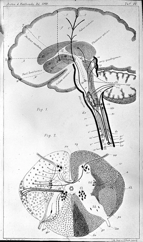
Basal Ganglia Connections
4 of 5